
Abstract
Introduction:
A total endoscopic approach to ventral hernia repair has advantages over conventional laparoscopy. The intraperitoneal cavity is not accessed and the extraperitoneal mesh placement minimizes the risk of adhesion formation. Also, chronic pain may be reduced since mesh fixation is not needed. Hernias with incarcerated content present a surgical challenge because of the possibility of visceral injury while reducing the contents of the hernia. By opening the posterior rectus sheath, the management of incarcerated contents during an endoscopic extraperitoneal approach is feasible without risk of visceral injury. This video shows this extraperitoneal technique for an incarcerated hernia.
Methods:
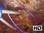
A 67-year-old man presented to the emergency room with abdominal distension and a bulge along a midline laparotomy scar. He had a history of a midline laparotomy for a right inguinal hernia that required intestinal resection and a laparoscopic repair of a left inguinal hernia. On physical examination, he presented with an M3W2 incisional hernia from the EHS classification (European Hernia Society—M3 = medial, periumbilical; W2 = width 4–10 cm) of 6 cm in diameter, partially reducible. An abdominal CT showed a left periumbilical defect of 6.2 cm width × 7.5 cm length with small bowel loops. A totally endoscopic retromuscular ventral hernia repair (e-TEP) was performed. A 2 cm incision below the left costal margin exposed the anterior left rectus sheath and this was incised along its lateral border. The retromuscular space was created using a balloon trocar. Two, one 5 mm and one 10 mm trocars were inserted into the left hypochondrium and left iliac fossa, respectively. The medial aspect of the posterior left rectus sheath was incised, and the posterior right rectus sheath was identified by incising it along its medial border. The hernia sac was opened, and the contents were reduced to fully expose the entire retromuscular space. The posterior sheaths were reapproximated to the midline using continuous nonabsorbable barbed suture, repairing the posterior aspect of the hernia defect at the same time. The anterior sheaths were approximated to reconstruct the midline. A lightweight polypropylene mesh was inserted through the 10 mm trocar and extended to completely cover the retromuscular space. The mesh was fixed with glue and a suction drain was inserted.
Results:
His postoperative course was uneventful. The drain was removed, and the patient was discharged on the first postoperative day. After 8 months of follow-up, the patient remains asymptomatic without a recurrent defect on CT scan.
Conclusions:
A totally endoscopic extraperitoneal approach to an incarcerated hernia with intestinal contents may be performed without visceral injury. This approach minimizes adhesion formation and reduces pain since mechanical fixation was not required.
No competing financial interests exist.
Runtime of video: 9 mins 0 secs


Get full access to this article
View all access options for this article.