
Abstract
Introduction:
During laparoscopy all viscera should be viewed. This is particularly true for the upper abdomen regarding gynecologic diseases such as ovarian cancer and endometriosis. Frequently, variants and abnormal findings of the liver occur. This video entails a tutorial video to facilitate assessment of such incidental liver findings.
Methods:
In the tertiary University Women's Hospital, Bern, Switzerland, video sequences of 29 patients with liver findings were collected during laparoscopic surgeries. The video material was discussed and interpreted with a hepatologist. A tutorial video of 29 liver findings and their interpretation is included. The local IRB statement is present (Kantonale Ethikkommission Bern, Req-2019-01004).
Results:
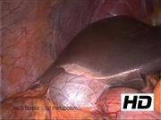
Surgeons performing laparoscopic operations should be familiar with such findings to allow for intraoperative decision-making. In summary, the video deals with the following findings: fibrosis is frequently found and work-up of these patients includes elastometry, biopsy, fat metabolism, alcohol consumption, and viral hepatitis. Patient number seven in the video shows a dilated hiatus of the falciform ligament, which possibly resulted from dilated embryonic vessels. Fatty liver is another common finding and shows histologic inflammation in about 10% of patients. The inflammation is divided into alcoholic and nonalcoholic steatohepatitis (ASH/NASH) with a cumulative risk of cirrhosis and hepatocellular cancer. Hemangioma are benign tumors that can be huge (e.g., patient number 14 in the video) but are typically asymptomatic. Hepatic cysts are similar and rarely cause any symptoms unless they compress adjacent organs. Patients 21 and 22 have peritoneal ovarian cancer metastases, liver metastases, and chemotherapy-associated steatohepatitis (CASH). The next patient shows diaphragmatic endometriosis, followed by a patient with “von Mayenburg complexes,” which are known to be biliary hamartoma. Focal nodular hyperplasia may enlarge with substantial dimensions and work-up with magnetic resonance imaging (MRI) shows contrast uptake of these lesions. If no contrast uptake occurs this argues for a lesion that may be consistent with an adenoma. If an adenoma is diagnosed, then oral contraceptives should be ceased and surgical resection may be necessary. The video concludes with trocar injuries to the liver after placement at the Palmer's point below the twelfth rib in the midclavicular line. Except from further investigation of the lesions no action had to be taken. The last sequence shows ascites in a patient with suspected ovarian cancer. During laparoscopy a cirrhotic liver was seen and found to be the cause of the ascites.
Conclusions:
In laparoscopic surgery, frequent variants and abnormal findings occur not only in the pelvis. This tutorial video of 29 patients shows liver findings that surgeons may confront. The video emphasizes the importance of viewing the whole abdominal cavity during every laparoscopy.
Selected Case Vignettes and Indications for the Respective Surgeries:
Patient #2 was operated to correct a symptomatic isthmocele of the uterus. Lateral to the falciform ligament a fatty degeneration of about 1 cm size was found. Further evaluation was suggested, but the patient followed up elsewhere. Patients #3–7: 1 Fibrotic lesions: Patients #3 and #5 had hysterectomies because of a symptomatic multimyomatous uterus. Patient #4 had a clear-cell carcinoma of the ovary (peritoneal biopsies taken during this laparocopy), received an open staging operation and adjuvant chemotherapy and is alive for three years now. Patient #6 had a cystadenofibroma of both ovaries. Patient #7 had laparoscopy because of a myomatous uterus and hypermenorrhea. Except from the fibrosis a dilated hiatus of the liver was seen around the falciform ligament possibly caused by dilated liver veins during fetal development. Patients #8–13: 2 Fatty livers: Patient #8 was operated because of a mesothelial cyst in the pouch of Douglas, #9 had an endometrioid adenocarcinoma of the uterus FIGO IA G2 treated with hysterectomy and bilateral salpingo-oophorectomy and pelvic sentinel lymphadenectomy. She had a body mass index of 33 kg/m2. Patient #10 had a symptomatic isthmocele of the uterus and #11 had the laparoscopy because of an invasive endometrioid grade 1 adenocarcinoma of the uterus. Patient #12 had surgery because of a borderline tumor of the ovary and further evaluation of the liver was suggested because of initiating cirrhosis (signs of a nutmeg liver). Patient #13 had a grade 1 adenocarcinoma of the uterus, too, and elevated liver function parameters (aspartate aminotransferase 41 U/L, alanine aminotransferase 60 U/L), for which reason further evaluation was suggested. Patient #14 3,4 was operated because of an ovarian cyst, showed a vascularized liver tumor where a hemangioma was suspected and was followed up with transabdominal ultrasonography, where a partially thrombosed hemangioma measuring 147 × 103 × 131 mm and multiple smaller hemangiomas were found. Hepatologic work-up included bloodwork and an MRI, where the diagnosis was confirmed. One year no changes were identified and no further visits were planned. Patient #15-20: 5 Cysts: Patient #15 had a uterine adenocarcinoma grade 2 and patient #16 was operated because of a myomatous uterus and endometriosis. Patient #17 had surgery because of symptomatic pelvic organ prolapse, patient #18 had a myomatous uterus and a cyst in segment III. Patient #19 suffered from a high-grade serous ovarian cancer and showed either liver cysts or hemangioma. Patient #20 had a borderline tumor of both ovaries. She had an autosomal dominant polycystic kidney disease and known cysts in the enlarged liver. Patient #21 had ovarian cancer FIGO IVa. Initially she had a diagnostic laparoscopy to verify the diagnosis histologically with a tumor marker CA125 of 4680 kU/L. Afterward she received three cycles chemotherapy (carboplatin, taxol) and in a second laparoscopy she was still considered to be inoperable, the CA125 being 1540 kU/L. She got another three cycles chemotherapy plus bevacizumab and then had this laparoscopy where the residual metastases on the diaphragm were seen and a staging laparotomy was carried out in November 2017 at a CA125 of 125 kU/L. She had a second-line chemotherapy with carboplatin and pegylated liposomal doxorubicin in 2018, the poly ADP ribose polymerase-inhibitor Niraparib in 2019 and another laparotomy for recurrent disease in September 2019. With progressive disease she received carboplatin and gemcitabine until January 2020, then Letrozol, and since December 2020 Hycamtin. Until December 2020 the CA125 always remained <140 kU/L, then it increased to 230 kU/L. Patient #22 suffered from a mucinous ovarian cancer having been treated with staging laparotomy and adjuvant chemotherapy in January 2017. She under laparoscopy because of a symptomatic lymphocele, where a CASH was found as well as peritoneal tumor regression and she was known to have multiple metastases in liver segments II, V, and VIII. Despite three further chemotherapeutic regimens she died in March 2018. Patient #23 6,7 was known to have endometriosis stage rASRM IV with extreme dysmenorrhea and right-sided shoulder pain. She was on oral dienogest. The endometriotic lesions of the diaphragm were excised and pleural nodule was resected. Patient #25 8 was operated because of a myomatous uterus. Because of the laparoscopic liver findings she received a postoperative MRI where seven hypervascularized nodules with contrast enhancement were detected, the largest measuring 106 × 65 mm. The radiologic diagnosis was focal nodular hyperplasia. Patient #28 9 had bilateral salpingo-oophorectomy because of bilateral ovarian tumors, which proved to be cystadenofibroma. She was known to have an alcohol-induced cirrhosis Child A5, MELD 7, and a liver stiffness of 25.7 kPa. Patient #29 had an alcohol-induced cirrhosis, too, and esophageal varices had been ligated previously.
No competing financial interests exist.
Runtime of video: 6 mins 07 secs
The Video was presented in the Section “Best Videos” during the 28th Annual ESGE Congress 2019, Thessaloniki, Greece.


Get full access to this article
View all access options for this article.