
Abstract
Introduction:
Trauma is the most common cause of death in the United States for those aged between 1 and 44 years. 1 Blunt trauma encompasses >90% of all injuries to children. 2 Specifically, blunt abdominal injury occurs in 6%–8% of pediatric trauma patients with common causes, including automobile vs pedestrian, bicycle accidents, motor vehicle accidents, and falls. 3 –6 Diagnostic laparoscopy has been used to identify and treat intra-abdominal injuries in the adult trauma patient for almost 30 years. 7 –9 Only recently has it evolved as a therapeutic option for pediatric blunt abdominal trauma. 10 We describe the case of a pediatric patient after blunt abdominal trauma that was effectively managed with diagnostic laparoscopy (run time: 5 minutes and 0 seconds).
Case:
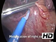
A 16-year-old boy presented as a restrained passenger in a motor vehicle collision. On secondary survey he had abdominal wall ecchymosis. Computed tomography demonstrated free fluid with associated right colonic wall thickening. Based on these findings, the patient was taken to the operating room for diagnostic laparoscopy. Trocars were placed in the standard manner at the umbilicus with two working ports. Hemoperitoneum was seen throughout the abdomen. There was a moderate-sized cecal hematoma with a serosal injury. In addition, a large bucket-handle mesenteric injury, a smaller mesenteric injury, and a small bowel enterotomy were identified. The right colon was fully mobilized laparoscopically in preparation for extracorporeal ileocectomy. The umbilical incision was extended 1 cm superiorly and inferiorly for a total of 4 cm. The small bowel was eviscerated through the enlarged umbilical incision with identification of the bucket-handle mesenteric injury. A small bowel resection was performed because of concerns of potential ischemia and future stricture. The second mesenteric injury was closed with interrupted sutures. The small bowel enterotomy was primarily repaired. The small bowel was returned to the abdomen and the cecum was easily eviscerated because of previous laparoscopic mobilization and an ileocecectomy with primary anastomosis was performed. The small bowel was inspected a final time from the Ligament of Treitz to the ileocolonic anastomosis. The bowel was returned to the abdomen. The fascia and port sites were closed. The patient's postoperative course was uncomplicated and he was discharged on postoperative day 4.
Results and Conclusions:
This case highlights the need for high clinical suspicion of small bowel injury during blunt traumatic work-up, even if hemodynamically stable; and the utilization of diagnostic laparoscopy to confirm blunt traumatic small bowel injury in a pediatric patient. Diagnostic laparoscopy with effective identification of traumatic injuries and full mobilization of the right colon prevented a traditional sub-xiphoid to pubis trauma laparotomy incision with its associated longer hospitalization, increased postoperative pain, and increased risk of future ventral hernia.
No competing financial interests exist.
Runtime of video: 5 mins


Get full access to this article
View all access options for this article.