
Abstract
Introduction:
Hydatid disease of liver is a rare endemic zoonotic infection caused by the echinococcus tapeworm. 1 Although open surgery remains the most widely used treatment, laparoscopy has been utilized with less postoperative pain, shorter hospital stay, and an early return to work. 2 Regardless of technique, spillage of the cystic contents should be avoided to reduce the risk of anaphylaxis, seeding, and further recurrence. 3 Various techniques and suction systems are described in the literature to prevent spillage 4,5 ; however, they are not easily available or completely reliable. This video uses a serrated 150 × 12 mm Endopath® XCEL™ (Ethicon Endo-Surgery LLC, Guaynabo, Puerto Rico) laparoscopic trocar for cyst puncture and aspiration with minimal spillage.
Materials and Methods:
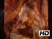
A 38-year-old man presented with pain in the right upper abdomen for 8 months with no associated complaints or comorbidities. On examination, a nontender swelling was palpated 4 cm below the right subcostal margin. Liver function tests were normal and hydatid serology was positive. Computed tomography of the abdomen revealed a 12 × 6 × 10 cm cystic lesion in segments V/VI of liver with daughter cysts. Based on the findings, the patient was taken for surgery and placed in left semilateral with reverse Trendelenburg position. After port placement, an adhesiolysis was performed between the cyst wall, gall bladder, and bowel. Three layers of 10% povidone–iodine-soaked gauze were used to isolate the cyst. A 12/150 mm XCEL® trocar was used to puncture the cyst and a 10 mm suction was used to evacuate the contents. Deroofing of the cyst wall was performed with an ultrasonic scalpel and a cystobiliary communication was noted. Three percent NaCl was installed in the cystic cavity and left for 20 minutes. The cystobiliary communication was sutured using a 3-0 polypropylene suture. The deroofed cyst wall and gauze were removed using an endobag and a povidone–iodine gauze was placed within the port site to maintain pneumoperitoneum and prevent recurrence. The cyst cavity was obliterated with omentum and a drain was placed.
Results:
The procedure was uneventful with no intraoperative complications. The patient was discharged on postoperative day 3. He was started on oral albendazole for 3 weeks and there was no recurrence at 24 months follow-up.
Conclusion:
This technique minimizes cystic spillage as the port serrations along the trocar provide a snug fit. The transparent conduit of the trocar enables continuous evaluation of the cyst cavity and its contents. The larger size of the trocar enables a 10 mm suction catheter to remove the cystic membranes and aggressively irrigates the cyst cavity with scolicidal solution. In addition, the three-layered 10% povidone–iodine-soaked gauze pieces isolate the cyst cavity before puncture to reduce intraperitoneal contamination. Contaminated contents should be removed using an endobag to prevent spillage or recurrence. Finally, laparoscopic deroofing of hydatid cyst using a 150 × 12 mm XCEL laparoscopic trocar may be a safe and feasible minimal option to prevent recurrence.
No competing financial interests exist.
Runtime of video: 8 mins 30 secs
A previous format of this video was presented at the SAGES 2019 conference on April 4, 2019 in Baltimore, MD.


Get full access to this article
View all access options for this article.