
Abstract
Introduction:
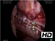
Adolescent idiopathic scoliosis (AIS) is a lateral rotated curvature of the spine >10° occurring in patients aged 10–16 years. 1 Vertebral body tethering (VBT) is an innovative and minimally invasive method that has been shown to have advantages over the more traditional open techniques. 2,3 This video demonstrates the technique for thoracoscopic exposure of vertebral bodies to facilitate VBT for correction of AIS.
Materials and Methods:
A total of 46 VBT cases have been performed at our institution. Eighteen cases underwent a combined thoracic and retroperitoneal lumbar approach. Ten of these cases underwent bilateral tethering for an S-shaped curve. The most common postoperative complaint seen in 17 cases was mild pain/paresthesias/numbness. Five patients experienced overcorrection as they grew and two of these underwent revision of their tethering. This video shows the thoracic technique used in one of our patients. The patient in this case was an otherwise healthy 16-year-old female. The patient was placed in the left lateral decubitus position. A double lumen endotracheal tube was placed, and the left lung was preferentially ventilated. One 5 mm trocar was placed just below the angle of the scapula to gain entry into the chest. Two 5 mm trocars were placed along the anterior axillary line for use during the exposure. The chest cavity was entered, and parietal pleura over the vertebral bodies was divided using hook electrocautery and blunt dissection. Division of the pleura was extended one level above and below the vertebrae being tethered. Intercostal vessels at the levels to be tethered were divided. Electrical stimulation testing was done between each level to confirm intact motor efferent nerves. The sympathetic chain overlying the vertebral bodies was divided. The insertion of the diaphragm on the vertebral bodies was gently dissected to gain access to the L1 vertebra from the chest. Several 15 mm trocars were placed in the midaxillary line to serve as working ports for orthopedic screw placements. Orthopedics then completed placement of the tethering hardware. The pleura was approximated over the hardware using a 2-0 V-lock suture and Endo Stitch autosuturing device. A 24F chest tube was placed through one of the 15 mm trocar sites and secured to the skin. The right lung was re-expanded under direct vision and the remaining ports were removed. Fascia, muscle, subcutaneous tissue, and skin were closed in the standard manner.
Results and Conclusions:
This patient had an uncomplicated 3-day hospital stay. The chest tube was placed to water seal on postoperative day no. 2 and removed on postoperative day no. 3. At her 8-week postoperative appointment, her thoracolumbar curve measured 18°. Her only complaint was mild incisional pain.
No competing financial interests exist.
Runtime of video: 8 mins 4 secs


Get full access to this article
View all access options for this article.