
Abstract
Introduction:
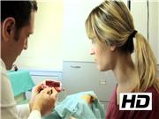
Minimal invasive training simulation laboratory has been developed to aid surgeons to overcome the challenging learning curve and included in our residents' curricula in 2012. Skills are trained in a programmed, progressive, and specific way, assessing performance. The first approach to endosurgery includes every aspect of minimally invasive surgery (MIS) such as knowledge of equipment and instruments, electrosurgery, ergonomy, and the art of MIS suturing.
Aim:
Present a versatile low-cost endotrainer for intermediate and advanced training in pediatric MIS before using the specific neonatal trainers.
Model:
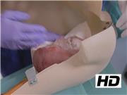
It consists in an adapted 10 year-old mannequin opened in half with the aid of hinges to maintain together. We call it “Gruyere” because of the multiple orifices strategically placed to perform abdominal pelvic procedures, esophageal and mediastinal procedures in the prone position and simulate left lobectomy in the lateral position. The retroperitoneal space was simulated and covered by an adhesive membrane. Commonly available components such as balloons, silicone, different type of fabrics, and siliconized vellon were used to simulate intra-abdominal pelvic organs, vessels, and nodes. Ex vivo porcine organs can also be used to resemble different procedures either in the abdomen or thorax. A simulated abdominal wall was created with synthetic materials such as sponge, cloth, nylon stockings, and silicone to resemble its components. This trainer allows the practice of trocar placement, lens management, and basic maneuvers as well as more complex procedures. Simulation of pelvic procedures such as ovarian cystectomy and lymphadenectomy can be achieved. Refined suturing practice can be done resembling an hepaticojejunostomy using porcine live tissue. We focus in training the whole team and the nurse role in handling the sutures to introduce them into the abdomen. We can see the practice of the shoeshine maneuver and suturing in a simulated Nissen. The prone position is meant to access the esophagus and mediastinum. Lymph nodes dissection is shown. A detailed tutorial of material, steps of each procedure and evaluation sheet has been designed to facilitate its use. We plan to use it in remote places with telementoring concepts.
No competing financial interests exist.
Runtime of video: 5 mins

Keywords
Get full access to this article
View all access options for this article.