
Abstract
Introduction:
Lymphangiomas are benign congenital cystic tumors. They are rare and occur secondary to failure of lymphatic vessels to form normal connections within the lymphatic system during development. 1,2 Retroperitoneal lymphangiomas are an extremely rare subset, accounting for ∼1% of these tumors. 3 Classically, these are thin-walled, unilocular, or multilocular cysts filled with a milky appearing fluid and lined by a layer of endothelial cells. 1 Excision is recommended in the setting of symptomatic, large (4–5 cm), or enlarging cystic lesions because of their propensity for increased complications and potential to invade other structures. 4,5 It is widely accepted that laparoscopic and open surgical excisions are safe and appropriate options for removal of retroperitoneal lymphangiomas. The robotic resection of retroperitoneal lymphangiomas is an excellent surgical option and provides outstanding exposure and observation.
Materials and Methods:
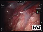
A 50-year-old woman presented with an incidentally found retroperitoneal mass on a CT scan of the abdomen and pelvis performed for hematuria and flank pain. Further questioning revealed epigastric pain and early satiety for 1 month. An MRI/magnetic resonance cholangiopancreatography confirmed the presence of a retroperitoneal mass and revealed no evidence of continuity with the duodenum, pancreas, kidney, or biliary systems. The patient elected to proceed with a robotic abdominal exploration and excision of the mass. On entering the abdomen, the cystic structure was observed. The lesion was safely dissected by mobilizing the transverse colon and hepatic flexure. Next, the duodenum was kocherized medially, exposing the lobulated mass in its entirety. Mobilization was subsequently carried out into the deeper planes of the retroperitoneal space. The excellent observation provided by the robot confirmed that the mass had no obvious connection to the duodenum, pancreas, or biliary system. The remainder of the dissection was carried out with a vessel sealer. Lymphatic vessels were divided with clips and a vessel sealer to ensure lymphostasis. The final connection between the cystic area and the retroduodenal space was performed using a stapler with a 2.5 mm vascular load.
Results:
The patient tolerated the operation well and had an uncomplicated postoperative course. Final pathology revealed a 5.6 × 3.7 × 1 cm fibromembranous retroperitoneal lymphangioma. Cytology was benign, containing small fragments of fibrous tissue, mixed inflammatory cells, macrophages, blood, and debris. She remained in good health at her follow-up visit.
Conclusions:
In patients with retroperitoneal lymphangiomas, the dissection and exposure may be more difficult than in other intra-abdominal locations. Totally robotic excision provides exceptional exposure and observation for dissection and provides a safe and effective removal. Robotic excision should be considered as an option in this patient population.
No competing financial interests exist.
Runtime of video: 7 mins 59 secs
An earlier version of this video was presented at SAGES conference in April of 2018.


Get full access to this article
View all access options for this article.