
Abstract
Introduction:
Polyps of the small intestine are uncommon, 1 and a vast majority of them are benign and asymptomatic. Nevertheless, some small bowel polyps may cause bleeding or intestinal obstruction. 2 Over the past two decades, diagnostic and therapeutic modalities have been used to diagnose and treat these polyps, including wireless endocapsule, double balloon enteroscopy, and multiplanar helical CT enterography. 3 –5 However, in low-income countries, these modalities may not be available. 6 When required, surgical intervention may be difficult and unrewarding if the lesion cannot be accurately localized. 7 With these considerations in mind, this video presents a useful and novel laparoscopic technique to fold the small bowel onto a gastroscope to localize and resect small bowel polyps.
Materials and Methods:
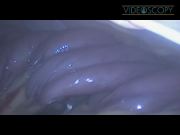
A female presented with two recent episodes of melena. She had no other associated symptoms and her medical history showed hypertension. She was on an antihypertensive medication and 100 mg ASA daily. She was hemodynamically stable with a hemoglobin of 6.7 mg/dL. She was transfused three units of packed red blood cells with an appropriate increase in her hemoglobin to 10 mg/dL. She underwent an upper and lower endoscopy, which were both normal. A CT angiography was also normal. A capsule endoscopy identified a small bowel polyp ∼2.30 m distal to the ligament of Treitz. A combined laparoendoscopic resection was planned. After general anesthesia, three 5 mm trocars were inserted into the right abdomen. The endoscope was inserted and traversed past the ligament of Treitz. Concurrently, the omentum and transverse colon were elevated in a cephalad direction and the endoscope was observed in the lumen of the jejunum. Using two atraumatic graspers, the small bowel was folded onto the endoscope in a coordinated maneuver with the gastroenterologist, who inflated and deflated the bowel to facilitate the folding movements. Endoscopy was performed using carbon dioxide. Ultimately, as the bowel was folded like an accordion onto the endoscope, the polyp was identified. The polyp was excised using a hot snare. A clip was applied and Indian ink tattooing was performed for postprocedure localization, if required.
Results:
The entire procedure lasted 40 minutes and the patient was released after 48 hours, without any complications and a stable hemoglobin. Histology analysis revealed an adenomatous 0.8 cm polyp with signs of recent bleeding. No malignancy was observed.
Conclusion:
Although not widely used, a combined laparoendoscopic resection of a small bowel lesion is an effective modality to treat these difficult lesions. By folding the small bowel like an accordion onto the endoscope, a completely endoscopic resection was possible and the patient avoided a formal small bowel resection. This is one of the first videos that documents the feasibility of this low-cost maneuver to treat small bowel polyps.
No competing financial interests exist.
Runtime of video: 6 mins 50 secs


Get full access to this article
View all access options for this article.