
Abstract
Introduction:
The herniation of the stomach along with other abdominal contents, such as small/large bowel, spleen, or pancreas, is classified as type 4 hiatus hernia. 1 These constitute about 5% of all hiatus hernias. Type 4 hernias are rare and torsion or volvulus of the stomach is known to occur in this anomaly. 2,3 These hernias are usually managed by open surgery. Laparoscopic treatment of type 4 hiatus hernia has been described. 4 We present a stepwise surgical approach to the treatment of a type 4 hiatus hernia with intrathoracic torsion in an infant.
Material and Methods:
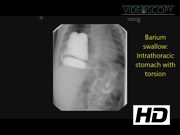
This 5-month-old child presented with a history of recurrent episodes of pneumonia. On examination, the child had a respiratory rate of 30/min and was hemodynamically stable. The chest X-ray revealed radiolucent opacities in the right hemithorax. Barium swallow showed the presence of stomach with torsion within the right hemithorax. A CT scan of the chest revealed the presence of small and large bowel in addition to the stomach.
Operative Technique:
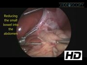
The child was placed at the foot end of the table after general anesthesia. The hips were abducted and knees flexed. The lower limbs were padded adequately. A No. 8 nasogastric tube was placed. Pneumoperitoneum was established by the Veress needle. The port positions are depicted in the video. The liver was retracted by using a 3-mm instrument. The widened esophageal hiatus was visualized with the herniation of the entire stomach, part of small bowel, and transverse colon. The contents of the hernia were reduced. The sac was excised taking care to preserve the vagal trunks. The fundic short gastric branches were divided. The widened hiatus was repaired by two 2-0 Ethibond sutures. A No. 16 NG tube was placed as a bougie after removing the previous No. 8 feeding tube. An umbilical tape was used to place traction on the gastroesophageal junction. A 360° floppy Nissens fundoplication was performed with three 2-0 Ethibond sutures. The stomach was fixed to the anterior abdominal wall by two 3-0 Vicryl sutures that were introduced from the anterior abdominal wall and sutured externally. The excised sac was retrieved through the umbilicus and all the 5-mm ports were closed with 3-0 Vicryl. The skin was closed with bioadhesive (Amcrylate) glue. Total blood loss was less than 5 mL. The operative time was 165 minutes. The nasogastric tube was placed to continuous dependent drainage overnight. Breastfeeding was commenced 12 hours after the surgery. The child was given paracetamol (intravenous initially and orally thereafter) and did not require opioid analgesics. The child was discharged 36 hours after the surgery.
Results:
The child has been followed up for 5 months and has had no further infections. Postoperative barium swallow confirmed the presence of stomach in the abdomen with smooth unobstructed flow of barium through the gastroesophageal junction.
Conclusion:
All types of hiatus hernias can be treated safely and effectively by laparoscopy.
No competing financial interests exist.
Runtime of video: 4 mins 58 secs

Keywords
Get full access to this article
View all access options for this article.
References
Supplementary Material
Please find the following supplemental material available below.
For Open Access articles published under a Creative Commons License, all supplemental material carries the same license as the article it is associated with.
For non-Open Access articles published, all supplemental material carries a non-exclusive license, and permission requests for re-use of supplemental material or any part of supplemental material shall be sent directly to the copyright owner as specified in the copyright notice associated with the article.