
Abstract
Introduction:
Internal hernia is a rare cause of intestinal obstruction, with paraduodenal hernias accounting for more than 50% of them. Two types of paraduodenal hernias are distinguished: left-sided paraduodenal hernias and the rarest (25% of cases) right-sided paraduodenal hernias. 1,2 The reported treatment is by a laparotomy. We report the case of a right-sided paraduodenal hernia that was treated by a laparoscopy.
Case Report:
A 28-year-old woman had complained about right upper abdominal pain for 2 years. The patient was previously operated on spec of a left upper abdominal wall hernia. On this occasion, no pathological findings appeared. Otherwise, the patient had no relevant pre-existing conditions. The discomfort appeared as acute colicky at intervals of several weeks and suspended in most cases spontaneously within hours. Within the scope of an intertemporal uncomplicated pregnancy, the discomfort appeared less frequent. During the pregnancy, the patient sought treatment twice in foreign medical centers with the same discomfort: first gynecological, and then in consultative treatment, both surgical and medicinal. During these ambulant examinations, a cholecystolithiasis could be excluded in each case based on both sonographic and laboratory chemical diagnosis. A gynecological cause was also excluded. Sonographically in the right middle abdomen, a small intestine loop with reduced motility was seen, and adhesions were suggested as possible diagnosis. In both cases, symptomatic therapies with analgesics and laxative measures followed. With continuing discomfort and the suspected single pathology in the small intestine, the patient's family doctor arranged for a magnetic resonance imaging (MRI) of the abdomen. Before realization, both gastroscopy and coloscopy were discussed, but later deferred as invasive diagnostics without clear objectives. The MRI showed a right-sided paraduodenal hernia. Most commonly seen signs of paraduodenal hernia are clustering of small-bowel loops and stretched, displaced, and engorged mesenteric vessels with displacement of other bowel segments. 3,4
Materials and Methods:
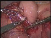
We performed an explorative laparoscopy under general anesthesia. The patient was placed in a supine position with her left arm tucked along her side and the right arm extended on an arm board. Four trocars were inserted. One 10 mm in inferior to the umbilicus, one 10 mm on the middle left abdominal wall, and two 5 mm trocars on the right and left iliac. The MRI finding was confirmed. The entire right colon was lying in the midline and in front of the small intestine. The small intestine entrapped in a peritoneal sac. An intestinal fibrous adhesion at the sac entrance severely confined the movement of the small intestine. The hernia sac was carefully dissected with aid of the 5-mm LigaSure system with care on preservation of the superior mesenteric artery and vein, followed by the incision of the adhesion, thus completely establishing the free movement of the small intestine. At the end, the intestine tract was placed in a nonrotation position with the entire small intestine behind and right of the ascending colon. To avoid unnecessary risks and complications among others postoperative adhesions, we chose not to perform an appendectomy. At the moment of the operation, there is no existing study that shows any relevant advantage when a prophylactic appendectomy—or a cholecystectomy—is performed at the cost of elevated complication risks.
Results and Conclusions:
The postoperative course of the patient was uneventful. The patient was released from the hospital at the second postoperative day. At that time, the patient was free of pain under pain medication. Bowel movement was regular, and the preoperative pain episodes did not appear any more. A 4-month follow-up revealed no changes. Diagnosis of congenital internal hernias remains a challenge for the clinical doctor, as they appear with various and untypical symptoms. Very few of them are diagnosed preoperative. 3 Traditionally, those diagnosed were treated by open surgery. 5,6 In literature, there appear a few cases of laparoscopic treatment of a paraduodenal hernia. 7 –10 This case adds up to them that treatment of uncomplicated similar cases is possible through a minimal-invasive laparoscopic surgery. The advances in laparoscopic surgery have made it possible for patients to enjoy its benefits in large-scale operations as well, including less pain, smaller infection, and overall complication risks, less overall cost due to quick recovery and release, and significantly better cosmetic results.
No competing financial interests exist.
Runtime of video: 9 mins 4 secs


Keywords
Get full access to this article
View all access options for this article.
References
Supplementary Material
Please find the following supplemental material available below.
For Open Access articles published under a Creative Commons License, all supplemental material carries the same license as the article it is associated with.
For non-Open Access articles published, all supplemental material carries a non-exclusive license, and permission requests for re-use of supplemental material or any part of supplemental material shall be sent directly to the copyright owner as specified in the copyright notice associated with the article.