
Abstract
Introduction:
Calyceal diverticula are cystic dilations within a renal collecting system and are prone to urinary stasis, stone formation, and recurrent infection. 1 Management of calyceal diverticula has evolved from open-surgical techniques to minimally invasive approaches, including endoscopic, percutaneous, and laparoscopic surgery. 2 We present a video of the surgical technique of laparoscopic calyceal diverticulectomy utilizing the surgical robot. This video details preoperative imaging and edited video of surgical technique.
Materials and Methods:
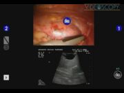
A 40-year-old woman with intermittent right renal colic was found to have a 3-cm upper-pole anterior calyceal diverticulum with significant stone burden on cross-sectional imaging. The patient had a remote history of a failed shockwave lithotripsy procedure. After the robot was docked, the kidney was mobilized inside the Gerota's fascia. Intraoperative ultrasonography confirmed the location of the diverticulum. The diverticulum was opened, and the stone fragments were removed. The diverticular infundibulum was suture ligated, and the diverticular lining was fulgurated using electrocautery. Renorraphy was performed, and a postoperative drain was left in place.
Results:
The patient tolerated the procedure well with no complications. The postoperative recovery was uneventful, and the patient was discharged on the postoperative day 2. In follow-up, her flank pain was resolved. The technique highlighted in this video has been successful for a series of patients with similar presentation at our institution. There have been no perioperative complications.
Conclusions:
Robotic calyceal diverticulectomy is a safe and effective alternative to laparoscopic and open diverticulectomy.
The authors of this abstract and video have no financial disclosures or conflicts of interest.
Runtime of video: 3 mins 46 secs


Get full access to this article
View all access options for this article.
References
Supplementary Material
Please find the following supplemental material available below.
For Open Access articles published under a Creative Commons License, all supplemental material carries the same license as the article it is associated with.
For non-Open Access articles published, all supplemental material carries a non-exclusive license, and permission requests for re-use of supplemental material or any part of supplemental material shall be sent directly to the copyright owner as specified in the copyright notice associated with the article.