
Abstract
Introduction:
Video-assisted thoracoscopic resection is a safe and effective method of treatment for intramural esophageal duplication in children. 1,2
Materials and Methods:
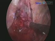
This method is demonstrated in this video of a 14-year-old patient with Crohn's disease with an incidental finding of intramural esophageal duplication by upper gastrointestinal contrast study identified during evaluation for a gastrointestinal stricture. After preoperative work-up, which included chest computed tomography to define the lesion, the patient underwent right thoracoscopic resection of the esophageal duplication. After intubation and positioning in the left lateral decubitus position, the patient underwent endoscopy to localize the lesion, and the endoscope was left in place for the duration of the case. The duplication was separated from the adjacent esophageal muscle fibers thoracoscopically, and the endoscope was then retracted past the point of resection. Air was insufflated into the esophagus while saline was instilled into the chest to assess for a leak. No chest tube was left postoperatively.
Results and Conclusions:
Postoperative pathology was consistent with a foregut duplication cyst. Clinically, the patient did well, and was tolerating clears and discharged home on the first postoperative day. Video-assisted thoracoscopic resection of intramural esophageal duplication is enhanced by the use of intraoperative endoscopy both through lesion localization and evaluation for leak at the time of resection. Postoperative recovery is enhanced secondary to short length of stay and elimination of need for chest tube or contrast study postoperatively due to the intraoperative endoscopic evaluation.
The authors have no conflicts of interest or disclosures to report.
Runtime of video: 3 mins 56 secs

Get full access to this article
View all access options for this article.
References
Supplementary Material
Please find the following supplemental material available below.
For Open Access articles published under a Creative Commons License, all supplemental material carries the same license as the article it is associated with.
For non-Open Access articles published, all supplemental material carries a non-exclusive license, and permission requests for re-use of supplemental material or any part of supplemental material shall be sent directly to the copyright owner as specified in the copyright notice associated with the article.