
Abstract
Introduction:
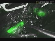
In this video, intraureteral indocyanine green (ICG) administration for identification of the ureter during robotic ureteral reconstructive procedures is presented.
Materials and Methods:
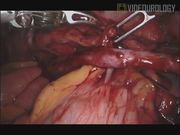
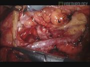
The first patient had retroperitoneal fibrosis. Florodeoxyglucose-positron emission tomography (FDG-PET) revealed high standardized uptake value (SUV) of the tissue surrounding right ureter. There was right grade II hydronephrosis on retrograde pyelography. Robotic ureterolysis was planned to release the ureter and to histopathologically evaluate the surrounding tissue. First, open-end catheter was inserted into the right ureter and taken into the surgical field. The procedure was performed with daVinci Xi system in modified left lateral decubitus position. Three 8 mm robotic ports and one 5 mm assistance port were used. Monopolar hot scissors were used in the right hand and Maryland and fenestrated bipolar forceps in the left hand. The ureter was identified after colon medialization. ICG was dissolved in 10 mL distilled water and applied through the open-end catheter. Then the open-end catheter was clamped. Fire-fly mode was operated and ureteral segments captured inside the fibrotic tissue were delineated. Dissection was carried on and ureter was freed. Fibrotic tissue surrounding the ureter was excised. Colonic serosa was sutured to the right abdominal wall beneath the ureter and ureter was intraperitonealized. A 4.8F Double-J stent catheter was inserted into the right ureter and 10 mm Jackson–Pratt drain was left in place. The second patient had an unsuccessful endoscopic right ureteral stone surgery at another center. Flexible ureterorenoscopy revealed right upper ureteral stricture and submucosal stone fragments. An open-end catheter was inserted like the first case. The procedure was performed with the daVinci Xi system in the same position with the same port configuration. Monopolar hot scissors were used in the right hand and fenestrated bipolar forceps in the left hand. The colon was medialized and the right kidney was identified. The ureter was identified and freed with blunt and sharp dissection. After release of the periureteral fibrotic tissue, ICG was applied through the open-end catheter. Fire-fly mode was operated and ureter and ureteral stricture were identified. Periureteral stone was seen at the level of the urethral stricture and removed. After transection of the ureter, first, proximal stenotic segment was resected and the kidney was mobilized. Then, distal stenotic segment was resected. After ureteral spatulation, ureteroureteral anastomosis was performed with 4/0 polyglactin. Open-end catheter was used to confirm the patency of the ureteral lumen during anastomosis. After completion of the anastomosis, the ureter was retroperitonealized. A Jackson–Pratt drain was left in place. At the end of the procedure, retrograde 4.8F Double-J stent catheter was inserted.
Results:
Histopathologic evaluation of the fibrotic tissue surrounding the ureter in the first patient revealed no malignancy. Postoperative period was uneventful in both patients. In the first patient, Double-J stent catheter was removed at postoperative fourth week. Postoperative sixth month FDG-PET revealed decreased SUV of the soft tissue around the ureter. Double-J stent catheter was removed at postoperative sixth week in the second patient. Right kidney was normal on postoperative second month CT pyelography.
Conclusions:
Intraureteral ICG administration facilitates identification of ureter during robotic ureteral reconstruction. 1,2
No competing financial interests exist.
Runtime of video: 4 mins 18 secs
Keywords
Get full access to this article
View all access options for this article.