
Abstract
Introduction:
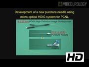
The indications for percutaneous nephrolithotomy (PCNL) have been a paradigm shift in the past decade. The advent of miniaturization of instruments such as smaller endoscopes, smaller retrieval devices, and energy sources has extended the indication of PCNL. 1 –4 The most important step is to make optimal percutaneous access, which facilitates to perform PCNL safely and effectively. We originally developed an “optical puncture needle” using ultrathin high-definition image guide (HDIG) system and applied it to making percutaneous access for PCNL.
Materials and Methods:
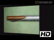
A 20-gauge puncture needle equipped with a 0.65 mm micro-optic, having an integrated light lead, was developed as part of a collaborative research with Takei Medical & Optical Co. Ltd. (Tokyo, Japan) and Sumita Optical Glass, Inc. (Saitama, Japan). The HDIG system consists of a 0.35 mm precise object lens and optical glass fiber, wherein real-time HD images can be seen through the digital image processing device. After evaluating safety, optical quality, and operation performance in an animal study using adult swine, a clinical study on the application of the optical puncture needle for PCNL, in patients with nephrolithiasis, was carried out at Okayama University Hospital. Before study initiation, the animal experiment and the clinical study were approved by the Animal Experiment Committee in Okayama University (OKU-2012632-444) and the Okayama University Institutional Review Board (Registration No. m14011), respectively.
Results:
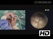
Between January 2014 and December 2016, 14 consecutive patients underwent percutaneous renal access using the optical puncture needle for PCNL. All patients have large volume nephrolithiasis, including staghorn calculi in four cases. Of the 14 patients, 3 patients underwent percutaneous renal access in the modified Valdivia position, otherwise in prone position. Eight patients did not have any hydronephrosis. In all cases, we succeeded in making optimal percutaneous renal access without any complications related to needle puncture. Median (range) operation time and estimated blood loss including urine were 178.5 (116–315) minutes and 10 (5–500) mL, respectively.
Conclusion:
This system potentially enables safe and effective placement of percutaneous renal access. Furthermore, it might allow initial puncture convert to PCNL seamlessly. This smallest endoscope could be applied for observation of upper urinary tract tumor or nonurologic condition, such as arthroscope or intravascular surgery.
No competing financial interests exist.
Runtime of video: 6 mins 44 secs
Previously presented by Hiromi Kumon and Koichiro Wada on November 9, 2013 at the 27th Annual Meeting of Japanese Society of Endourology, held in Nagoya, Japan.
Get full access to this article
View all access options for this article.