
Abstract
Purpose:
To introduce a modified technique of balloon dilation in X-ray-free ultrasound-guided percutaneous nephrolithotomy (PCNL) for staghorn calculi.
Materials and Methods:
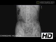
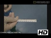
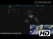
From March 2014 to April 2015, 21 consecutive patients (22 renal units) with staghorn stones underwent X-ray-free ultrasound-guided PCNL with balloon dilation. 1 Under the guidance of ultrasound, the superior calix (as the first choice) was punctured by an 18-gauge hollow needle with its stylet; if urine spouts from the hollow after removing the stylet, the access to the renal collecting system could be confirmed. The needle had length marker in its body and the needle length between skin and the collecting system was recorded and referred to as dilation depth. Then a 0.038-in zebra guidewire (Urovision) was introduced into the collecting system and a 10F fascial dilator with sheath (Urovision) was used to dilate the tract along the guidewire after making a 1-cm skin incision. Through the 10F sheath, a 6F ureterscope (Olympus) was passed into the collecting system to adjust the guidewire into the ureter to function as both safety and working guidewire. Subsequently the 6F nephrostomy balloon (Cook) was placed over the guidewire until the balloon length was equal to dilation depth, which was marked by a marker pen. Under ultrasound monitoring, the location of the balloon was further secured and whether the balloon was inflated uniformly without the waist was confirmed when the pressure at 20 atm was held for 5 minutes. Then the balloon was removed and a 22F Amplatz dilator with sheath was placed into the collecting system along the 7F guide catheter over the guidewire. After removing the dilator, the Amplatz sheath was left as the work sheath. Lithotripsy was performed using a 20.8F nephroscope (Wolf) and EMS ultrasound lithotripsy (Nyon). After stone extraction, a 7F Double-J catheter was placed antegradelly and an 18F catheter as nephrostomy tube was placed, which was removed 2 days after the operation. The Double-J catheter was removed 4 weeks later. For stones in the calices uncovered by the superior calix approach, additional tracts were necessary.
Results:
The average age of the 21 patients (11 males and 10 females) was 48.8±10.7 years. The average stone size was 4.8±1.8 cm. All of the procedures were effectively performed with balloon dilation under X-ray-free ultrasound guidance. Of them, two patients needed two tracts whereas one patient needed three tracts. The average lithotripsy time was 92.4±26.5 minutes. The mean hemoglobin before operation and 2 days after operation was 13.1±2.6 and 12.0±2.2 g/dl, respectively, whereas the mean hematocrit before operation and 2 days after operation was 39.7±7.0% and 36.5±5.5%, respectively. The average hospitalization after operation was 5.3±1.9 days. No patient needed transfusion. Also there was no severe complication. Immediately after operation, the stone-free rate was 81.8%.
Conclusion:
For nonobesity patients, balloon dilation is feasible in X-ray-free ultrasound-guided PCNL. With modified techniques, balloon dilation makes X-ray-free ultrasound-guided PCNL for treating staghorn calculi easy and safe.
Tie Zhou, Guanghua Chen, and Wei Zhang contributed equally to this work.
No competing financial interests exist.
Runtime of video: 3 mins 34 secs
Get full access to this article
View all access options for this article.