
Abstract
Introduction:
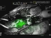
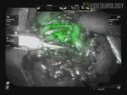
In addition to enhanced magnification, the da Vinci® Surgical System offers the ability to display near-infrared fluorescence (NIRF) imaging. NIRF imaging has previously been described in conjunction with the administration of the fluorophore indocyanine green (ICG). The principle urologic application of NIRF imaging has been to distinguish normal parenchyma from tumor during partial nephrectomy with intravenous ICG. We describe a novel use of NIRF imaging without ICG to identify intraluminal areas of interest during robotic-assisted laparoscopic (RAL) surgery marked by the white light (WL) of endoscopes.
Materials and Methods:
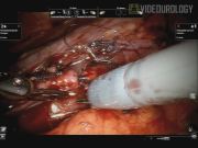
A single surgeon performed RAL surgery on six patients aided by NIRF imaging to visualize the areas of interest marked by the WL of endoscopic instruments. Patient 1 underwent RAL partial ureterectomy with a psoas hitch and Boari flap with left ureteroscopy for ureteral stricture. Patients 2 and 3 underwent RAL diverticulectomies with cystoscopy for urothelial cell carcinoma (UCC) limited to bladder diverticula. Patient 4 underwent RAL partial cystectomy with cystoscopy for focally invasive UCC. Patient 5 underwent RAL nephroureterectomy with cystoscopy for upper tract UCC. Patient 6 underwent RAL pyelolithotomy with ureteroscopy for a staghorn calculus.
Results:
The use of NIRF imaging with marking by the WL from endoscopic instruments enabled more precise identification of important areas and successful completion of RAL surgery in these six patients. The light source from the endoscopes could not be seen well with the standard RAL imaging. Turning the intra-abdominal RAL light source off eliminates the surrounding field of vision. By filtering light wavelengths below near-infrared, NIRF imaging caused the WL of the endoscopes to illuminate green while allowing simultaneous vision of the surrounding tissues. The increased precision enabled by this technique maximized the healthy tissue preserved in these procedures.
Conclusions:
Our case series represents, to the best of our knowledge, the first description of a novel technique using NIRF imaging to identify otherwise obscured intraluminal areas of interest marked by the WL of endoscopic instruments and aid in successful completion of RAL surgery. This technique has been effectively utilized to improve precision in four disparate types of urologic RAL surgery.
The authors declare that no competing financial interests exist.
Runtime of video: 7 mins 45 secs
Keywords
Get full access to this article
View all access options for this article.