
Abstract
Abstract
Introduction:
Indocyanine green dye, which is generally used as a diagnostic agent, becomes fluorescent under near-infrared light after binding to serum proteins. In most recent studies, indocyanine green dye with near-infrared fluorescent (ICG-NIRF) imaging has been used effectively to identify tumoral tissue and its resection margins.1,2 This technique has been popularized for various robotic interventions;2,3 however, its use in robotic total adrenalectomy has not been reported yet. In this report, we aimed to describe our initial experience with ICG-NIRF imaging during robotic left adrenalectomy in a patient with primary hyperaldosteronism (pHA).
Case Presentation:
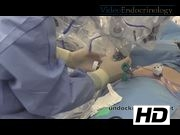
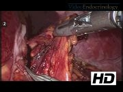
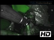
A 28-year-old female patient was referred for adrenalectomy from Endocrinology with a diagnosis of pHA. She had severe hypertension and hypokalemia. Her plasma aldosterone level was 76.5 ng/dL (range: 3.1–35.4 ng/dL) and renin activity was 1.3μg/L/h (range: upright=0.8–5.8 and supine=0.5–1.8). Computed tomography imaging of the adrenal gland revealed a 16×9 mm sized adrenal mass on the left. Robotic lateral transabdominal adrenalectomy for the left adrenal mass was planned. The procedure was performed under general endotracheal anesthesia. She was positioned in the left lateral decubitus position. The abdomen was entered with an optical trocar and the splenocolic and splenorenal ligaments were divided. Intraoperative ultrasound was performed and the adrenal gland was identified. The robot was docked and using a robotic vessel sealer and Maryland grasper, the gland was circumferentially dissected (our previous report describes the procedure in detail 4 ). One 1.5 mL (2.5 mg/mL) dose of ICG solution (0.25 μg/mg; Pulsion Medical Systems AG, Munich, Germany) was then injected intravenously and the robot toggled to NIRF mode. Seconds later, the adrenal parenchyma became fluorescent at intensity significantly less than the liver with a longest duration of 5 minutes. Three different injections of ICG at a total dose of 3.5 mL were administrated during operation with an interval of 20 minutes to assist in visualization during the procedure. The adrenal vein was isolated, clipped, and divided. Robotic adrenalectomy was successfully performed with no fluorescent tissue left behind. After undocking, tumor removal using a specimen bag was performed laparoscopically. The total procedure time was 160 minutes. Estimated blood loss was 10 mL. Pathology showed a 14×3 mm sized adrenocortical adenoma consistent with aldosteronoma. The patient's blood pressure and plasma aldosterone level normalized (5.2 ng/mL) and she was taken off antihypertensives in the postoperative period.
Conclusions:
This video illustrates the efficacy and utility of the ICG-NIRF imaging during robotic total left adrenalectomy. An increased visualization through real-time image guidance helps identify the contours of the adrenal gland thus allowing both mass localization and complete resection.
All authors declare no conflicts of interest.
Runtime of video: 6 mins 17 secs
Keywords
Get full access to this article
View all access options for this article.
References
Supplementary Material
Please find the following supplemental material available below.
For Open Access articles published under a Creative Commons License, all supplemental material carries the same license as the article it is associated with.
For non-Open Access articles published, all supplemental material carries a non-exclusive license, and permission requests for re-use of supplemental material or any part of supplemental material shall be sent directly to the copyright owner as specified in the copyright notice associated with the article.