
Abstract
Natural infection of dengue virus (DENV) in bats is an unexplored field in Colombia. To detect the presence of DENV in bats, a descriptive prospective study using a nonprobabilistic sampling was carried out; 286 bats in 12 sites were caught. Sample tissues of different animals were obtained; the RNA was obtained from tissues and a nested-RT-PCR was carried out and detected amplicons of 143 fragment of the NS5 gene were sequenced by the Sanger method. In nonhematophagous bats
Introduction
Emerging and re-emerging infectious diseases have now become among the most serious threats to public health. Approximately 75% of the diseases that have emerged during the past two decades have wild reservoirs (Woolhouse and Gowtage 2005, Jones et al. 2008). In this sense, bats are hosts of high viral diversity with high zoonotic potential worldwide (O'Shea et al. 2014, Calderon et al. 2016).
Some flavivirus such as dengue virus (DENV), Koyose virus (YOKV), Tamana virus (TABV), Rio Bravo virus (RBV), Japanese encephalitis virus (JEV), and West Nile virus (WNV) have been detected in bats (Bunde et al. 2006, Jiang et al. 2015, Tajima et al. 2015, Thompson et al. 2015, Abudes-Gallegos et al. 2018). Many flaviviruses can cause diseases in humans such as from a nonspecific febrile syndrome, triggering encephalitis, to hemorrhagic fever and death (Feitoza et al. 2017, Hall et al. 2017, Mattar et al. 2017). Dengue fever is a disease of great prevalence in tropical countries. Although bats can harbor many infectious viruses, they do not develop apparent signs of disease from any virus (Calderon et al. 2016). It is believed that the increase in the body temperature of bats as a result of flight increases the metabolic rate, the mitochondrial activity that triggers the immunological cascade, and the production of interleukins and prostaglandins, which would prevent them from being infected (Krysko et al. 2011, Wang et al. 2011, O'Shea et al. 2014, Mattar and Gonzalez 2016). The objective of this study was to establish a natural infection of DENV in bats from departments of Córdoba and Sucre (Colombia).
Materials and Methods
Study type and sampling
A descriptive prospective study using a nonprobabilistic sampling was carried out; 286 bats in 12 sites were caught: 8 in Córdoba and 4 in Sucre, both departments included the main ecosystems; these departments are located in the Caribbean area of Colombia (Fig. 1). The study was approved by the ethics committee of the Faculty of Veterinary Medicine of the University of Cordoba, Colombia; the committee took into account the instructions for researching with non-commercial purposes animals of the National Environmental Authority of Colombia. The bats were captured with the use of mist nets; they were later identified using dichotomous taxonomic keys (Linares 1998). Specimens listed as endangered species and pregnant or lactating females were released. Euthanasia was performed using an overdose of sodium pentobarbital. Brain, heart, lung, liver, kidney, and spleen were collected. The organs were stored in cryovials with Trizol™ (Invitrogen, Carlsbad, CA) and kept in liquid nitrogen.
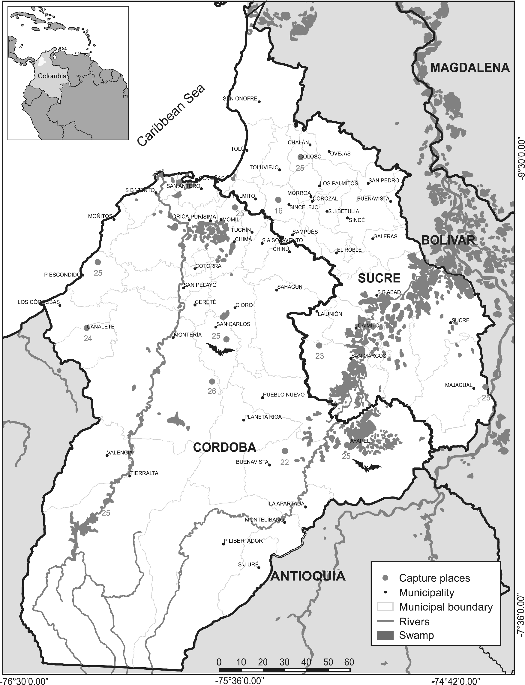
Capture sites of bats in Córdoba and Sucre (Colombia).
Molecular detection
RNA extraction was performed with Trizol (Invitrogen). The aliquots were suspended in 150 μL of nuclease-free water. The concentration of the RNA obtained from each organ was then measured using the NanoDrop 2000 (Spectrophotometers™). The cDNA synthesis was carried out with the reverse transcriptase enzyme M-MLV (Invitrogen) using random primers, as recommended by the manufacturers.
A nested RT-PCR using in the first round (Flavi 1+: 5′-GAYYTIGGITGYGGIGIGGIRGITGG-3′ and Flavi 1−: 5′-TCCCAICCIGCIRTRTCRTCIGC-3′) was performed; the nested with by Flavi 2+ (5′-YGYRTIYAYAWCAYSATGGG-3′) and Flavi 2− (5′-CCARTGITCYKYRTTIAIRAAICC-3′) was performed. As previously described, the set of primers amplifies products of 1360bp and, 143bp respectively (Sanchez et al. 2005). As a control of species, complementary primers were used to sequence a mitochondrial gene
Phylogenetic analysis
Using Clustal W, 2 sequences obtained in this study and 38 partial sequences of the NS5 gene were aligned with four DENV serotypes registered in GenBank. For the phylogenetic construction, the maximum likelihood method was used. The substitution model was Kimura of two parameters and to estimate the branch supports of each group, 10,000 pseudoreplications were produced; in total 100 positions were analyzed. The phylogenetic analysis was performed with MEGA X.
Results
Table 1 shows the distribution of bats species by found sources.
Distribution of Bats Species by Food Sources
Amplicons with a size of 143 bp of the NS5 gene of DENV in brain, lung, liver, and kidney of
The sequence MG011655.1 showed a similarity of 95.1% and coverage of 98.41% with the sequence FJ392595 of serotype-2 dengue virus (DENV-2) NS5 gene. The sequence MG011656.1 presented a similarity of 96.1% with coverage of 100% compared with the sequence FJ392595 of the GenBank. The results of the phylogenetic analysis show that the consensus sequence showed a high percentage of similarity with sequences of the gene coding for the NS5 protein of DENV-2. The phylogenetic analysis suggested the presence of DENV-2 in these tissues.
Four monophyletic clades were obtained, clearly differentiated that correspond to the four serotypes of DENV, each clade group individuals of the same serotype, and are separated by high values of branch support (Fig. 2). The sequences obtained, MG011655.1 and MG011656.1 (between two red circles), were integrated with sequences homologous to DENV-2 (shown with a single red circle).
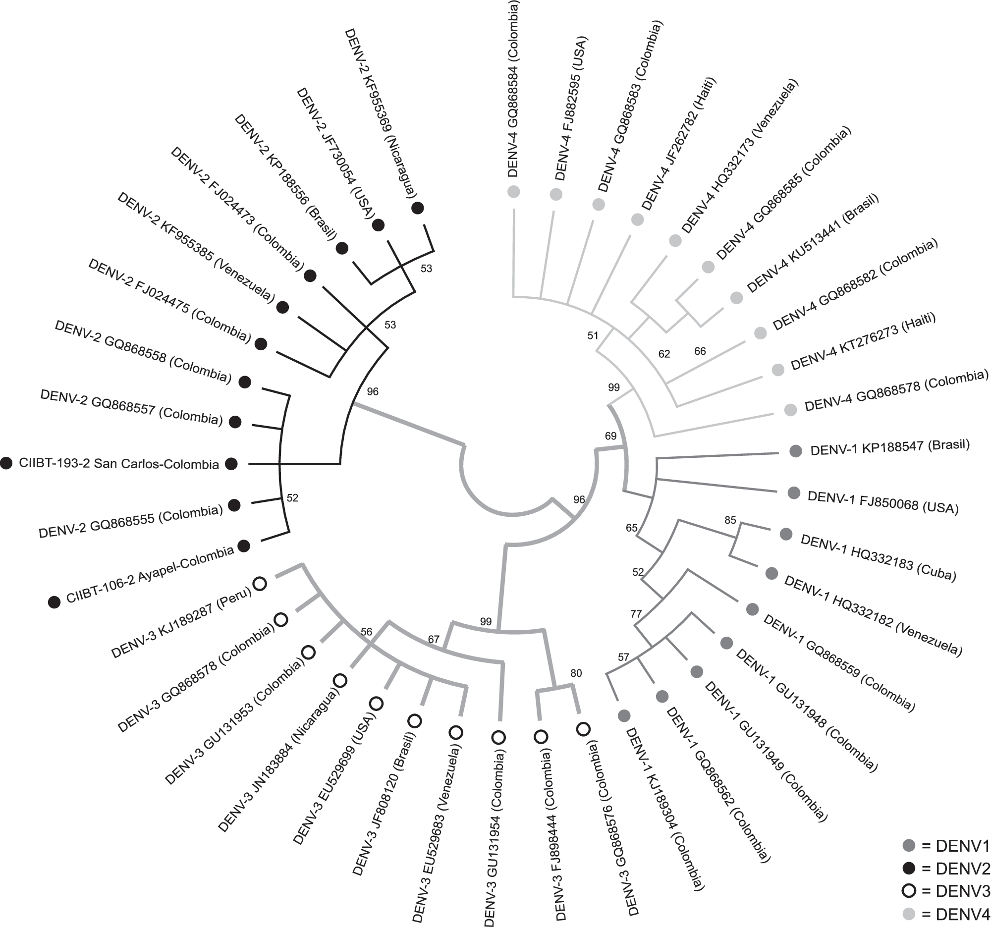
Phylogenetic tree by ML method; four clades are shown and the obtained sequences from Cordoba and Sucre bats are shown with additional
Discussion
Several investigations (Platt et al. 2000, Aguilar-Setien et al. 2008, Lavergne et al. 2009, Machain et al. 2013, Sotomayor-Bonilla et al. 2014, Vicente-Santos et al. 2017, Abudes-Gallegos et al. 2018) have demonstrated the presence of DENV in bats. In contrast, a study involving 240 wild bats from Campeche and Morelos (Mexico) indicated the absence of serological or molecular evidence of the DENV, the authors suggest that American bats could not be reservoirs or amplification host for DENV infection (Cabrera-Romo et al. 2016). That study included 19
The molecular evidence in this study agree with the conclusions of Marinkelle (1996), Platt et al. (2000), Aguilar-Setien et al. (2008), Lavergne et al. (2009), Machain et al. (2013), Hayman et al. (2013), Sotomayor-Bonilla et al. (2014), Schountz (2014), Moratelli and Calisher (2015), and Vicente-Santos et al. (2017) who clearly suggest that bats can be involved with different pathogens that affect human health.
In rural areas of Córdoba and Urabá (Colombia), mosquitoes
The transmission of DENV by mosquitoes could be limited in wild ecosystems, where urban species of
The transmission of DENV to bats should involve the feeding of
Further investigations are still needed to determine whether the DENV detected in
Conclusion
This study presents the first molecular evidence of the infection natural in bats captured in the Colombian Caribbean.
Footnotes
Author Disclosure Statement
No competing financial interests exist.