
Abstract
Introduction:
Sindbis virus (SINV) is a mosquito-borne Alphavirus known to infect birds and cause intermittent outbreaks among humans in Fenno-Scandia. In Sweden, the endemic area has mainly been in central Sweden. Recently, SINV infections have emerged to northern Sweden, but the vectorial efficiency for SINV of mosquito species in this northern region has not yet been ascertained.
Objective:
Mosquito larvae were sampled from the Umeå region in northern Sweden and propagated in a laboratory to adult stage to investigate the infection, dissemination, and transmission efficiency of SINV in mosquitoes.
Materials and Methods:
The mosquito species were identified by DNA barcoding of the cytochrome oxidase I gene. Culex torrentium was the most abundant (82.2%) followed by Culex pipiens (14.4%), Aedes annulipes (1.1%), Anopheles claviger (1.1%), Culiseta bergrothi (1.1%), or other unidentified species (1.1%). Mosquitoes were fed with SINV-infected blood and monitored for 29 days to determine the viral extrinsic incubation period. Infection and dissemination were determined by RT-qPCR screening of dissected body parts of individual mosquitoes. Viral transmission was determined from saliva collected from individual mosquitoes at 7, 14, and 29 days. SINV was detected by cell culture using BHK-21 cells, RT-qPCR, and sequencing.
Results:
Cx. torrentium was the only mosquito species in our study that was able to transmit SINV. The overall transmission efficiency of SINV in Cx. torrentium was 6.8%. The rates of SINV infection, dissemination, and transmission in Cx. torrentium were 11%, 75%, and 83%, respectively.
Conclusions:
Cx. torrentium may be the key vector involved in SINV transmission in northern Sweden.
Introduction
S
In Sweden (Lundstrom et al. 1991), SINV was first recognized in the 1960s in the central region of the country from patients presenting exanthema and arthralgia accompanied by fever. Since then, the virus has been isolated from samples of both humans and vectors (Kurkela et al. 2004). SINV disease has historically had the highest incidence in the central parts of Sweden (Lundstrom et al. 1991). However, the SINV seroprevalence in the north of the country has increased during the past 25 years (Ahlm et al. 2014) and in 2013 an outbreak occurred in northern Sweden close to the Gulf of Bothnia (Bergqvist et al. 2015). In neighboring Finland, the virus was initially detected in 1980s and since then the virus has caused large intermittent outbreaks in different parts of the country (Brummer-Korvenkontio et al. 2002).
Culex torrentium has been identified as an enzootic vector of SINV in Sweden (Lundstrom et al. 1990, Hesson et al. 2015), whereas Cx. univittatus mosquitoes has been identified to be a SINV vector in South Africa, Israel, and Saudi Arabia (McIntosh et al. 1978, Wills et al. 1985, Samina et al. 1986). Furthermore, in Kenya we detected SINV mainly in Aedes mcintoshi (Sang et al. 2017). Because of the recent expansion of SINV to northern Sweden, we were interested to determine which mosquito species were responsible for SINV transmission in this area. Through SINV vector competence studies and application of a DNA barcoding approach, we determined the transmission efficiency and accurately identified the mosquito species competent for SINV. This fosters understanding of the maintenance and spread of SINV in northern Sweden.
Materials and Methods
Study area
The mosquito sampling was conducted during September and October 2017 in the northern part of Sweden. The sampling was performed close to a bird conservation site and along the coastline of the Gulf of Bothnia in Umeå, where a previous outbreak of SINV disease was reported to have occurred in northern Sweden (Bergqvist et al. 2015).
Virus strain
The SINV Lovanger strain (GenBank KF737350.1) used in this study was isolated during the 2013 SINV outbreak in northern Sweden from Culiseta morsitans (Bergqvist et al. 2015). The virus stock was obtained after a single passage on BHK-21 cells in Dulbecco's minimum essential medium (DMEM; Gibco®) supplemented with 2% fetal bovine serum (FBS; GE Healthcare Life Sciences, Cramlington, United Kingdom), 2% HEPES, 2% penicillin–streptomycin (PEST; GE Healthcare Life Sciences, South Logan, UT), and 2%
Larvae sampling and rearing
The rearing of the mosquito larvae and laboratory experiments were conducted at an insectary at Swedish Defense Research Agency (FOI), Umeå. Mosquito larvae were sampled using a larvae dipper with a telescope handle. The larvae (n = 400) were separated from the debris using sieves before being placed in tins filled with water from the same source. At the insectary, the larvae were transferred from the containers and placed in larval trays supplied with dechlorinated water containing 0.1 mg per larva Tetramin® (Melle, Germany). The larvae were monitored daily until maturity to pupal stage. The pupae (n = 380) were then transferred using Pasteur pipettes to containers of ∼100 mL water, and placed into mosquito cages (BugDorm insect rearing cages 30 × 30 × 30 cm; NHBS, United Kingdom). The adult mosquitoes were kept at room temperature and 50% humidity and fed daily with 10% filter sterilized sucrose solution in distilled water.
Infection of mosquitoes
One-week-old adult mosquitoes (n = 185) were starved for 24 h before being exposed to infectious blood meals containing defibrinated horse blood spiked with SINV (106 plaque-forming units/mL). For control purposes, larvae sampled from the same location and period as those tested for SINV were propagated in the laboratory and adult mosquitoes (n = 180) were analyzed for the presence of naturally (transovarially) acquired SINV. All the mosquitoes were negative for SINV.
The female mosquitoes were aspirated into containers and fed for 30 min using artificial feeding containers wrapped with parafilm membrane. After blood feeding, the female mosquitoes were immobilized briefly at −20°C for 2–5 min and fully engorged females (n = 95) were selected and kept up to 29 days. Blood-fed mosquitoes (n = 5) were harvested at day 0 (1-h postfeeding) (all Cx. torrentium), and the entire bodies were tested individually by PCR confirming the presence of the SINV RNA from the blood meals. The remaining 90 mosquitoes (depending on the survival rate per incubation container) were collected at days 4 (n = 21), 7 (n = 23), 14 (n = 30), and 29 (n = 16). Individual mosquitoes were dissected and head, wings and legs, and thorax and abdomen were removed and tested for the presence of SINV RNA. In addition, saliva was collected on days 7, 14, and 29.
Vector competence experimental assays
The thorax and abdomen, head, wings, and legs of mosquitoes were dissected using hypodermic needles (26G–27G size) and a stereo microscope. Each body part was placed in separate 1.5 μL Eppendorf tubes, containing 300 μL of DMEM supplemented with 2% fetal FBS (GE Healthcare Life Sciences), 2% HEPES, 2% PEST (GE Healthcare Life Sciences), and 2%
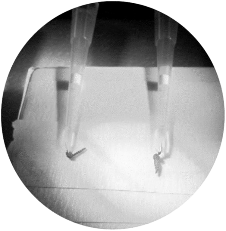
Saliva collection from mosquitoes analyzed in the study.
Amplification of the cytochrome oxidase subunit I gene
For genetic typing of mosquitoes, a barcoding region of the cytochrome oxidase subunit I (COI) gene was amplified and sequenced, as described before (Engdahl et al. 2014). DNA extractionof samples from thorax and abdomen of each mosquito (n = 95) was performed using a DNA Extraction Kit (DiaSorin).
The primers used for COI gene amplification were as follows: GB_1358_83F, 5′-ACTCAAGAAAGAGGTAAAAAGGAAAC-3′ and TL2-N-3014R, 5′-TAATATGGCAGATTAGTGCATTGGA-3′. The PCR amplification was performed by using the KAPA Taq PCR Kit (KAPA Biosystems, Boston, MA). The preferred cycling conditions after an initial denaturation at 95°C for 3 min were as follows: 35 cycles at 95°C for 3 s, annealing at 55°C for 30 s, and extension at 72°C for 1 min, before standby at 4°C. The PCR products were visualized on 1.2% agarose gels (containing GelRed; Biotium, Inc.) before purification by NucleoSpin Gel, PCR Clean-up kit (Macherey-Nagel) and sequencing (GATC Sequencing Company). The obtained sequences were compared with available sequences in GenBank to determine the identity of the mosquito species.
Data analysis
Microsoft Excel 2010 was used to perform the calculations and comparisons based on the standardized formulas for infection rate, dissemination rate, transmission rate, and transmission efficiency. The infection rate in percent was calculated as the proportion of engorged females with PCR-positive thoraces and abdomens in relation to all engorged females. The dissemination rate was estimated in a similar way as the proportion of PCR-positive heads, legs and wings in relation to all infected samples with positive abdomen. The species-specific transmission efficiencies were assessed as the proportion of disseminated females with SINV nucleic acid-positive saliva, and transmission rates were estimated as the proportion of engorged females with PCR-positive saliva.
Results
Mosquito species analyzed
In total, 90 female mosquitoes were analyzed in this study. Five species of mosquitoes were found among the samples, and identified by molecular barcoding as Cx. torrentium 74 (82.2%), Cx. pipiens 13 (14.4%), and one mosquito each of Culiseta bergrothi, Aedes annulipes, and Anopheles claviger. Of the tested mosquitoes, only Cx. torrentium was found to be susceptible to, and able to disseminate and transmit SINV. Viral RNA was detected in thorax and abdomen, wings and legs, and head. Four mosquitoes with PCR-positive saliva also showed CPE in BHK-21 cells at 4 days postinfection, implying that viable virus was present in the saliva. To rule out naturally acquired transovarial infection, the viral sequences obtained from the saliva were found to be identical to the SINV strain Lovanger that was used to experimentally infect the mosquitoes.
Infection rate, dissemination rate, transmission rate, and transmission efficiency of Cx. torrentium
The calculations were based on Cx. torrentium mosquitoes because they were the most abundant (82.2%) and was the only species that was infected by SINV.
The overall SINV infection rate for the Cx. torrentium mosquitoes that were infected during the study was 11% with a dissemination rate of 75% and a transmission efficiency of 83%. The transmission rate was 6.8%.
SINV infection was determined in samples from thorax and abdomen and at day 14 most mosquitoes showed infection with a drop thereafter (Table 1). SINV infection rate in Cx. torrentium for days 4, 7, 14, and 29 were 5.2%, 5%, 20%, and 13.3%, respectively.
A cut-off cycle threshold value of 32 was applied.
SINV, Sindbis virus.
There was no evidence of dissemination to the head, wings and legs at day 4, although the infection rate was 5.2%. At day 7, SINV had disseminated in three mosquitoes, and later in two at day 14, and one at day 29 (Table 2). The dissemination rate for SINV was 50% at days 14 and 29 (Table 2).
A cut-off cycle threshold value of 32 was applied.
The dissemination rate was 300% on day 7 because two additional mosquitoes negative for SINV infection at the thorax and abdomen had positive head, wings, and legs.
When analyzing the mosquito saliva by infecting BHK-21 cells, we found that SINV transmission increased from day 7 to 14 with a slight decline on day 29. The transmission rate was 33.3% on day 7, three mosquitoes transmitted SINV on day 14 and 100% of mosquitoes transmitted SINV on day 29 (Table 3).
Transmission rate was 150% on day 14 because an additional mosquito that was negative for SINV at the head, wings, and legs had SINV-positive saliva.
Confirmed by cell culture, RT-PCR and sequencing.
ND, not determined.
Cx. torrentium/Cx. pipiens hybrid mosquitoes
When the mosquitoes were sequenced 22 single-nucleotide polymorphisms (SNPs) or signatures were found in the COI gene (nucleotides 669–1356), and used in this study to distinguish Cx. torrentium from Cx. pipiens. Interestingly, of all mosquitoes (n = 90) 82.2% were identified as Cx. torrentium and many also showed Cx. pipiens signatures at some of the SNP positions (Table 4). Fifty-two (57.8%) that were identified as Cx torrentium had one to two Cx. pipiens SNPs, suggesting hybrid mosquitoes. No signatures of Cx. torrentium were found in the corresponding sequence of Cx. pipiens. Only one Cx. torrentium/Cx. pipiens hybrid mosquito was infected in the thorax and abdomen but did not disseminate and transmit. The rest of the infected mosquitoes were Cx. torrentium.
G
Underlined bold letters indicate the nucleotide found in (the COI gene of) Cx. torrentium, Cx. pipiens and hybrid mosquitoes. The SNP position and mosquito number is also specified for each hybrid mosquito.
A conserved nt change among the captured mosquitoes (not toward Cx. pipiens or Cx. torrentium): was found in positions 732 (A→G) #48, 57, 95, 104, 108, 109, 114, 115, 117, 118.
Differences in nt sequence between Cx. pipiens and Cx. torrentium; and also numbers 2–3, 5, 29, 32, 34–37, 40–41, 57, 61–64, 67, 68, 70, 76, 80, 84, 85, 88, 95, 96, 99, 104, 105, 109–111, 113–115, 117, 118.
SNP, single-nucleotide polymorphism.
Discussion
This study showed that Cx. torrentium sampled in the field in northern Sweden could transmit SINV. It is only recently that SINV has appeared in northern Sweden, and it is important to determine which mosquito species function as competent vectors in regions where the virus emerges. Vector competence for SINV has been examined previously for mosquitoes in Sweden and Cx. torrentium sampled in Sundsvall, 220 km south of our study, could transmit SINV between chickens (Lundstrom et al. 1990). Both Cx. pipiens and Cx. torrentium have the ability to transmit the virus, although Cx. torrentium was the more efficient vector, also over a broad spectrum of temperatures (Turell et al. 1990). Aedes mosquitoes including Aedes cinereus, Aedes communis, and Aedes excrucians have also been demonstrated to be susceptible to SINV infection after ingestion of blood from viremic chicken (Turell et al. 1990). During the 2013 outbreak in northern Sweden we isolated SINV from adult Cs. morsitans (Bergqvist et al. 2015) and the year after in Aedes mosquito larvae (Ae. communis, Aedes punctor, and Aedes diantaeus) (Tingstrom et al. 2016). These results indicate that members of the Aedes and Culiseta genera may act as additional or bridging vectors of SINV in Sweden and hint at the possibility of vertical/transovarial transmission through the egg. Our study demonstrated the capacity of Cx. torrentium in northern Sweden to transmit SINV and further, SINV persisted over a period of 29 days in the mosquitoes in the laboratory settings. However, the importance of that persistence in nature needs to be investigated. According to Hesson et al. (2014), Cx. torrentium is highly abundant in the North and Central Europe, which contributes to its importance as a SINV vector (Hesson et al. 2014).
Genetic barcoding based on sequencing of the COI gene, as suggested by BOLD (Barcode of Life Database) (Hajibabaei et al. 2005), is a taxonomic method that uses one or more standardized short genetic markers in an organism's DNA to identify different species. The genetic typing approach by Engdahl et al. (2014) was useful in identification of many mosquito species and the method was able to distinguish between Cx. torrentium and Cx. pipiens (Engdahl et al. 2014).
Interestingly, some of the mosquitoes in our study with Cx. torrentium SNPs also had Cx. pipiens SNPs at specific positions in the COI gene. This finding suggested the presence and circulation of Cx. torrentium/Cx. pipiens hybrids. This technique, using the COI gene, has previously identified Culex mosquito hybrids and could be used in situations where the vectors are sympatric (Danabalan et al. 2012). The detection of Cx. torrentium/Cx. pipiens hybrids in northern Sweden may suggest creation of bridge vectors for SINV from birds to humans (Farajollahi et al. 2011, Zittra et al. 2016). Ecologically distinct forms of Cx. pipiens with different feeding preferences have been identified; Cx. pipiens pipiens that mainly feeds on birds and Cx. pipiens molestus that feeds on mammals including humans (Farajollahi et al. 2011, Hesson et al. 2016, Lindström 2017).
Cx. torrentium tends to have predilection for birds and is confirmed to be an enzootic vector for not only SINV but also West Nile virus (Lundstrom et al. 1990a, 1990b, Lundstrom 1999, Hesson et al. 2015, Zittra et al. 2016). This finding suggests a role for Cx. torrentium as one of the key vectors in the transmission cycle of SINV in northern Sweden.
Conclusions
To better understand the vector capacity of Cx. torrentium, additional factors including host preference, host feeding patterns, abundance, longevity, dispersal, and reproductive capacity need to be assessed together with the vector competence. In addition, knowledge of the exact habitats facilitating hybridization and/or the factors contributing to hybridization may provide insights in assessing the risk of SINV transmission in Sweden.
Footnotes
Acknowledgments
This work was supported by the Swedish Research Council Formas (grant no. 221-2014-1556) and the Medical Faculty, Umeå University. The authors thank Barbro Ekstrand-Hammarström for her technical support, Prof. Eva Veronesi and Prof. Alexander Mathis of University of Zurich, Switzerland for transfer of skills and knowledge in conducting vector competence experiments.
Author Disclosure Statement
No conflicting financial interests exist.