
Abstract
It has been demonstrated that the diameters of porous particles are underestimated by Coulter measurements. This phenomenon has also been observed in hydrogel particles, but not characterized. Since the Coulter principle uses the displacement of electrolyte to determine particle size, electrolyte contained within the swelled hydrogel microparticles results in an underestimate of actual particle diameters. The increased use of hydrogel microspheres in biomedical applications has led to the increased application of the Coulter principle to evaluate the size distribution of microparticles. A relationship between the swelling ratio of the particles and their reported Coulter diameters will permit calculation of the actual diameters of these particles. Using polyethylene glycol diacrylate hydrogel microspheres, we determined a correction factor that relates the polymer swelling ratio and the reported Coulter diameters to their actual size.
Introduction
H
Particle analyzers using the Coulter principle send an electrolyte diluent through an aperture, in which a small electrical current is passed. An aspirated particle will have its own voltage drop, which alters the impedance measured at the aperture. This impedance change is directly proportional to the electrolyte volume displaced by the passing particles: for solid nonconducting particles, the displaced electrolyte volume is identical to the particle size; for porous nonconducting particles, the displaced electrolyte volume is less than the particle size due to retention of electrolyte within particle pores.10,11 Hydrogels swollen with electrolyte are highly permeable to ionic species when within an external electric field, which results in a much smaller impedance change, a lower calculated displaced volume, and hence, an underestimation of particle size. 12 Others have observed this phenomenon with alginate microspheres, 13 and we observed a similar size underestimation with our polyethylene glycol diacrylate (PEGDA) hydrogel microspheres and set out to determine a correction factor and relate it to hydrogel physical properties. This correction factor is relevant to multiple applications, since a variety of investigations have employed the Coulter method to estimate the size of their hydrogel microparticles.14–24
Materials and Methods
Microsphere formation
Hydrogel precursor solutions were formed as previously described. 4 Briefly, three aqueous precursor solutions were formed with the following molecular weight PEGDA: 5, 10, and 20 kDa. Each solution was prepared by combining the respective PEGDA (10% w/v) with triethanolamine (1.5% v/v), Pluronic acid F68 (1.0% v/v), 37 mM 1-vinyl-2-pyrrolidinone, and 0.1 mM eosin Y in HEPES-buffered saline (pH 7.4). A hydrophobic photoinitiator solution was produced by combining 2,2-dimethoy-2-phenyl acetophenone with 1-vinyl-2-pyrrolidinone (300 mg/mL), which was in turn mixed with mineral oil (3 μL/mL). Microspheres were formed by combining the polymer precursor solution with the mineral oil solution (200 μL/mL) and generating a vortex-induced emulsion (2 s vortex) under white light (20 s exposure) to photopolymerize the suspended droplets. Resulting microspheres were then isolated from the oil through centrifugation at 325 g for 5 min. Following centrifugation, microspheres were filtered to concentrate their distribution to the 100–250 μm range.
Microsphere particle analysis
Poly-
Histogram comparisons
Horák et al. described the underestimation of porous microparticle diameters by a factor f given by the following:
where dc and do are the Coulter and observed diameters, respectively, and p is the porosity in terms of volume fraction of the electrolyte-filled pores. 10 Since the porosity of solid porous particles does not correlate to the swelling of hydrogel microspheres, we sought to first determine a factor that would correct for the underestimation and then to relate the factor to a physical parameter of the hydrogel microspheres. Coulter and ImageJ histograms were input into a MATLAB code that compared the two histograms using the chi-square two sample test. The code numerically solved for f by minimizing the chi-squared distance between the Coulter and ImageJ histograms.
Hydrogel swelling ratio and mesh size calculation
Precursor solution (750 μL) containing 10% 5, 10, or 20 kDa PEGDA was photopolymerized with white light in a glass mold to form 5-mm-thick hydrogel sheets measuring ∼25 × 75 mm. Disks 1 cm in diameter were punched from sheets, placed in phosphate-buffered saline (PBS) containing 0.05% azide, and allowed to swell for 24 h in a humidified incubator at 37°C. The weight of each disk was recorded after swelling to equilibrium (Weq) and drying by lyophilization (Wdry) to determine the swelling ratios (S), given by the following:
Since it was not known whether polymer mesh size would also correlate to Coulter measurements, a dextran release study was performed as previously described to determine the mesh size of the various molecular weight hydrogels.
25
Briefly, solutions of fluorescently labeled charge-neutral dextrans (5% w/v; Sigma) were prepared by combining various dextran molecular weights (10, 20, 40, 70, and 140 kDa) with the PBS-azide solution at 0.05 mg/mL. The previously formed hydrogel disks were then swelled in 1 mL of each dextran molecular weight solution in a humidified incubator at 37°C to allow dextran diffusion into the gel. After 24 h, the gels were removed from well plates, blotted dry, and placed in fresh PBS-azide solutions without dextran. Dextran effusion from the gels was measured after 24 h with fluorescent readings at ex/em 530/490. Fluorescent signals were converted to concentrations by comparison to dextran standard curves. Concentrations were first normalized by dividing values with gel weight, then normalized concentrations were plotted against published values for each molecular weight dextran hydrodynamic radius.
26
The area (A) under this curve is reported to give a quantitative measure of hydrogel permissivity for the range of hydrodynamic radii assayed.
25
With the values for hydrogel permissivity, the relative mesh size can be determined by the following equation:
where ξx is the relative mesh size of a hydrogel with molecular weight x and permissivity Ax. 25 Since the relative mesh size for 10%10 kDa hydrogels formulated according to our methods has been determined to be ∼280 Å, 27 this value was used for Aknown to estimate the mesh sizes of the remaining hydrogels.
Results
Diameters measured by the Coulter counter were consistently smaller than those determined by imaging for every molecular weight PEGDA (Fig. 1a–c). Once a correction factor (f) had been determined numerically (Table 1), Coulter distributions were multiplied by f and overlaid with ImageJ distributions (Fig. 1d–f). Values for hydrogel swelling ratio, molecular weight, and mesh size were calculated to determine whether there was any correlation with f (Table 1). For PEG hydrogels, the correction factor f was quadratically related to hydrogel mesh size and linearly related to hydrogel molecular weight and swelling ratio. Alginate was used as a test material to evaluate whether the fits of f could be used to predict values for f in materials other than PEGDA. There was a correlation to the swelling ratio, but not to molecular weight or mesh size (Fig. 2), which predicted an f of 0.144 compared to the actual numerically determined value of 0.125. The empirical equation that relates the correction factor to the swelling ratio is as follows:
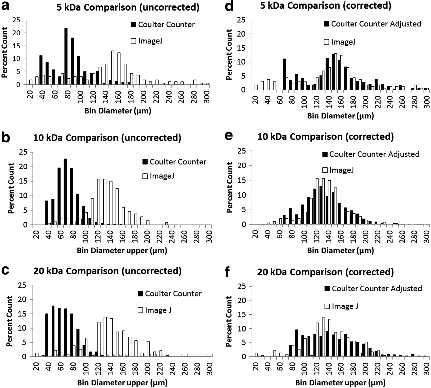
Size distributions of polyethylene glycol diacrylate (PEGDA) hydrogel microspheres as measured by a Coulter counter and image analysis using NIH ImageJ. Identical samples are measured in each graph.
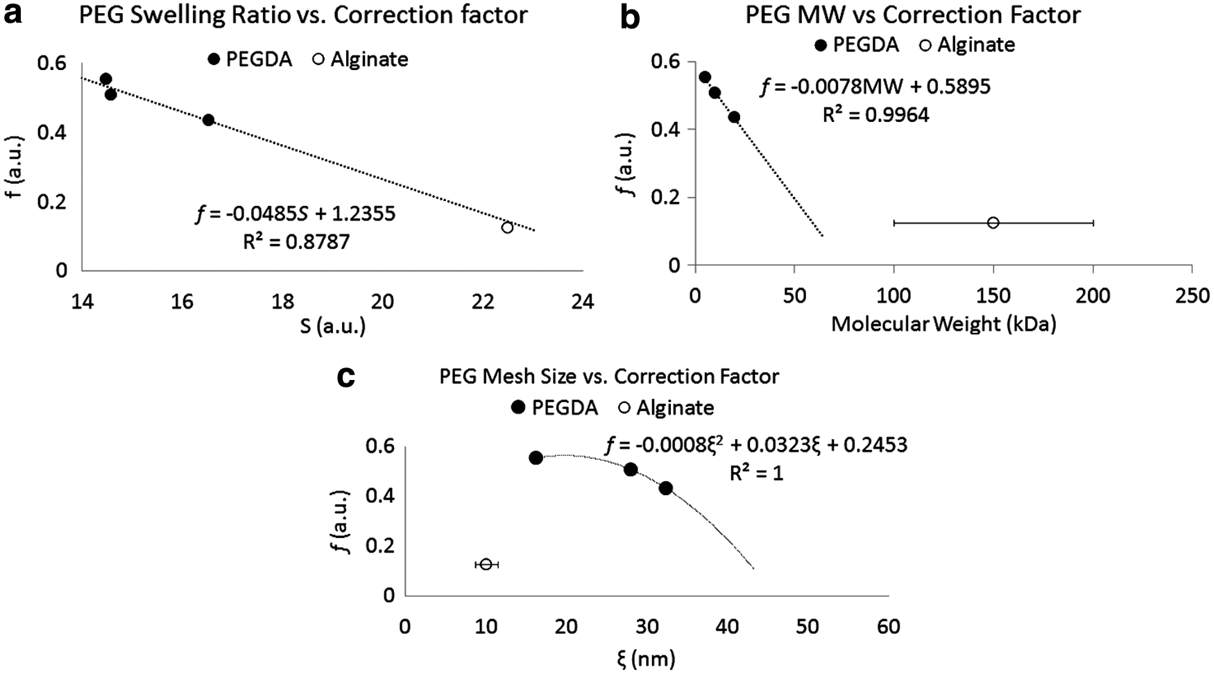
Correction factor, f, plotted against hydrogel physical properties. Trendlines were fit only to PEGDA data, then alginate was plotted on the same graph to evaluate whether fit equations would predict alginate physical properties.
PEGDA, polyethylene glycol diacrylate.
Discussion
The results indicate that when using the Coulter principle to measure hydrogel microparticles, it is important to account for the effects of hydrogel swelling in electrolyte, which results in an apparent transparency of these microparticles to devices that use the Coulter principle. An f correction factor of 1 would indicate that no correction was necessary and that Coulter and imaged microparticle measurements were identical. A correction factor >1 would indicate that Coulter measurements overestimated hydrogel microsphere sizes, while factors <1 indicate the underestimation of the microsphere size. Our results demonstrate decreasing f correction values with an increasing swelling ratio, which show that as the hydrogels absorb more fluid the Coulter measurement becomes more inaccurate. As the particles swell more, greater amounts of electrolyte contribute to the volume of the microspheres, and hence, the Coulter measurements increasingly underestimate these particles. Equation (4) permits the correction of the Coulter measurements for PEGDA and alginate hydrogel microparticles. Future studies should evaluate Equation (4) against other molecular weight alginates and other hydrogel materials.
Conclusions
This correction factor has not been exhaustively evaluated against all of the different types of hydrogels that have been measured with the Coulter principle, which is a limitation of this study. However, the correction factor is important because it is applicable to both PEGDA and a common range of molecular weight alginates used to form microparticles. For alginates of different molecular weight, but with similar swell ratios, the correction factor should still be applicable. Since the majority of studies of microencapsulating mammalian cells use alginate, this correction factor is relevant to these studies. Coulter counting devices are convenient and less time consuming than image analysis; this correction factor enables researchers to continue to use their devices and obtain accurate measurements of their microparticles.
Footnotes
Acknowledgments
The first author was supported by a fellowship from the NASA sponsored NJ Space Grant Consortium. Alginate microspheres were made by Ileana Marrero Berríos of the Yarmush laboratory.
Disclosure Statement
No competing financial interests exist.