
Abstract
Bone marrow stromal cells (BMSCs) include cells with multidirectional differentiation potential described as mesenchymal stem cells. For clinical use, it is important to develop a way to isolate BMSCs from bone marrow in a closed system without centrifugation. After screening 200 biomaterials, we developed a device containing a nonwoven fabric filter composed of rayon and polyethylene. The filter selectively traps BMSCs among mononuclear cells in bone marrow based on affinity, not cell size. The cells are then recovered by the retrograde flow. Using canine and human bone marrow cells, the biological properties of BMSCs isolated by the device were compared with those obtained by conventional methods using centrifugation. The total number isolated by the device was larger, as was the number of CD106+/STRO-1+ double-positive cells. The cells showed osteogenic, chondrogenic, and adipogenic differentiation potential in vitro. Finally, the direct transplantation of cells isolated by the device without in vitro cultivation accelerated bone regeneration in a canine model of osteonecrosis in vivo. The proposed method is rapid and efficient, does not require a biological clean area, and will be useful for the clinical application of mesenchymal stem cells in bone marrow.
Introduction
To avoid the need for centrifugation, we have attempted to develop a device that can isolate BMSCs by filtration, and have selected nonwoven fabrics as biomaterials. Nonwoven fabrics are engineered materials that provide specific functions such as absorbency, liquid repellency, resilience, stretch, softness, strength, flame retardancy, washability, and cushioning. 5 As for clinical applications, nonwoven fabrics are used mainly for two purposes, as scaffolds and filters. A number of studies have used nonwoven fabrics as scaffolds for tissue regeneration.5–7 The material and diameter of the fabric are important for cell attachment.6,8 For example, polyethylene terephthalate with fibers 9.0 μm in diameter is ideal for the osteogenic differentiation of MSCs. 6 As filters, nonwoven fabrics have been used to sterilize medical materials. 9 Lymphocytapheresis is a form of treatment for patients with autoimmune diseases, in which lymphocytes in peripheral blood are removed to reduce immunoreactions. To replace the centrifugation process in lymphocytapheresis, a filtration method using nonwoven fabrics has been developed. 10 The trapping effect is attributed to the affinity of the material for cells and the size of the material's fibers, with almost all leukocytes trapped when the fibers are less than 3 μm in diameter.10,11
As pure mechanical trapping based on cell size can damage cell membranes, we have tried to develop a filter that traps MSCs through affinity and here show that a new device composed of rayon–polyethylene nonwoven fabrics can isolate BMSCs from bone marrow aspirates, which contain cells compatible with multipotent cells in vitro and in vivo.
Materials and Methods
Screening of materials
The basic concept behind our approach is to trap BMSCs among bone marrow aspirates on a filter, and collect them using a retrograde flow (Fig. 1A). Biomaterials for the filter were screened based on microscopic structure (evenness), diameter (over 10 μm), weight (over 50 g/m2), bio-safety, and availability (as the first screening). Potentially suitable biomaterials were further screened by conducting colony-forming unit (CFU) assays described later using swine bone marrow cells in three steps (as the second, third, and fourth screening; Supplemental Fig. S1, available online at
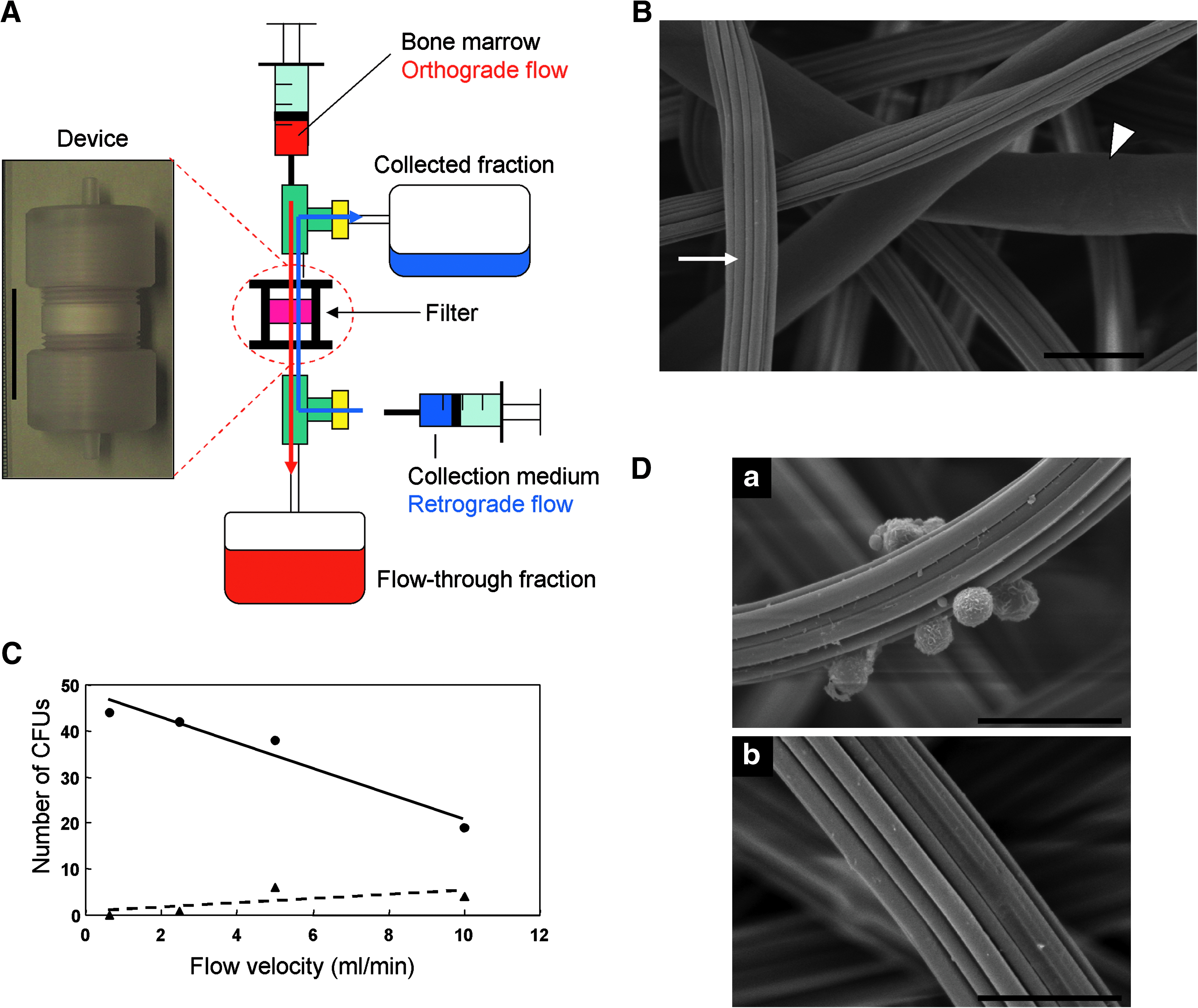
Device containing a nonwoven fabric filter. (
Electron microscopic analyses
After bone marrow had permeated through it, the filter was treated with 2.5 wt% glutaraldehyde in saline for 2 days at 4°C. The samples were dried by passing them through a series of graded-alcohol-saline solutions. Finally, the solution was changed to 100% t-butanol, and the samples were freeze-dried for 10 h at −5°C. The dried membranes were coated with gold–palladium and examined using a S-3000N scanning electron microscope (Hitachi, Tokyo, Japan).
Harvesting of bone marrow
Humans
Ten milliliters of bone marrow was taken from iliac crests of donors who received orthopedic operative procedures requiring autologous bone grafts from iliac crests. The donors had no history of concurrent illness or of medication that could affect bone metabolism. The Ethics Committee of the Faculty of Medicine, Kyoto University, approved the procedure, and informed consent was obtained from each donor according to the declaration of Helsinki.
Dogs
Ten milliliters of bone marrow was aspirated from iliac crests of male beagle dogs (10–13 kg) under intravenous anesthesia. The experiments with animals were approved by the institutional animal research committee, and performed according to the Guidelines for Animal Experiments of Kyoto University.
Preparation of MNCs
Human marrow samples were divided equally into three aliquots (3 mL each). One aliquot was applied to the device (method D), which had been saturated with saline. After washing the filter with 10 mL of saline, trapped cells were collected by a retrograde flow of the culture medium into a collection bag (Fig. 1A). The second aliquot was suspended in 5 mL of α-minimum essential medium GlutaMAX (Invitrogen, Carlsbad CA,) with 10% fetal bovine serum (Hyclone, South Logan, UT). The suspension was centrifuged at 1200 rpm for 5 min. The supernatant and the buffy coat were re-suspended in another tube with 5 mL of α-minimum essential medium GlutaMAX with 10% fetal bovine serum. This method is hereafter designated method S (simple centrifugation).4,12 The final aliquot of bone marrow was fractionated by centrifugation over a density cushion using a Ficoll gradient (GE Healthcare Life Sciences, Piscataway, NJ), as described. 3 The interface layer was harvested and washed twice with 10 mL of Hank's buffer solution. This method is hereafter designated method F (Ficoll gradient).
Collection efficacy
Cells trapped on the filter and collected by retrograde flow in the collection bag were designated the collected fraction, and cells flowing through the filter into the flow-through bag were designated the flow-through fraction (Fig. 1A). Collection efficacy was determined as the ratio of the number of cells in each fraction to that in the applied bone marrow.
CFU assay
For the CFU assay, the MNCs were diluted and plated in 60-mm cell culture dishes at a density of 2.5 × 10⁵ cells/dish and incubated for 14 days (n = 3). The cells were fixed with methanol and stained with a 0.05% crystal violet solution. We counted the number of colony with a diameter more than 4 mm.
Flow cytometry
Cells were stained at room temperature for 30 min with the following antibodies: CD10-phycoerythrin (PE), CD34-allophycocyanin (APC), CD45-peridinin chlorophyll protein (PerCP-Cy5.5), CD106-fluorescein isothiocyanate (FITC), CD166-PE (BD Biosciences, San Jose, CA), CD90-FITC (Beckman Coulter, Fullerton, CA), CD271-APC (Miltenyi Biotec, Bergisch Gladbach, Germany), and STRO-1-PE (Santa Cruz Biotechnology, Santa Cruz, CA). They were subsequently subjected to flow cytometry using a FACS Calibur instrument (BD Biosciences).
Induction of differentiation and histochemical evaluating
Differentiation was induced using the standard method.
13
The differentiation potential was evaluated as below.
Osteogenic differentiation: Calcified nodules were evaluated by alizarin red staining, and calcium content was quantified. Adipogenic differentiation: Intracellular lipid droplets were stained with oil red-O, and the amount of triglyceride was quantified. Chondrogenic differentiation: Cartilage matrix was evaluated by alcian blue staining, and the amount of glycosaminoglycan was quantified. These analyses were described previously.
14
Reverse transcription–polymerase chain reaction
Total RNA was extracted using RNeasy Kit (Qiagen, Hilden, Germany). All reverse transcription reactions were performed using the Super Script First Strand Synthesis System (Invitrogen). Polymerase chain reaction amplification was carried out using rTaq polymerase (Toyobo, Osaka, Japan). All polymerase chain reactions were performed using GeneAmp 9700 (PE Applied Biosystem, Foster City, CA) and specific primers for each gene. 14
Preparation of the canine model of osteonecrosis and cell transplantation
A canine model of scapho-lunate osteonecrosis was prepared as described previously. 15 Briefly, scapho-lunates on one side were exposed dorsally, and a cortical window of 5 × 10 mm was made, through which as much cancerous bone as possible was removed. After the curettage, the cavity was filled with liquid nitrogen for 10 min, and the frozen bone was thawed at room temperature for 10 min. This freeze–thaw procedure was repeated three times. On one side, the bone cavity was filled with beta-tricalcium phosphate (β-TCP) granules (200 μg, 0.2 cm3) (Osferion®; Olympus, Tokyo, Japan) (hereafter designated the control side). On the other side, canine BMSCs (6.0 × 10⁷ ± 1.5 × 10⁷) isolated by the device from 10 mL of bone marrow were mixed with the same amount of β-TCP and transplanted into the bone cavity (hereafter designated the BMSC side). Finally, the bone window was plugged with 5 × 10 mm cortical bone. Animals were sacrificed and evaluated at 4 weeks after the operation (n = 6).
Imaging analyses
X-ray
Antero-posterior and lateral views of both wrists were obtained. The carpal height ratio was calculated from the antero-posterior view.15,16 The Ståhl index was calculated from the lateral view.15,17
Micro-computed tomography
Canine carpal bones were scanned by computed tomography (CT) (SMX-100CT-SV3 type; Shimazu, Kyoto, Japan). The image consisted of 800 slices with a voxel size of 68.224 mm in all three axes. Coronal and sagittal cross-sectional views of the scapho-lunate were reconstructed using adjunctive software. The same setting was used for all samples. The image reconstruction and quantification of the scapho-lunate were performed with VG Studio MAX software (Nihon Visual Science, Tokyo, Japan). 15
Macroscopic and microscopic assessments
The volume and weight of extracted scapho-lunate bones were actually measured using a scale. The samples were fixed in a 10% formalin solution, decalcified with Plank-Rychlo solution, and then embedded in paraffin. Serial sections were cut at 4 μm, and stained with hematoxylin–eosin and silver impregnation.
Using a light microscope, the absorption of β-TCP granules and the formation of bone were graded as follows. 18 For hematoxylin–eosin staining, granules were not surrounded by cells or newly forming bone (grade 1); granules were surrounded by osteoblasts and/or osteoclasts (grade 2); and granules were surrounded by newly forming bone (grade 3). For silver impregnation staining, granules were not completely surrounded by collagen fibrils (grade 1); granules were completely surrounded by collagen fibrils (grade 2); and granules were completely eroded with reticulated collagen fibrils (grade 3). All the β-TCP granules in a whole section were classified according to the three grades. The rates of each grade were then calculated.
Statistics
Results are expressed as the mean ± SE. The statistical analysis was first performed by the analysis of variance (ANOVA), and if there was a significant difference among samples, Turkey's post hoc test was performed for multiple comparisons. The Student's t-test was carried out to compare individual data. A significant difference was accepted at p < 0.05.
Results
Screening of materials
The first screening, of 200 potential biomaterials for the filter, was based on the parameters described in the Materials and Methods section. Forty materials were then selected for the second screening using swine bone marrow cells (n = 2). Ten biomaterials showed CFU counts of more than 10, and were selected for the third screening by CFU assay (n = 3), of which six preceded to the fourth and last screening (n = 3). Finally, a rayon–polyethylene nonwoven fabric was selected as the material for the filter (Supplemental Fig. S1, available online at
Selective collection of BMSCs by the device
To analyze the device's selectivity, cultured human BMSCs or peripheral leukocytes were applied to it, and the cells in the collected fraction and flow-through fractions were enumerated (Fig. 2B). More than 90% of BMSCs were collected in the collected fraction, compared to only 28% of peripheral leukocytes (Fig. 2B). This selectivity was also observed when equal numbers (1 × 10⁶) of BMSCs and peripheral leukocytes were mixed and then applied to the device (Fig. 2C). Because BMSCs and peripheral leukocytes differ in size, we could distinguish between them in the cell counting chamber (Fig. 2A). Approximately 80% of BMSCs were collected in the collected fraction, and an almost equal percentage of peripheral leukocytes were collected in the flow-through fraction (Fig. 2C). More than 90% of CD44+ cells were collected with the device, leaving almost no CD44+ cells in the flow-through fraction, whereas less than 20% of CD45+ cells were collected (Fig. 2D). These results suggested the selective collection of BMSCs by the device.
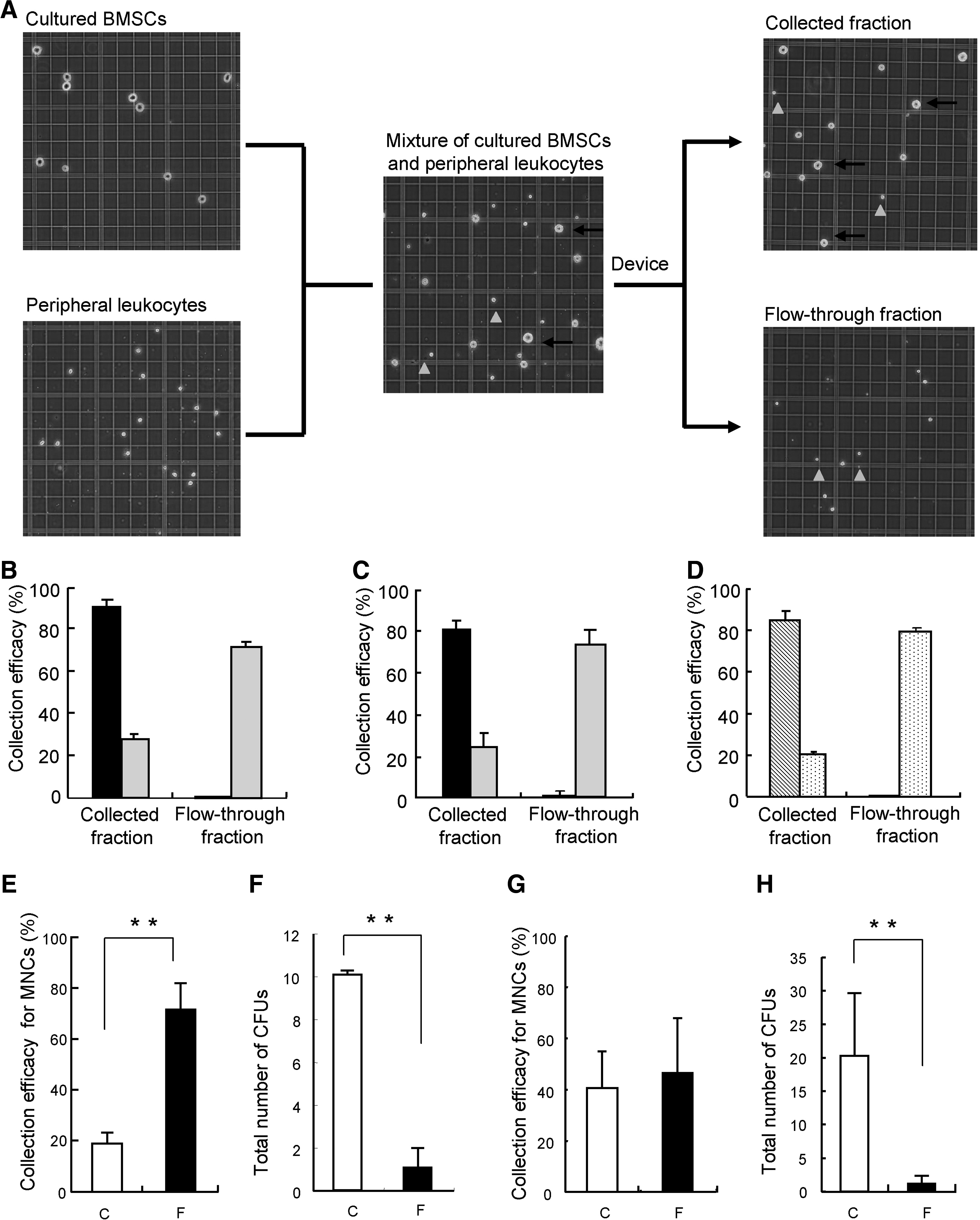
Selective collection of bone marrow stromal cells (BMSCs) with CFUs by the device. (
Selective collection of colony-forming cells from bone marrow by the device
Next, the selectivity of the device was analyzed using cells derived from canine (Fig. 2E, F) and human (Fig. 2G, H) bone marrow. In both cases, most erythrocytes passed through the filter and were collected in the flow-through fraction. Only a few were found in the collected fraction (data not shown). For canine bone marrow, the number of MNCs was significantly smaller in the collected fraction than in the flow-through fraction (p = 0.002; Fig. 2E). The number of CFUs, however, was much higher in the collected fraction (p < 0.001; Fig. 2F). For human bone marrow, the number of MNCs in the collected and flow-through fraction did not differ (Fig. 2G). However, the number of CFUs was much higher in the collected fraction than in the flow-through fraction (p < 0.001; Fig. 2H). These results indicated the selective collection of CFUs by the device.
Comparison of method D with methods S and F
The biological properties of cells isolated by each of the three methods were compared. First, we compared the collection efficacy of method D with that of method S or F. ANOVA revealed significant differences among the collection efficacies for MNCs (canine, p = 0.024; human, p = 0.0002). Comparisons between individual methods were then carried out using Turkey's post hoc test. For canine bone marrow, the collection efficacy for MNCs was significantly lower with method D (20.7 ± 4.2%) than method S (41.6 ± 1.6%) (p < 0.01; Fig. 3A), and the same between method D and method F (16.8 ± 14.0%) (Fig. 3A). As for contamination by erythrocytes, method S showed larger numbers than method D or F. For human bone marrow, there was no difference in collection efficacy between method D (40.5 ± 14.5%) and method S (43.9 ± 10.8%) (Fig. 3B), and method F (7.3 ± 1.8%) was significantly less efficient than method D (p < 0.01; Fig. 3B). The number of contaminating RBCs was again high with method S (Fig. 3B). Next, the MNCs collected by each method were seeded on plastic dishes, and the number of CFUs was counted 14 days later. ANOVA indicated no significant difference among the numbers of CFUs of MNCs prepared by the three methods in canine (p = 0.40) and human bone marrow (p = 0.95) (Fig. 3C, D). From the results on collection efficacy and number of CFUs, the total number of CFUs isolated from bone marrow was calculated for each method. In the case of canine bone marrow, the total number of CFUs with method D (10.1 ± 0.2) was equivalent to that with method S (11.7 ± 4.7) and much larger than that with method F, but not significant (6.6 ± 0.1) (Fig. 3E). Similar results were obtained with human bone marrow cells. The total number of CFUs with method D (20.3 ± 9.4) was equivalent to that with method S (22.3 ± 4.4), and much larger than that with method F (3.6 ± 1.0) (p < 0.01; Fig. 3F). These results indicated that the device collected colony-forming cells from bone marrow with an efficacy equal to that of current methods requiring centrifugation. In addition, the processing time for method D (8.4 ± 1.1 min; Fig. 3G) was significantly shorter than that for the other methods (method S, 18.8 ± 2.9 min; method F, 56.5 ± 3.4 min) (ANOVA, p < 0.001; Turkey's post hoc test, p < 0.01; Fig. 3G).
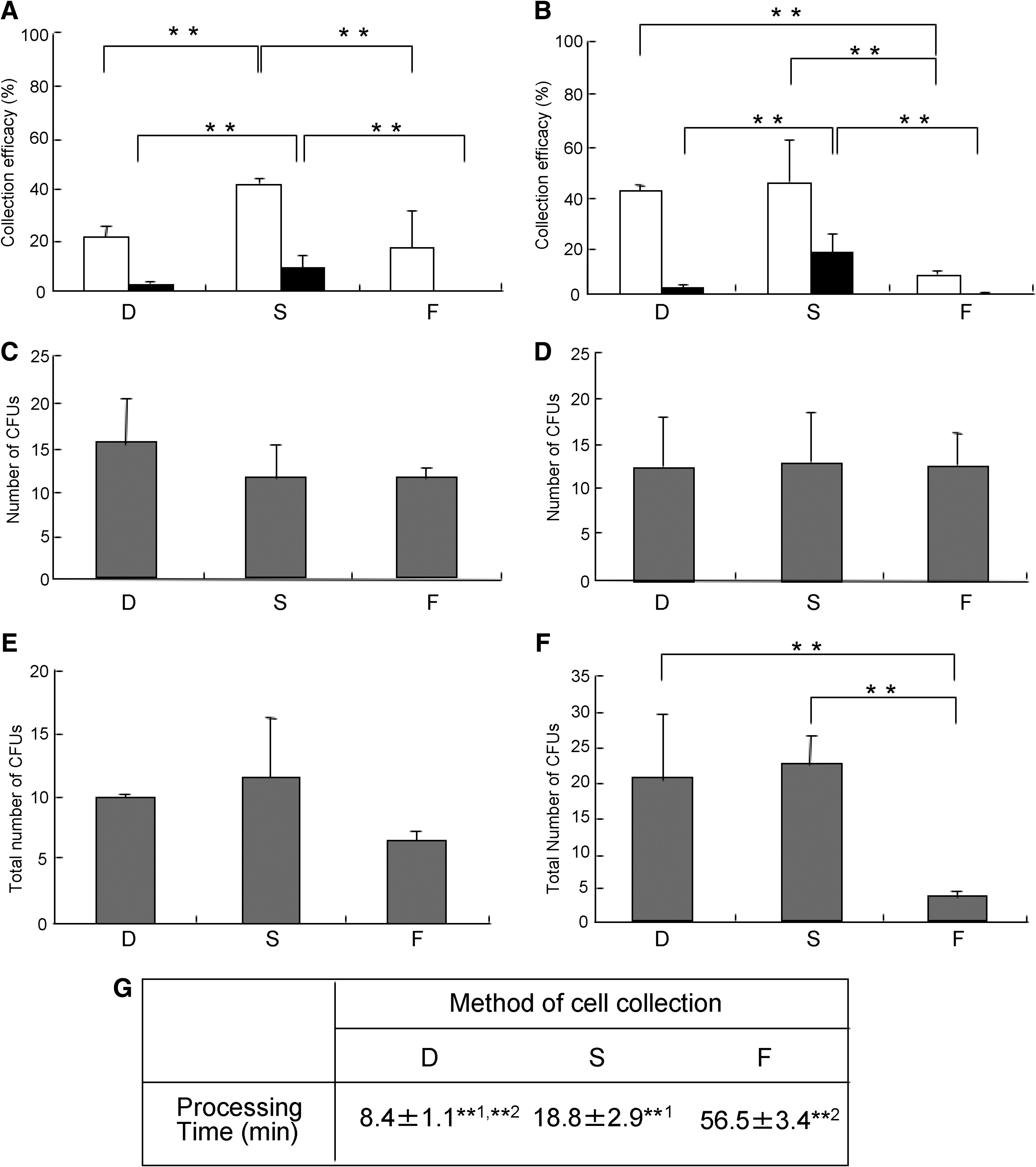
Device selectively prepares MNCs with CFUs. Collection efficacy for MNCs (white box) and erythrocytes (black box) isolated with each method from canine (
Expression of MSC markers in isolated BMSCs
To compare the cell population isolated by each of the three methods, the expression of cell surface markers was analyzed by fluorescence-activated cell sorting (FACS). Because a combination of markers is more reliable, we focused on CD10+/CD271+, CD90+/CD271+, or CD106+/STRO-1+ double-positive cells. The CD90+/CD271+ and the CD106+/STRO-1+ populations were significantly larger among cells collected by method D than those collected by method S or method F (ANOVA, p < 0.001; Turkey's post hoc test, p < 0.01 for CD90+/CD271+, p < 0.05 for CD106+/STRO-1+, respectively; Fig. 4A, B).
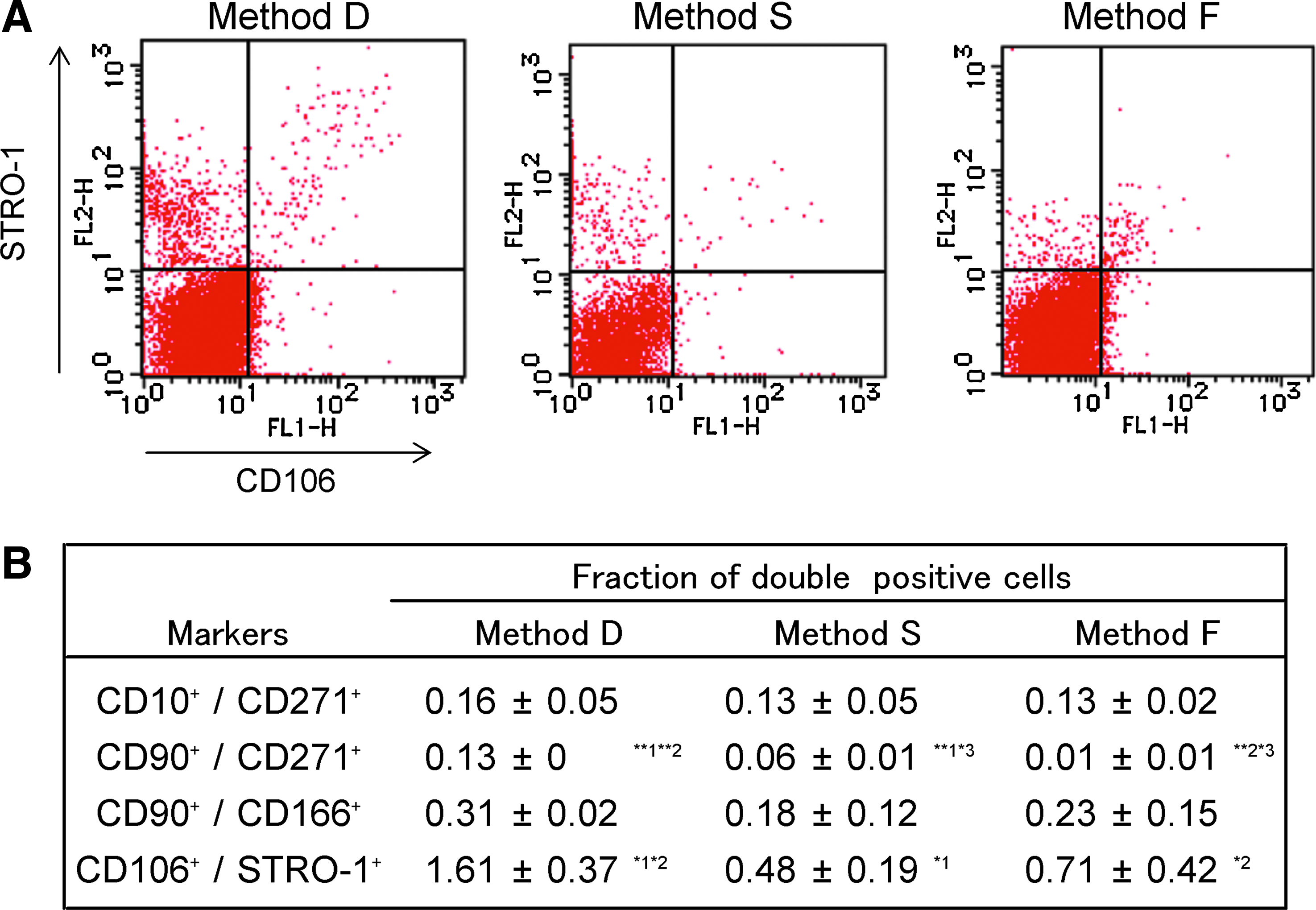
Device selectively prepares MNCs, which express mesenchymal marker. (
Differentiation potential of isolated BMSCs
To analyze their potential to differentiate, the BMSCs isolated by each of the methods were propagated on culture dishes and incubated for 2 weeks under standard growth conditions for MSCs. They were then induced to undergo osteogenesis, adipogenesis, and chondrogenesis. The morphology of the cells isolated by each of the methods was almost the same for both canine (Supplemental Fig. S2A, available online at
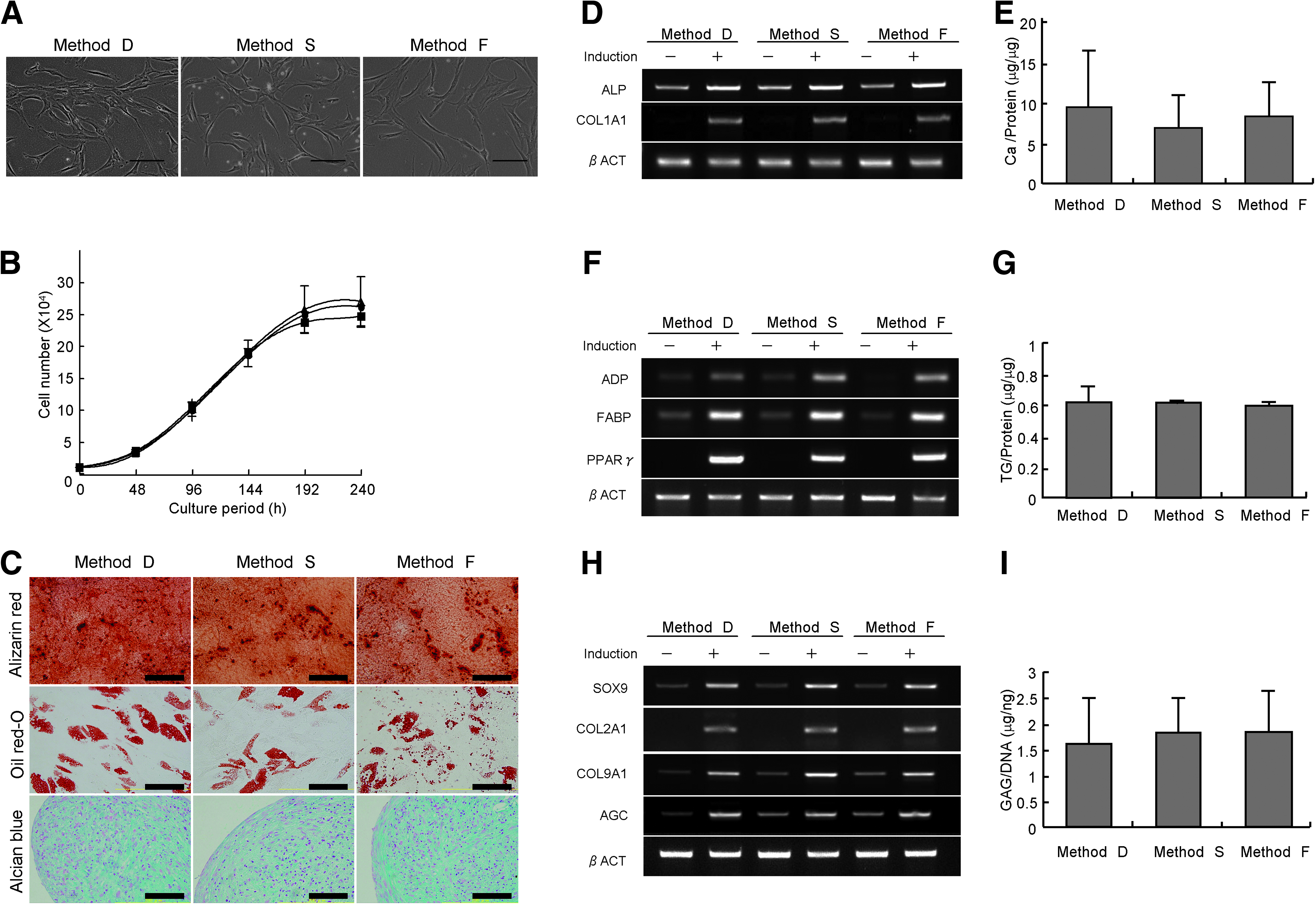
Growth and differentiation potential of human BMSCs isolated by each method. The same numbers of BMSCs isolated by the three methods from human bone marrow were propagated on culture dishes for 2 weeks under standard conditions. (
Regeneration of bone tissues by cells isolated with the device
To confirm the in vivo osteogenic potential of cells collected with the device, we used the cells to regenerate bone tissues in a model of osteonecrosis using canine scapho-lunates, which we established previously. 15 Micro-CT findings at 4 weeks after the operation indicated bone regeneration to be more prominent on the BMSC side than control side in all cases (Fig. 6A, B). Carpal height ratio and the Ståhl index calculated from plain X-rays show the degree of collapse of scapho-lunate bone. Both parameters were significantly higher on the BMSC side than control side (Fig. 6C). Total volume and bone volume were analyzed from the micro-CT data. Bone volume and therefore bone ratio were greater on the BMSC side (Fig. 6C). Both actual weight and volume were also significantly higher on the BMSC side (Fig. 6C). These quantitative analyses suggested that the application of BMSCs prepared by the device contributed significantly to bone regeneration.
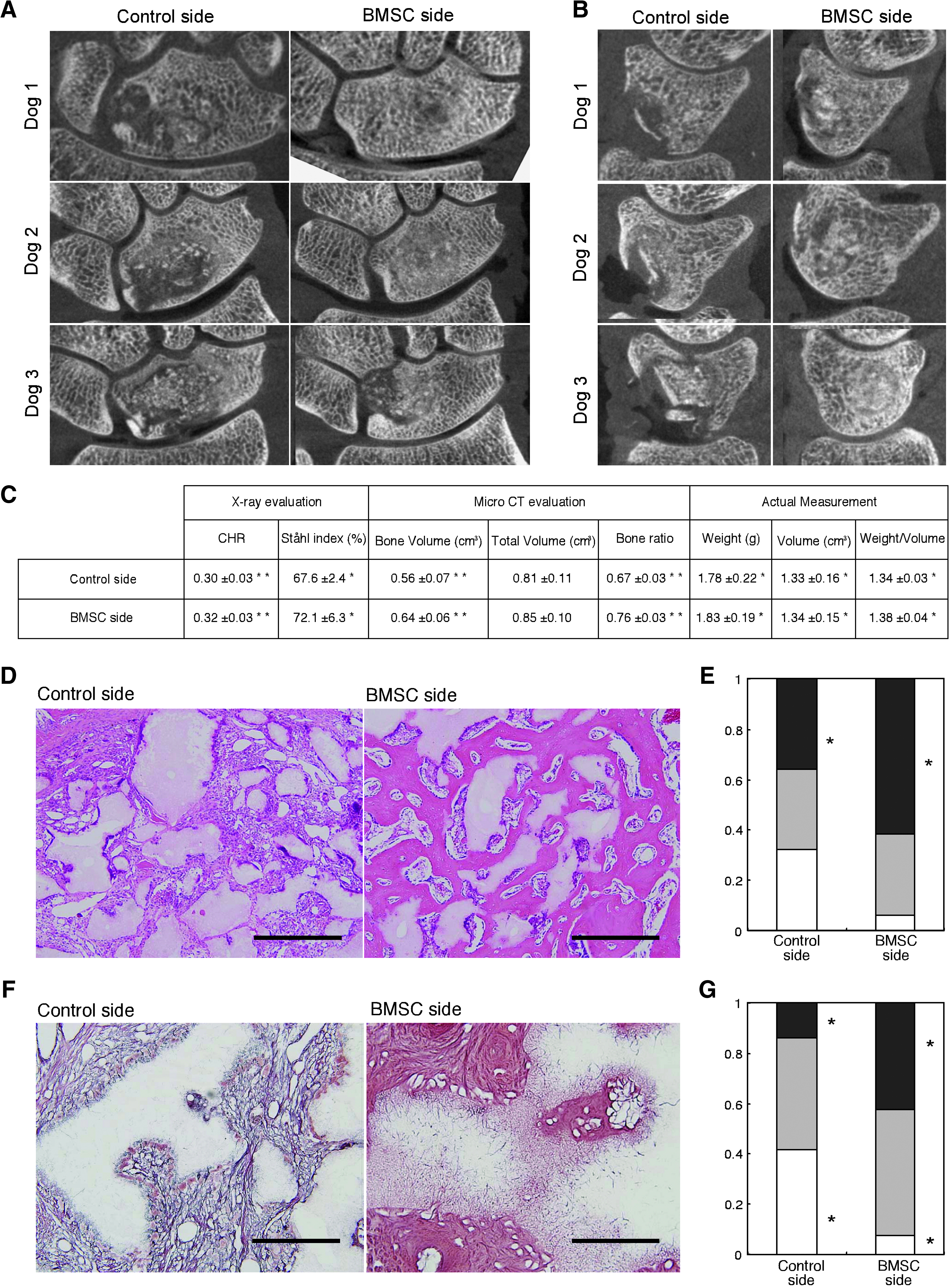
Regeneration of bone tissues by cells isolated with the device. Data for six dogs at 4 weeks after the operation are presented. Frontal (
After the physical and radiological examinations, samples were processed for histological analyses. Microscopic findings of samples taken from the control side showed that a large amount of β-TCP remained unabsorbed, although multinucleated giant cells were found adjacent to β-TCP. In contrast, abundant new bone with the active absorption of β-TCP by multinucleated giant cells was found on the BMSC side (Fig. 6D). The extent to which β-TCP was absorbed as new bone formed was scored using criteria described in the Materials and Methods section. The grade 3 fraction was significantly larger on the BMSC side (p = 0.032; Fig. 6E).
To analyze the interaction between the prepared cells and β-TCP, silver impregnation staining was performed (Fig. 6F). The staining shows the invasion of collagen fibers into biodegradable materials. Collagen fibers in β-TCP granules adjacent to regenerated bone tissue were abundant on the BMSC side, but rare on the control side (Fig. 6F). Quantitative analyses showed the extent of the invasion to be significantly greater on the BMSC side (p = 0.034; Fig. 6G). These results suggested that BMSCs prepared using the device enhanced bone regeneration in vivo.
Discussion
Bone marrow cells consist of two types of MNCs: hematopoietic cells including hematopoietic stem cells, and stromal cells including MSCs. Approximately 99% of MNCs in bone marrow belong to the former population, and among the stromal cells, less than 5% have multidirectional differentiation potential compatible with the concept of MSCs. Therefore, the proportion of MSCs among bone marrow MNCs is estimated at less than 0.05%. 19 To obtain BMSCs including MSCs from bone marrow, it is essential to separate the hematopoietic MNCs.
Takenaka developed a novel filter composed of nonwoven fabrics to trap peripheral leukocytes for the treatment of autoimmune disease. 10 Because erythrocytes are deformed, which allows them to pass through small pores and have low adhesiveness, they can be separated from MNCs by the filter. 10 They used fibers 1.7 μm in diameter, because all the leukocytes were trapped when the fiber was less than 3 μm in diameter. 10 This filter, however, is not applicable to the separation of MSCs from hematopoietic MNCs, both of which will be captured, making it difficult to separate them by cell size. Therefore, we used fibers 15 μm in diameter with greater affinity for BMSCs than hematopoietic MNCs. MSCs are more adherent than hematopoietic cells.4,19 MSCs adhere much more tightly to highly hydrophilic and rough surfaces than to hydrophobic and smooth surfaces. 20 The contact angle of the nonwoven fabric in the current device is estimated to be 20°, which is highly hydrophilic and may make it possible to trap MSCs in the device. This adherent trapping method may prevent excess shearing stress caused by the flow. Cells isolated by the device showed no increase in bradykinine or lactate dehydrogenase (LDH) (data not shown). In addition, mRNA expression of the p16 and p53 genes was not increased, and the karyotype showed no abnormalities (data not shown).
In terms of its potential clinical applications, the device has several advantages. The filter is composed of rayon and polyethylene. Rayon is used for the dialysis membranes of hemodialyzers and known to decrease platelet activity. 21 Polyethylene is a biomaterial employed in total hip replacement and as a scaffold for tissue engineering. 22 Therefore, all the materials are safe for clinical use. The isolation of BMSCs in a closed system is a major advantage of this device. Centrifugation requires mixing with an appropriate solution, which has to be performed in an open system. Current guidelines require the centrifugation process to be performed in an isolated area with specialized equipment. Because all steps can be done in a closed system, the device can isolate BMSCs from bone marrow on the operation table. Further, processing time is shorter than for the other two methods (Fig. 3G), which may help to reduce cell death during the isolation procedures. In addition, the method does not require special skills, which will help to obtain constant results and also be an advantage for clinical application.
We have shown that in terms of differentiation potential, the cells isolated using the device were equivalent to those prepared by current methods using centrifugation. The total number of CFUs obtained from bone marrow was equal to that achieved with method S and higher than that obtained with method F, suggesting that the device recovers almost all cells with the ability to form CFUs. FACS analyses showed that the method was superior to others in terms of the recovery of the MSC marker-positive population. CD10, CD44, CD90, CD106, and STRO-1 are considered surface markers of MSCs,13,23 and, recently, CD271 was identified as a marker of MSCs. 24 The fractions of CD90+/CD271+ cells were larger among cells collected using the device than those isolated with the other two methods (Fig. 4). The CD106+/STRO-1+ fraction (1.61 ± 0.37%; Fig. 4) was even larger than that collected by FACS (1.4 ± 0.3%), 25 which has a risk of microbial contamination and unfavorable effects. We have no clear explanation for this selective collection. Because CD106+/STRO-1+ cells attach to plastic dish efficiently and have high colony-forming activity, 26 one possibility is that CD106+/STRO-1+ cells have high affinity to the filter materials used in the device. Finally, we have shown that the direct application of the trapped cells without ex vivo cultivation assisted the regeneration of bone tissue in vivo (Fig. 6), although the role of transplanted cells may not be simply to reconstitute bone tissues by themselves but also to produce cytokines stimulating host cells, 15 suggesting that BMSCs collected with the new device on the operating table could be used for conditions requiring an acceleration of bone regeneration such as the nonunion of bone fractures.
Footnotes
Acknowledgments
We thank Dr. H. Iwata for suggestions and Drs. M. Neo, S. Fujibayashi, M. Takemoto, H. Ito, and H. Yoshitomi for clinical samples. This work was supported by Grants-in-aid for Scientific Research from the Japan Society for the Promotion of Science; from the Ministry of Education, Culture, Sports, Science, and Technology; from the Ministry of Health, Labor, and Welfare; and from the New Energy and Industrial Technology Development Organization.
Disclosure Statement
No competing financial interests exist.
References
Supplementary Material
Please find the following supplemental material available below.
For Open Access articles published under a Creative Commons License, all supplemental material carries the same license as the article it is associated with.
For non-Open Access articles published, all supplemental material carries a non-exclusive license, and permission requests for re-use of supplemental material or any part of supplemental material shall be sent directly to the copyright owner as specified in the copyright notice associated with the article.