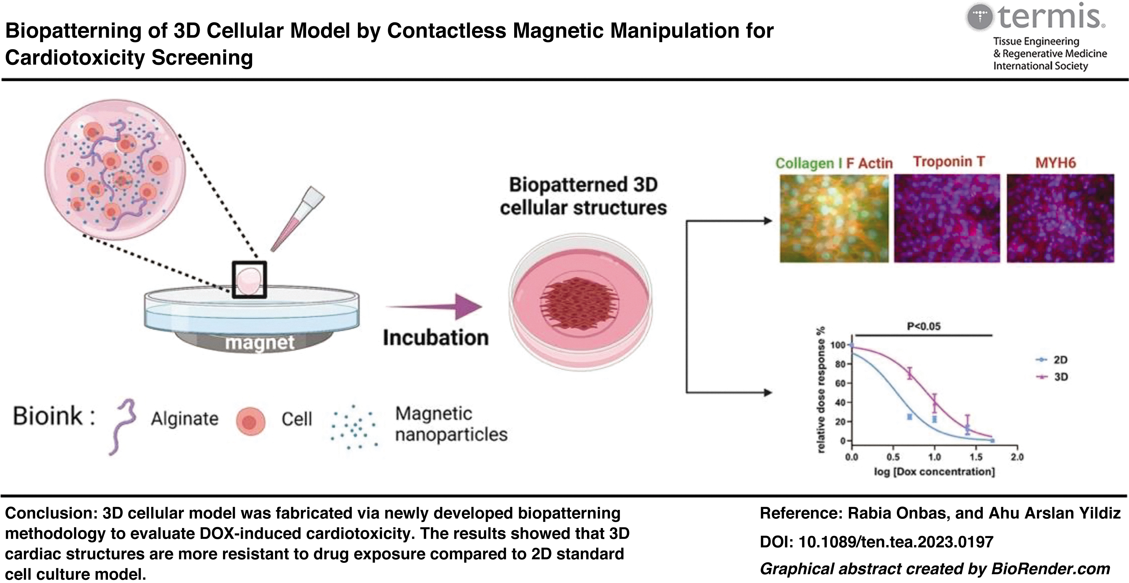
Patterning cells to create three-dimensional (3D) cell culture models by magnetic manipulation is a promising technique, which is rapid, simple, and cost-effective. This study introduces a new biopatterning approach based on magnetic manipulation of cells with a bioink that consists alginate, cells, and magnetic nanoparticles. Plackett-Burman and Box-Behnken experimental design models were used to optimize bioink formulation where NIH-3T3 cells were utilized as a model cell line. The patterning capability was confirmed by light microscopy through 7 days culture time. Then, biopatterned 3D cardiac structures were formed using H9c2 cardiomyocyte cells. Cellular and extracellular components, F-actin and collagen Type I, and cardiac-specific biomarkers, Troponin T and MYH6, of biopatterned 3D cardiac structures were observed successfully. Moreover, Doxorubicin (DOX)-induced cardiotoxicity was investigated for developed 3D model, and IC50 value was calculated as 8.1 μM for biopatterned 3D cardiac structures, which showed higher resistance against DOX-exposure compared to conventional two-dimensional cell culture. Hereby, developed biopatterning methodology proved to be a simple and rapid approach to fabricate 3D cardiac models, especially for drug screening applications.
Impact statement
Contactless manipulation and cell patterning techniques provide rapid and cost-effective three-dimensional (3D) cell culture model formation for tissue engineering applications. The present study introduces a new methodology that comprised alginate-based bioink to pattern cells via contactless magnetic manipulation to fabricate 3D cardiac structures. The developed cardiac model was evaluated in terms of Doxorubicin-induced cardiotoxicity and biopatterned 3D cardiac structures were found more resistant to drug exposure compared to two-dimensional control.
Get full access to this article
View all access options for this article.
References
1.
EschenhagenT, EderA, VollertI, et al.Physiological aspects of cardiac tissue engineering. Am J Physiol Heart Circ Physiol, 2012; 303(2):133–143; doi: 10.1152/AJPHEART.00007.2012
2.
LiX, ZhangR, ZhaoB, et al.Cardiotoxicity screening: A review of rapid-throughput in vitro approaches. Arch Toxicol, 2016; 90(8):1803–1816; doi: 10.1007/S00204-015-1651-1
3.
FosterNC, HallNM, Haj AJEl. Two-dimensional and three-dimensional cartilage model platforms for drug evaluation and high-throughput screening assays. Tissue Eng Part B Rev, 2021; 28(2):421–436; doi: 10.1089/ten.teb.2020.0354
4.
TakedaM, MiyagawaS, FukushimaS, et al.Development of in vitro drug-induced cardiotoxicity assay by using three-dimensional cardiac tissues derived from human induced pluripotent stem cells. Tissue Eng Part C Methods, 2018; 24(1):56–67; doi: 10.1089/ten.tec.2017.0247
5.
PradhanS, HassaniI, ClaryJM, et al.Polymeric biomaterials for in vitro cancer tissue engineering and drug testing applications. Tissue Eng Part B Rev, 2016; 22(6):470–484; doi: 10.1089/ten.teb.2015.0567
6.
BrackenMB. Why animal studies are often poor predictors of human reactions to exposure. J R Soc Med, 2009; 102(3):120–122; doi: 10.1258/jrsm.2008.08k033
7.
ZhangYS, AlemanJ, ArneriA, et al.From cardiac tissue engineering to heart-on-a-chip: Beating challenges. Biomed Mater, 2015; 10(3):034006; doi: 10.1088/1748-6041/10/3/034006
8.
BarnardND, KaufmanSR. Animal research is wasteful and misleading. Sci Am, 1997; 276(2):80–82; doi: 10.1038/SCIENTIFICAMERICAN0297-80
9.
GurskiLA, PetrelliNJ, JiaX, et al.3D matrices for anti-cancer drug testing and development. Oncology Issues, 2010; 25(1):20–25; doi: 10.1080/10463356.2010.11883480
10.
WareMJ, ColbertK, KeshishianV, et al.Generation of homogenous three-dimensional pancreatic cancer cell spheroids using an improved hanging drop technique. Tissue Eng Part C Methods, 2016; 22(4):312–321; doi: 10.1089/ten.tec.2015.0280
11.
ShiW, KwonJ, HuangY, et al.Facile tumor spheroids formation in large quantity with controllable size and high uniformity. Sci Rep, 2018; 8(1):6837; doi: 10.1038/s41598-018-25203-3
12.
GuoW, ChenZ, FengZ, et al.Fabrication of concave microwells and their applications in micro-tissue engineering: A review. Micromachines (Basel), 2022; 13(9):1555; doi: 10.3390/mi13091555
13.
HeW, HalberstadtCR, GonsalvesKE. Lithography application of a novel photoresist for patterning of cells. Biomaterials, 2004; 25(11):2055–2063; doi: 10.1016/j.biomaterials.2003.08.055
14.
ShriraoAB, HussainA, ChoCH, et al.Adhesive-tape soft lithography for patterning mammalian cells: Application to wound-healing assays. Biotechniques, 2012; 53(5):315–318; doi: 10.2144/000113928
15.
HwangH, KangG, YeonJH, et al.Direct rapid prototyping of PDMS from a photomask film for micropatterning of biomolecules and cells. Lab Chip, 2009; 9(1):167–170; doi: 10.1039/B810341K
16.
RothEA, XuT, DasM, et al.Inkjet printing for high-throughput cell patterning. Biomaterials, 2004; 25(17):3707–3715; doi: 10.1016/J.BIOMATERIALS.2003.10.052
17.
ChenP, GüvenS, UstaOB, et al.Biotunable acoustic node assembly of organoids. Adv Healthc Mater, 2015; 4(13):1937–1943; doi: 10.1002/adhm.201500279
18.
WuY, AoZ, ChenB, et al.Acoustic assembly of cell spheroids in disposable capillaries. Nanotechnology, 2018; 29(50):504006; doi: 10.1088/1361-6528/aae4f1
19.
InoK, ItoA, HondaH. Cell patterning using magnetite nanoparticles and magnetic force. Biotechnol Bioeng, 2007; 97(5):1309–1317; doi: 10.1002/bit.21322
20.
FrascaG, GazeauF, WilhelmC. Formation of a three-dimensional multicellular assembly using magnetic patterning. Langmuir, 2009; 25(4):2348–2354; doi: 10.1021/la8030792
21.
SouzaGR, TsengH, GageJA, et al.Magnetically bioprinted human myometrial 3D cell rings as a model for uterine contractility. Int J Mol Sci, 2017; 18(4):683; doi: 10.3390/ijms18040683
22.
Abdel FattahAR, MishrikiS, KammannT, et al.3D cellular structures and co-cultures formed through the contactless magnetic manipulation of cells on adherent surfaces. Biomater Sci, 2018; 6(3):683–694; doi: 10.1039/c7bm01050h
23.
ItoA, HayashidaM, HondaH, et al.Construction and harvest of multilayered keratinocyte sheets using magnetite nanoparticles and magnetic force. Tissue Eng, 2004; 10(5/6):873–880.
24.
ItoA, InoK, HayashidaM, et al.Novel methodology for fabrication of tissue-engineered tubular constructs using magnetite nanoparticles and magnetic force. Tissue Eng, 2005; 11(9/10):1553–1561.
25.
RenT, MaitusongM, ZhouX, et al.Programing cell assembly via ink-free, label-free magneto-archimedes based strategy. ACS Nano, 2023; 17(13):12072–12086; doi: 10.1021/acsnano.2c10704
26.
GuptaT, AithalS, MishrikiS, et al.Label-free magnetic-field-assisted assembly of layer-on-layer cellular structures. ACS Biomater Sci Eng, 2020; 6(7):4294–4303; doi: 10.1021/acsbiomaterials.0c00233
27.
GuptaT, SahuRP, DabaghiM, et al.Biophysical and biochemical regulation of cell dynamics in magnetically assembled cellular structures. ACS Omega, 2023; 8(22):19976–19986; doi: 10.1021/acsomega.3c02052
28.
MishrikiS, AithalS, GuptaT, et al.Fibroblasts accelerate formation and improve reproducibility of 3D cellular structures printed with magnetic assistance. Research, 2020; 2020:3970530; doi: 10.34133/2020/3970530
29.
XiaY, RogersJA, PaulKE, et al.Unconventional methods for fabricating and patterning nanostructures. Chem Rev, 1999; 99(7):1823–1848; doi: 10.1021/cr980002q
30.
QinD, XiaY, WhitesidesGM. Soft lithography for micro- and nanoscale patterning. Nat Protoc, 2010; 5(3):491–502; doi: 10.1038/nprot.2009.234
31.
Martinez-RivasA, González-QuijanoGK, Proa-CoronadoS, et al.Methods of micropatterning and manipulation of cells for biomedical applications. Micromachines, 2017; 8(12):347; doi: 10.3390/MI8120347
32.
ZhangM, KrishnamoorthyS, SongH, et al.Ligament flow during drop-on-demand inkjet printing of bioink containing living cells. J Appl Phys, 2017; 121(12):124904; doi: 10.1063/1.4978744
33.
InoK, OkochiM, HondaH. Application of magnetic force-based cell patterning for controlling cell-cell interactions in angiogenesis. Biotechnol Bioeng, 2009; 102(3):882–890; doi: 10.1002/bit.22104
34.
TsengH, GageJA, HaislerWL, et al.A high-throughput in vitro ring assay for vasoactivity using magnetic 3D bioprinting. Sci Rep, 2016; 6(1):30640; doi: 10.1038/srep30640
35.
TsengH, GageJA, ShenT, et al.A spheroid toxicity assay using magnetic 3D bioprinting and real-time mobile device-based imaging. Sci Rep, 2015; 5:13987; doi: 10.1038/srep13987
36.
PisanicTR, BlackwellJD, ShubayevVI, et al.Nanotoxicity of iron oxide nanoparticle internalization in growing neurons. Biomaterials, 2007; 28(16):2572–2581; doi: 10.1016/j.biomaterials.2007.01.043
37.
BuyukhatipogluK, ChangR, SunW, et al.Bioprinted nanoparticles for tissue engineering applications. Tissue Eng Part C, 2010; 16(4):631–642; doi: 10.1089 = ten.tec.2009.0280
38.
BerryCC, WellsS, CharlesS, et al.Cell response to dextran-derivatised iron oxide nanoparticles post internalisation. Biomaterials, 2004; 25(23):5405–5413; doi: 10.1016/j.biomaterials.2003.12.046
39.
MatijeviE, ScheinerP. Ferric Hydrous Oxide Sols II1. Preparation of Uniform Particles by Hydrolysis of Fe(lll)-Chloride, -Nitrate, and -Perchlorate Solutions. J Colloid Interface Sci, 1978; 63(3):509–524; doi: 10.1016/S0021-9797(78)80011-3
40.
CavazzutiM. Optimization Methods: From Theory to Design Scientific and Technological Aspects in Mechanics. Springer-Verlag: Berlin, Heidelberg;, 2013; doi: 10.1007/978-3-642-31187-1_1
41.
MyersRH, MontgomeryDC, Anderson-CookCM. Response Surface Methodology: Process and Product Optimization Using Designed Experiments. 4th Edition. 2016. pp. 865. SBN: 978-1-118-91601-8.
42.
ZhouN, LiuC, LvS, et al.Degradation prediction model and stem cell growth of gelatin-PEG composite hydrogel. J Biomed Mater Res A, 2016; 104(12):3149–3156; doi: 10.1002/jbm.a.35847
43.
Malakpour-PermlidA, BuzziI, HegardtC, et al.Identification of extracellular matrix proteins secreted by human dermal fibroblasts cultured in 3D electrospun scaffolds. Sci Rep, 2021; 11(1):6655; doi: 10.1038/s41598-021-85742-0
44.
YangJJ, ChenYM, LiuJF, et al.Spontaneous redifferentiation of dedifferentiated human articular chondrocytes on hydrogel surfaces. Tissue Eng Part A, 2010; 16(8):2529–2540; doi: 10.1089/ten.tea.2009.0647
45.
TareRS, HowardD, PoundJC, et al.Tissue engineering strategies for cartilage generation-micromass and three dimensional cultures using human chondrocytes and a continuous cell line. Biochem Biophys Res Commun, 2005; 333(2):609–621; doi: 10.1016/j.bbrc.2005.05.117
46.
TasogluS, YuCH, LiaudanskayaV, et al.Magnetic levitational assembly for living material fabrication. Adv Healthc Mater, 2015; 4(10):1469–1476; doi: 10.1002/adhm.201500092
47.
DeynouxM, SunterN, DucrocqE, et al.A comparative study of the capacity of mesenchymal stromal cell lines to form spheroids. PLoS One, 2020; 15(6):e0225485; doi: 10.1371/journal.pone.0225485
48.
ShichiY, GomiF, SasakiN, et al.Epithelial and mesenchymal features of pancreatic ductal adenocarcinoma cell lines in two- and three-dimensional cultures. J Pers Med, 2022; 12(5):746; doi: 10.3390/jpm12050746
49.
IdaY, HikageF, ItohK, et al.Prostaglandin F2α agonist-induced suppression of 3T3-L1 cell adipogenesis affects spatial formation of extra-cellular matrix. Sci Rep, 2020; 10(1):7958; doi: 10.1038/s41598-020-64674-1
50.
AhmmadB, LeonardK, Shariful IslamM, et al.Green synthesis of mesoporous hematite (α-Fe2O 3) nanoparticles and their photocatalytic activity. Adv Powder Technol, 2013; 24(1):160–167; doi: 10.1016/j.apt.2012.04.005
51.
PalmA. Raman spectrum of polystyrene. J Phys Chem, 1951; 55(8):1320–1324.
52.
HongistoV, JernströmS, FeyV, et al.High-throughput 3D screening reveals differences in drug sensitivities between culture models of JIMT1 breast cancer cells. PLoS One, 2013; 8(10):e77232; doi: 10.1371/journal.pone.0077232
RichardS, SilvaAKA, MaryG, et al.3D magnetic alignment of cardiac cells in hydrogels. ACS Appl Bio Mater, 2020; 3(10):6802–6810; doi: 10.1021/acsabm.0c00754
56.
TimmDM, ChenJ, SingD, et al.A high-throughput three-dimensional cell migration assay for toxicity screening with mobile device-based macroscopic image analysis. Sci Rep, 2013; 3:3000; doi: 10.1038/srep03000
57.
ItoA, TakizawaY, HondaH, et al.Tissue engineering using magnetite nanoparticles and magnetic force: Heterotypic layers of cocultured hepatocytesand endothelial cells. Tissue Eng, 2004; 10(5/6):833–840.
58.
ItoA, HibinoE, KobayashiC, et al.Construction and delivery of tissue-engineered human retinal pigment epithelial cell sheets, using magnetite nanoparticles and magnetic force. Tissue Eng, 2005; 11(3/4):489–496.
59.
ElliottNT, YuanF. A review of three-dimensional in vitro tissue models for drug discovery and transport studies. J Pharm Sci, 2011; 100(1):59–74; doi: 10.1002/jps.22257
60.
SardãoVA, OliveiraPJ, HolyJ, et al.Morphological alterations induced by doxorubicin on H9c2 myoblasts: Nuclear, mitochondrial, and cytoskeletal targets. Cell Biol Toxicol, 2009; 25(3):227–243; doi: 10.1007/S10565-008-9070-1
61.
ImamuraY, MukoharaT, ShimonoY, et al.Comparison of 2D- and 3D-culture models as drug-testing platforms in breast cancer. Oncol Rep, 2015; 33(4):1837–1843; doi: 10.3892/OR.2015.3767/HTML
62.
LiuH, ZhangX, LiuJ, et al.Vascularization of engineered organoids. BMEMat, 2023; 1(3):e12031; doi: 10.1002/bmm2.12031
63.
CukiermanE, PankovR, StevensDR, et al.Taking cell-matrix adhesions to the third dimension. Science, 2001; 294(5547):1708–1712; doi: 10.1126/science.1064829
64.
PampaloniF, ReynaudEG, StelzerEHK. The third dimension bridges the gap between cell culture and live tissue. Nat Rev Mol Cell Biol, 2007; 8(10):839–845; doi: 10.1038/NRM2236
65.
GriffithLG, SwartzMA. Capturing complex 3D tissue physiology in vitro. Nat Rev Mol Cell Biol, 2006; 7(3):211–224; doi: 10.1038/nrm1858
66.
DuvalK, GroverH, HanLH, et al.Modeling physiological events in 2D vs. 3D cell culture. Physiology (Bethesda), 2017; 32(4):266–277; doi: 10.1152/PHYSIOL.00036.2016
67.
HuX, LiS, PengP, et al.Prosthetic heart valves for transcatheter aortic valve replacement. BMEMat, 2023; 1(2):e12026; doi: 10.1002/bmm2.12026
Supplementary Material
Please find the following supplemental material available below.
For Open Access articles published under a Creative Commons License, all supplemental material carries the same license as the article it is associated with.
For non-Open Access articles published, all supplemental material carries a non-exclusive license, and permission requests for re-use of supplemental material or any part of supplemental material shall be sent directly to the copyright owner as specified in the copyright notice associated with the article.