
Abstract
Acute chest syndrome (ACS) is a frequent cause of acute lung disease in children with sickle cell disease (SCD). Patients may present with ACS or may develop this complication during the course of a hospitalization for acute vaso-occlusive crises (VOC). ACS is associated with prolonged hospitalization, increased risk of respiratory failure, and the potential for developing chronic lung disease. ACS in SCD is defined as the presence of fever and/or new respiratory symptoms accompanied by the presence of a new pulmonary infiltrate on chest X-ray. The spectrum of clinical manifestations can range from mild respiratory illness to acute respiratory distress syndrome. The presence of severe hypoxemia is a useful predictor of severity and outcome. The etiology of ACS is often multifactorial. One of the proposed mechanisms involves increased adhesion of sickle red cells to pulmonary microvasculature in the presence of hypoxia. Other commonly associated etiologies include infection, pulmonary fat embolism, and infarction. Infection is a common cause in children, whereas adults usually present with pain crises. Several risk factors have been identified in children to be associated with increased incidence of ACS. These include younger age, severe SCD genotypes (SS or Sβ0 thalassemia), lower fetal hemoglobin concentrations, higher steady-state hemoglobin levels, higher steady-state white blood cell counts, history of asthma, and tobacco smoke exposure. Opiate overdose and resulting hypoventilation can also trigger ACS. Prompt diagnosis and management with intravenous fluids, analgesics, aggressive incentive spirometry, supplemental oxygen or respiratory support, antibiotics, and transfusion therapy, are key to the prevention of clinical deterioration. Bronchodilators should be considered if there is history of asthma or in the presence of acute bronchospasm. Treatment with hydroxyurea should be considered for prevention of recurrent episodes. This review evaluates the etiology, pathophysiology, risk factors, clinical presentation of ACS, and preventive and treatment strategies for effective management of ACS.
Introduction
A
Etiology and Pathophysiology
ACS is a form of acute lung injury in SCD. The development of ACS represents a vicious cycle of lung infarction, inflammation, and atelectasis leading to ventilation–perfusion mismatch, hypoxemia, and acute increases in pulmonary artery and right ventricular pressures. 3 At the cellular level, in the presence of low alveolar oxygen tension, abnormal rheology of the sickled red blood cells (sRBCs) facilitates adhesion to each other, leukocytes, and the vascular endothelium, resulting in vaso-occlusion and tissue hypoxia. These interactions also cause the release of inflammatory cytokines which promote acute and chronic inflammation in the airways by virtue of being in close proximity with the vasculature. 4 Evidence for this pathophysiological mechanism comes from markedly elevated plasma levels of vascular cell adhesion molecule-1, an adhesion receptor on endothelial cells, in patients with ACS along with significantly lower levels of cytoprotective factors such as nitric oxide (NO) metabolites. 5 The three major identifiable etiologies for the development of ACS are pulmonary infections, pulmonary fat embolism from necrotic bone marrow, and pulmonary infarction as outlined in Fig. 1. 6
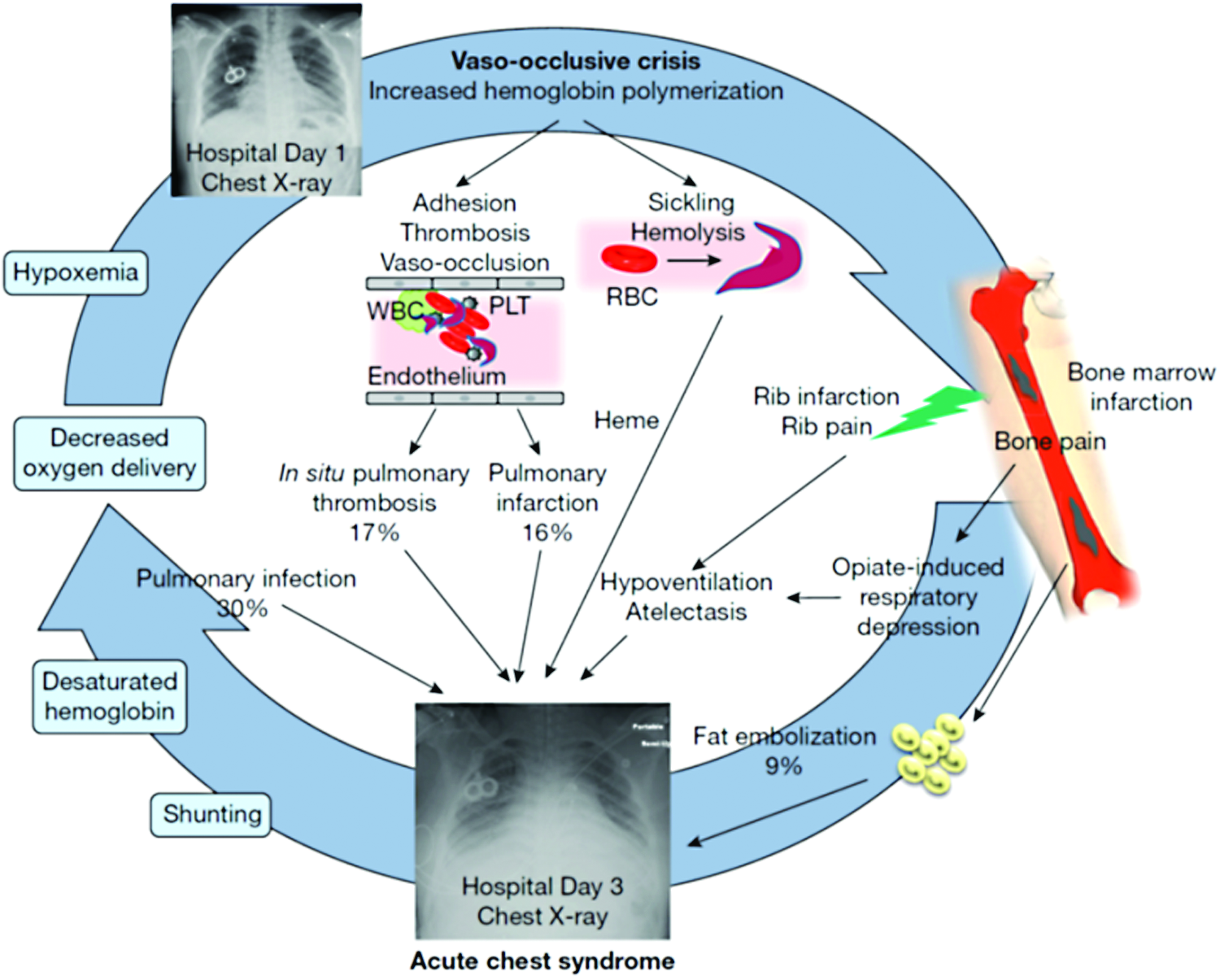
A vicious cycle in the pathogenesis of ACS, involving RBC sickling, cellular adhesion, hemolysis, and vaso-occlusion. Common causes for the development of ACS include fat embolism, pulmonary infection, pulmonary infraction, and hypoventilation. Reprinted from Chest, 149 (4), Enrico M. Novelli and Mark T. Gladwin, Crises in Sickle Cell Disease, 1082–1093, Copyright (2016), with permission from Elsevier. ACS, acute chest syndrome; PLT, platelet; RBC, red blood cell; WBC, white blood cell. Color images available online at www.liebertpub.com/ped
Pulmonary infection
The multicenter National Acute Chest Syndrome Study (NACSS) evaluated 671 ACS episodes in 538 patients and nearly half of the patients were children and adolescents. 6 A specific cause for ACS was found in only 38% of episodes with infection as a leading etiology in children. The infectious organisms which were identified in the study included Chlamydia pneumoniae, Mycoplasma pneumoniae, respiratory syncytial virus (RSV), Staphylococcus aureus, Streptococcus pneumoniae, and other pathogens in decreasing order of frequency. Viral infections were more common in children <10 years of age. Infections can precipitate an ACS episode in a susceptible SCD patient by inducing an excessive inflammatory lung injury response in the presence of a damaged lung microvasculature.5–8 This has been further demonstrated in transgenic mouse models of HbSS, which develop lung injury at low endotoxin levels that do not adversely affect the wild-type mice.9,10 The incidence of S. pneumoniae infection in the recent years has decreased as more effective vaccines are available and widely used. Influenza, notably the H1N1 strain, also produces seasonal outbreaks of ACS and may cause severe disease. 11 Parvovirus B19 through its association with bone marrow infarction/necrosis and potentially pulmonary fat embolism can also result in ACS. 12
Pulmonary fat embolism
Pulmonary fat embolism, with or without pulmonary infection, can cause ACS in both adults and children. 6 During acute pain episodes, infarcted and necrosed bone marrow (especially of the pelvis and femur) release fat droplets in the bloodstream, which embolize to the lungs. Fat emboli in the lung vasculature are metabolized to free fatty acids, including secretory phospholipase A2 (sPLA2), which mediate alveolar inflammation and endothelial injury. 13 Diagnosis of pulmonary fat embolism in NACSS was based on quantitative evaluation of oil red-positive fat-laden alveolar macrophages in bronchoalveolar lavage (BAL) or induced sputum specimens. Although this method is sensitive for the diagnosis of pulmonary fat embolism, its specificity and clinical usefulness is questionable since fat-laden alveolar macrophages are also associated with microaspiration, gastroesophageal reflux-related respiratory disease, and hyperlipidemia.14–16 The presence of these additional factors can influence the detection of the fat-laden macrophages.
Pulmonary infarction and in situ thrombosis
Pulmonary infarction is another major contributor to the development of ACS and was determined to be the cause in 16% of ACS episodes in NACSS. This occurs as a result of increased adhesion of sRBCs to the endothelium causing vaso-occlusion, which leads to ventilation–perfusion mismatch and exacerbation of hypoxemia. 17 In a small subset of patients, computed tomography (CT)–pulmonary angiography identified subsegmental thromboembolism in 17% of ACS episodes without associated peripheral thrombosis, possibly suggestive of in situ thrombosis leading to infarction. 18 Furthermore, there is a high prevalence of pulmonary embolism in patients with SCD associated with thrombosis in the distal venous circulation.19,20 This underappreciated entity may contribute to the pulmonary symptoms associated with ACS and also increase the risk of mortality.6,21
Hypoventilation
Splinting caused by rib infarction can lead to decreased respiratory effort resulting in hypoventilation and atelectasis. 22 This may be further exacerbated by excessive opioid administration or following general anesthesia.6,23 As a result, pooling of secretions at lung bases occurs and predisposes to ventilation–perfusion mismatch and tissue hypoxia.
Biomarkers of ACS
Some investigators have sought to define objective biomarkers to predict the occurrence of ACS based on the understanding of the pathophysiology. Serum concentration of sPLA2 was shown to be elevated in a small group of patients with ACS and also correlated with clinical severity. 13 In a follow-up study, elevation was detected even 24–48 h before clinical onset of ACS in children during hospitalization for VOC. 24 These findings have been variably replicated with elevated levels found in 80% of subjects with ACS participating in a multicenter trial, 25 but with a poor positive predictive value of 24% in another cohort of patients hospitalized for acute pain episodes. 26 Moreover, the time it takes to perform the test makes it impractical for clinical use as enzyme-linked immunosorbent assay-based assays are not widely available in clinical laboratories. 24
C-reactive protein, an acute-phase reactant, was suggested as a surrogate for sPLA2 and was found to have serum concentrations that paralleled those of sPLA2 during the hospital course of 20 patients with SCD admitted for VOC and ACS. 27 Whether CRP can be used as a substitute for predicting development of ACS in patients with VOC is not clear. Another acute-phase reactant, pentraxin-related protein (PTX3), has been found to be elevated in SCD patients with VOC, similar to the levels seen in other inflammatory and ischemic states. 28 Patients with higher initial PTX3 concentrations have been found to be more likely to develop ACS and were observed to have further increase in PTX3 levels upon progression to ACS. 29 But there is a lack of feasibility in using these biomarkers in the clinical setting to predict occurrence of ACS as these tests are not widely available.
Clinical Presentation
A classical episode of ACS is defined as an acute illness characterized by the finding of a new segmental pulmonary infiltrate consistent with consolidation, but not atelectasis along with one or more new respiratory symptoms or signs, such as cough, chest pain, fever (>38.5°C), hypoxemia, and tachypnea.8,30 Various other definitions of ACS have been used in clinical practice and research, but none considers rate of decline of respiratory status as a diagnostic criteria and thus lack specificity. 31 ACS is often a clinical diagnosis especially in the presence of hypoxemia as radiographic changes may lag behind clinical findings. It can mimic bacterial pneumonia, asthma exacerbation, or bronchiolitis at presentation. It can also progress rapidly from mild hypoxemia to acute respiratory failure within 24 h of onset of respiratory symptoms, which in itself constitutes a distinct clinical phenotype of ACS.6,30,32 This form of rapidly progressive ACS is seen more commonly in adults as compared with children with SCD, and is frequently associated with thrombocytopenia and multiorgan failure.32–34 The etiology and underlying pathophysiological mechanisms of this subgroup of ACS are unclear, but shares resemblance with other thrombotic microangiopathic syndromes.32,34,35
The presentation, causal mechanisms, and natural course of ACS differ considerably with age. The highest incidence of ACS is in children <10 years of age 36 with frequently occurring triggers in the form of pulmonary infections. 6 As a result, they usually present with fever, cough, and wheeze. Older children and adults more frequently present with dyspnea and chest pain and tend to follow a more severe course.6,30 Adults also tend to present with lower mean oxygen saturations because of a higher prevalence of pulmonary fat embolism. 6 Children usually present with isolated upper or middle lobe involvement as compared with bilateral lower or multiple lobes in adults. 6
A close temporal relationship exists between ACS and VOC. Nearly half of all ACS episodes occur between 1 and 3 days after admission for severe VOC, 6 suggesting that VOC may be a prodromal event for the development of ACS. A retrospective report from a single institution revealed that approximately a third of patients who developed ACS had sought acute care for painful VOCs (mostly chest and back) within 2 weeks of hospitalization for ACS. 37 Buchanan et al. 38 have suggested that opioid selection for the treatment of VOC episodes may have an impact on the development of ACS, with patients receiving Nubain (Nalbuphine) less likely to develop ACS; however, their results were confounded by the use of continuous infusion analgesia. This observation has not been studied in a prospective clinical trial.
Neurological complications, such as infarctive stroke, silent cerebral infarcts, and posterior reversible leukoencephalopathy syndrome, have been shown to be higher after severe episodes of ACS in children.39–41 These neurological events have also been seen with platelet counts <200,000 per cumm in children hospitalized with ACS. 6
Risk Factors
Several prospective and retrospective studies have attempted to identify risk factors for the prediction of development, recurrence, and severity of ACS. Based on these data, Table 1 lists risk factors that have been found to be associated with the development of ACS episodes.
Hb, hemoglobin; HbF, fetal hemoglobin; HbS, sickle hemoglobin; HbSS, homozygous SCD; VOC, vaso-occlusive crises; WBC, white blood cell.
In the prospective multicenter Cooperative Study of Sickle Cell Disease (CSSCD), younger age, lower fetal hemoglobin (HbF) concentration, higher steady-state Hb, and higher steady-state white blood cell (WBC) counts were found to be significant risk factors for ACS. 36 Those with more severe genotypes (HbSS and HbSβ0 thalassemia) had a greater risk of developing ACS than the milder ones (HbSC and HbSβ+ thalassemia). 36 In a retrospective study on 175 pediatric patients hospitalized for VOC, a significantly higher WBC (19.1 ± 4.8/cmm versus 13.2 ± 5.2/cmm) and lower admitting Hb level from baseline (7.7 ± 1.2 g/dL versus 9.3 ± 2.0 g/dL) were found in those who developed ACS. 38 These data suggest that a higher steady-state Hb level likely promotes VOC by increasing blood viscosity, and acute hemolysis following VOC may contribute to the development of lung injury.
Higher levels of free heme (nonprotein bound) in plasma, a byproduct of hemolysis, were associated with increased odds of developing ACS [odds ratio (OR) = 2.56, P = 0.016] in a cohort of children with SCD. 42 This observation is supported by the recent findings of a polymorphism in the promoter region of the heme oxygenase-1 gene, involved in catabolism of heme, to be associated with degree of hemolysis and severity of ACS. 43 A recent study in sickle cell mouse model suggests that extracellular hemin (the oxidized prosthetic moiety of Hb) may contribute to ACS pathogenesis through toll-like receptor and provides a proof of principle for using recombinant hemopexin (a scavenger protein of free heme) as a therapeutic strategy for ACS in mice. 44
In a large cross-sectional study, Pulmonary Hypertension and the Hypoxic Response in SCD, involving pediatric patients with HbSS and other sickle cell genotypes (N = 503), acute pulmonary events (either ACS or pneumonia) were found to be independently associated with >3 severe pain episodes in the preceding year irrespective of the genotype. 45 In patients with HbSS, significant associations for development of acute pulmonary events were also found with history of asthma, higher steady-state tricuspid regurgitant velocity (TRV), and higher WBC count. The prognostic significance of higher TRV in children is unclear as it has not been found to be particularly associated with ACS in other studies.46–48
Another risk factor that has been shown to predict recurrence of ACS episodes is a history of ACS in younger age, especially in the first 3 years of life, as reported by two independent cohorts of pediatric patients.49,50 Moreover, the highest incidence rate of recurrence has been reported to be within 6 months from the last event. 51 Perhaps pulmonary injury from previous ACS episodes predisposes to increased susceptibility to recurrent events or alternatively a genetic predisposition underlies the development of frequent ACS episodes. 52 A genetic association with ACS in patients with SCD, particularly in the younger age group, has been identified in the form of a single nucleotide involving COMMd7, which is a gene highly expressed in the lung that interacts with nuclear factor-κB signaling to control inflammatory responses. 53
Certain risk factors are danger signals for a severe disease course, such as an arterial:alveolar oxygen gradient of >30 (on room air) as reported from a small series in children. 54 Multilobar involvement, platelet count of <200,000/cumm at diagnosis, and history of cardiac disease were found to be independent predictors of respiratory failure in the NACSS study. 6 A decrease in platelet count of more than 10% from steady state when admitted for VOC was also reported to be a significant predictor for developing ACS (OR = 6.90) in a retrospective study of adult patients with SCD. 55
General anesthesia, asthma/airway hyperreactivity, and smoking may act as triggers for the development of ACS in SCD. Patients undergoing an abdominal surgery, such as splenectomy and/or cholecystectomy, are especially at risk for developing ACS postoperatively secondary to hypoventilation/atelectasis. 56 Tobacco smoke exposure (TSE) either primary in adolescents and adults or secondary (at all ages) has been shown to be associated with increased rates of VOC and ACS.57–60 The mechanisms underlying the association between TSE and ACS, particularly in young children, could be related to impairment of lung growth/development in utero from secondhand TSE, increased viral respiratory illnesses, airway inflammation, and increased expression of adhesion markers. Secondhand smoke exposure can also cause pulmonary function abnormalities such as lower airway obstruction in children with SCD. 61
Asthma/airway hyperreactivity and ACS
Asthma is common in children with SCD with occurrence estimates of 15%–28%62–64 and at least 55% have airway hyperreactivity without a clinical diagnosis of asthma.65–68 Asthma exacerbations in children with SCD have been associated with increased incidence of ACS episodes at a young age (median age of 2.4 years).62,65,69–75 Those with asthma are five times more likely (OR = 4.9, 95% confidence interval = 2.2–10.7) to present with respiratory symptoms at the time of a VOC. 71 Several plausible explanations for the relationship between asthma and ACS or VOC are, ventilation–perfusion mismatch, synergistic inflammatory response, and vascular leak. Although the importance of comorbid asthma in SCD outcomes is being increasingly recognized, there still remain substantial gaps in diagnosis and management.
Management
All patients with ACS should be hospitalized for careful monitoring of their oxygenation and clinical status. Patients benefit from early, aggressive intervention especially in the presence of risk factors such as multilobar involvement, coexisting respiratory diseases, and for whom blood is not readily available. Table 2 offers guidance for management based on an expert panel report from National Heart, Lung, and Blood Institute (NHLBI), 76 which can be effectively put into clinical practice and lead to rapid resolution in majority of patients. As part of the initial workup, a child with SCD presenting with fever should have a CBC, reticulocyte count, blood culture, basic biochemistry, type and crossmatch, and a chest X-ray, irrespective of respiratory symptoms. A screening algorithm for ACS in pediatric patients with SCD is presented schematically in Fig. 2. Rapid screening tests in nasopharyngeal aspirates for RSV, influenza A (including H1N1 subtype), and influenza B, in season should be performed.
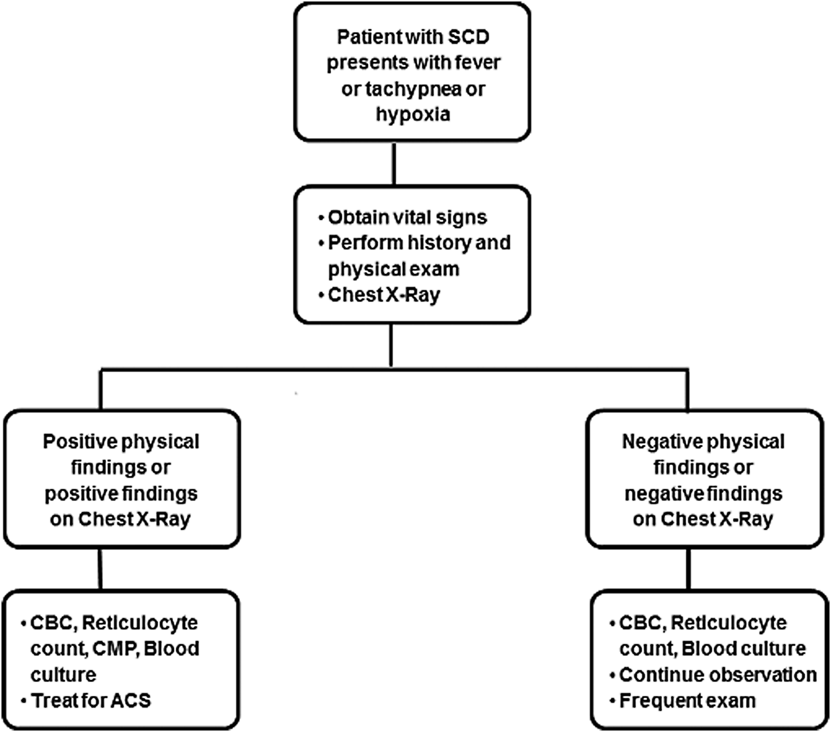
Screening algorithm for ACS in pediatric patients with SCD. CBC, complete blood count, CMP, comprehensive metabolic panel; SCD, sickle cell disease.
NSAID, nonsteroidal anti-inflammatory drug; pRBC, packed red blood cell.
General principles of management in all patients with ACS are supportive care with hydration, antibiotics, pain control, and maintenance of oxygenation and ventilation. Hydration is generally given with intravenously administered hypotonic fluids at approximately three-fourths of a maintenance rate as patients with ACS may be at risk for pulmonary edema. 77
The use of aggressive incentive spirometry while awake to prevent atelectasis is an important aspect of management.78,79 Alternatively, the use of mechanical chest physiotherapy maneuvers, such as positive expiratory pressure devices, may be considered in younger children who may not be able to coordinate incentive spirometry or is limited by chest wall pain.80,81 The use of respiratory therapy warrants further study in patients with SCD at high risk for ACS. Supplemental oxygen should be given for oxygen saturation ≤94% and/or tachypnea. It is important to note that patients might have chronic hypoxemia as a result of conditions such as pulmonary hypertension and chronic sickle lung disease, but any deterioration in oxygen saturation from baseline steady state warrants prompt action. Noninvasive ventilation such as bilevel positive airway pressure may be used to improve oxygenation and decrease work of breathing in patients with ACS. These benefits may prevent progression to respiratory failure and reduce the need for intubation and mechanical ventilation, but there is currently limited evidence regarding its precise role in managing children with ACS. 82 Indications for more intensive respiratory support include worsening hypoxemia, severe dyspnea, and respiratory acidosis (pH <7.35).
All patients with ACS should be started on empiric antibiotic coverage for pneumococcal infection, commonly with third-generation cephalosporins. Vancomycin should be added for coverage of Methicillin-resistant S. aureus infection in severely ill patients. Coverage for atypical bacteria with macrolide or another antibiotic is also indicated in the treatment for ACS, considering that these are the most common infectious agents and also contribute to severe disease.6,83,84 There are no clinical trials comparing the various antibiotic regimens. 85 In the influenza season, empiric treatment with antiviral medication oseltamivir should be included, ideally started within 48 h of onset of symptoms.
Pain arising from thoracic bone infarction (ribs, sternum, and thoracic spine) leads to alveolar hypoventilation 86 and is highly correlated with development of ACS. 87 The goal of effective pain management should be to achieve a balance between analgesia to reduce respiratory splinting and hypoventilation, while avoiding respiratory depression. 88 The presence of chest pain in adults with SCD, as compared with nonchest pain in SCD, has been shown to be associated with the development of shallower breathing patterns along with reduced tidal volumes and resolution of the differences in tidal volumes after instituting opioid analgesia. 89 Evidence from several randomized controlled trials (RCTs) and observational studies supports the use of around-the-clock administration of nonsteroidal anti-inflammatory drugs (NSAIDs) and opioids to achieve adequate analgesia.90–95 Recommendations regarding types of analgesics and administration protocols may be referred to those developed by NHLBI based on expertise of the study panel and some were adapted from published guidelines from American Pain Society.76,96 NSAIDs solely can be used to treat mild–moderate pain with rapid addition of opioids if there is progression to severe pain. The NSAID, Ketorolac, is widely used, and is administered parenterally, but can only be used for 5 days. Morphine and Hydromorphone are commonly used as first-choice opioid analgesics. Patients with ACS should be monitored for exacerbation of pain episodes and pain scores should be recorded along with vital signs to titrate analgesia. While not typical, the input of pain management specialists may be considered to guide pain management, particularly for patients who are sensitive to the sedative effects of opioids, or have chronic pain with tolerance to opioids.
Early and aggressive packed RBC (pRBC) transfusions, simple or exchange, (extended matching to D, C, E, and Kell antigens) result in improvements in oxygen saturation and A-a oxygen gradient.6,97 Transfusions have considerably improved outcomes as demonstrated in the DeNOVO trial in which none of the 30 patients who received pRBCs required mechanical ventilation. 98 Recommendation for transfusion in ACS from the NHLBI expert panel is to administer simple transfusion (10 mL/kg pRBCs) if Hb is more than 1 g/dL below baseline, especially if baseline Hb is <9 g/dL. 76 The goal should be to either increase the Hb concentration to 9–11 g/dL or reduce Hb S levels to <30%. It has been noted in observational studies that simple transfusion is as effective as exchange transfusion in improving clinical outcomes and is adequate in most circumstances as front-line therapy for ACS.6,56 However, exchange transfusion should be considered in patients with severe or rapidly progressive illness requiring mechanical ventilation.
Children with SCD with history of ACS have worse pulmonary lung functions than age-matched children with SCD who have not experienced ACS. Various studies have reported a significant decrease in the percentage predicted FEV1 and FEV1/FVC ratio suggesting lower airway obstructive lung disease to be a common abnormality in children with ACS.67,99–101 Thus, for patients with airway hyperreactivity/asthma, use of bronchodilator therapy every 4 h is recommended irrespective of the presence of wheezing, even though its efficacy has not been tested in any RCTs. 102 A short course of low-dose steroids has shown efficacy in attenuating the course of ACS 103 and also decrease requirement for transfusion therapy. 104 However, routine corticosteroid therapy for ACS is controversial in patients with SCD as its use has been associated with rebound pain crisis, hospitalization,103,105–108 and hemorrhagic stroke. 109 It is noteworthy that risk of readmission for pain with steroids was reduced if patients had received transfusion therapy during the episode. 105
If a patient does not improve with supportive care and empiric antibiotics, consider diagnostic bronchoscopy with BAL to evaluate for other sources of infection and possible thoracocentesis if large effusions are present. In the presence of unexplained hypoxemia with absent clinical and X-ray findings, consider obtaining CT chest to evaluate pulmonary vasculature as well as lung parenchyma. In the presence of pulmonary embolism, treatment guidelines are the same as for the general population, although clinical research is ongoing to establish the role of anticoagulation for in situ thrombosis. The effectiveness of vasodilators such as inhaled NO has not been fully ascertained in clinical trials, but has shown some benefit in several case reports.110–112 Doppler echocardiogram should be performed in every patient admitted to the intensive care unit with ACS to diagnose right ventricular failure secondary to pulmonary hypertension which is associated with high mortality. 113 Although this is more common in the adult population, but may present in late adolescence.
Prevention
ACS is the second most common cause for hospitalization after VOC in patients with SCD. Recurrent episodes of ACS in childhood may lead to sequelae such as obstructive and/or restrictive abnormalities, fibrosis, chronic hypoxemia, and early mortality.2,100 Thus, it is imperative to reduce the frequency of future episodes by implementing preventive interventions.
Prophylactic antibiotic administration with Penicillin VK, annual influenza (including H1N1) vaccination from 6 months of age, 23-valent pneumococcal polysaccharide vaccine (≥2 years of age), 13-valent pneumococcal conjugate vaccine (shortly after birth), and avoidance of TSE may be beneficial in decreasing risk of developing ACS. All hospitalized patients with SCD should be carefully examined for respiratory symptoms and be continuously monitored by pulse oximetry to detect changes in oxygen saturation. The routine use of incentive spirometry at a prescribed regimen of 10 puffs every 2 h (while awake), in the setting of acute VOC and postsurgical state, has been shown to be effective in preventing ACS.79,114 Patients hospitalized for chest and back pain often require opioids to achieve analgesia to reduce respiratory splinting, doses of which should be optimally titrated to avoid causing oversedation and atelectasis. The secondary effects of hypoventilation and atelectasis resulting from opioids can be overcome by incentive spirometry. Preoperative blood transfusion is generally recommended in patients undergoing major surgery involving general anesthesia to prevent ACS postoperatively. 115
Asthma, chronic obstructive pulmonary disease, restrictive lung disease, and obstructive sleep apnea are prevalent in 20%–48% in SCD individuals.62,69,72 These respiratory disorders are associated with increased morbidity such as higher rates of ACS and decreasing lung function over time.62,72,73,116–118 There is also strong evidence for asthma and decreased FEV1 percent to be independent risk factors for mortality in SCD.117,119–121 Keeping this in mind, expert panel report from NHLBI recommends assessing for signs and symptoms of respiratory problems by history and physical in patients with SCD during routine clinic visits. If found to be symptomatic, objective assessment using pulmonary function tests should be done to determine the cause and institute appropriate treatment of the identified problem. 76
A single episode of ACS at any age should prompt discussion with the family regarding therapy with hydroxyurea, a potent inducer of HbF. It has been shown to be safe in both adults and children, including infants, and higher HbF levels have been inversely correlated with the development of ACS.122–126 In addition, hydroxyurea decreases the WBC count, which likely has a positive effect on decreasing endothelial inflammation and vaso-occlusion.127–131 Chronic transfusion therapy may also have a protective effect for ACS especially in those children with recurrent episodes of ACS, defined as two or more episodes within a 2-year period, and who do not respond or are not compliant with hydroxyurea. 132 This effect has been demonstrated during the randomized Stroke Prevention Trial in which a dramatic reduction in ACS events was seen in the chronically transfused versus observation group (2.2 versus 15.7 events per 100 patient years, P = 0.0001). 133 However, the use of chronic transfusions is also associated with additional morbidity of transfusional hemosiderosis, potential for alloimmunization, and includes risks of transmission of infections, and may not be an optimal long-term strategy. Lastly, stem cell transplant may be considered for recurrent ACS that is refractory to hydroxyurea while weighing in the risks versus benefits, donor availability, and patient age.134,135
Conclusion
ACS is a common complication of SCD necessitating frequent hospital admissions. Physicians caring for patients with SCD should be vigilant for the clinical features of early ACS so that optimal preventive and management strategies can be instituted. Families of affected patients should be educated about the seriousness of the complication and the need to seek immediate medical attention. Early and aggressive supportive care and transfusion therapy has been associated with a marked reduction in ACS-associated mortality. Hydroxyurea is a key preventive intervention. In the future, therapeutics using anti-inflammatory, antipolymerization, and antiadhesive medications offer potential new approaches to the treatment of ACS.
Footnotes
Author Disclosure Statement
The authors have no relevant financial relationships to disclose.