
Abstract
Emerging evidence suggests that repeated blast exposure (RBE) is associated with brain injury in military personnel. United States (U.S.) Special Operations Forces (SOF) personnel experience high rates of blast exposure during training and combat, but the effects of low-level RBE on brain structure and function in SOF have not been comprehensively characterized. Further, the pathophysiological link between RBE-related brain injuries and cognitive, behavioral, and physical symptoms has not been fully elucidated. We present a protocol for an observational pilot study, Long-Term Effects of Repeated Blast Exposure in U.S. SOF Personnel (ReBlast). In this exploratory study, 30 active-duty SOF personnel with RBE will participate in a comprehensive evaluation of: 1) brain network structure and function using Connectome magnetic resonance imaging (MRI) and 7 Tesla MRI; 2) neuroinflammation and tau deposition using positron emission tomography; 3) blood proteomics and metabolomics; 4) behavioral and physical symptoms using self-report measures; and 5) cognition using a battery of conventional and digitized assessments designed to detect subtle deficits in otherwise high-performing individuals. We will identify clinical, neuroimaging, and blood-based phenotypes that are associated with level of RBE, as measured by the Generalized Blast Exposure Value. Candidate biomarkers of RBE-related brain injury will inform the design of a subsequent study that will test a diagnostic assessment battery for detecting RBE-related brain injury. Ultimately, we anticipate that the ReBlast study will facilitate the development of interventions to optimize the brain health, quality of life, and battle readiness of U.S. SOF personnel.
Introduction
The use of explosives in military combat and training spans a history of at least 1000 years. 1 In recent combat settings such as Operation Enduring Freedom (OEF), Operation Iraqi Freedom (OIF), and Operation New Dawn (OND), traumatic brain injury (TBI) related to explosive blasts was a leading cause of mortality and morbidity in military personnel. 2 -4 Military personnel also experience repeated blast exposure (RBE) 5 in the absence of overt TBI symptoms or a TBI diagnosis. For these warfighters, it remains unclear whether RBE causes neuropsychological symptoms, affects combat performance, or causes long-term adverse effects on brain health. 6 -8 Emerging evidence suggests that RBE may be associated with a broad spectrum of cognitive, behavioral, and physical symptoms, including memory loss, mood changes and headaches. 9,10 However, neuroimaging 11 -13 and blood biomarker 12,13 studies indicate a complex and uncertain relationship between RBE and alterations in brain structure and function.
Similarly, the precise pathophysiological mechanisms by which blast waves affect the human brain are not fully understood. It is believed that the shockwave penetrates the intracranial vault via the acoustic canals, optic canals, nasal sinuses, and/or foramen magnum. 7,14,15 The potential injury to neurons, glia, and cerebrovasculature may be heterogeneous with respect to anatomic distribution, cell type, and severity. 16 This pathophysiological heterogeneity may explain the broad range of mutually exacerbating symptoms experienced by highly exposed military personnel. 17 –21
Some United States (U.S.) Special Operations Forces (SOF) personnel experience higher rates of blast exposure during training and combat than most other military personnel 22 –25 and are thus at an elevated risk for RBE-related brain injury. Yet the effects of RBE on brain structure and function in SOF have not been comprehensively characterized, and reliable diagnostic biomarkers are lacking. 9,26 As a result, SOF personnel with undetected brain injuries may continue to undergo training or be deployed to combat, placing themselves at risk of additional RBE, delaying recovery, and potentially affecting career longevity. Identifying whether there are biomarkers that detect RBE-related brain injury is critically important to SOF brain health and combat readiness.
To address these knowledge gaps, we convened a multi-disciplinary team of neurologists, neuropsychologists, radiologists, medical physicists, chemists, and neuroscientists to investigate the long-term effects of RBE in active-duty SOF personnel. The study, Long-Term Effects of Repeated Blast Exposure in U.S. SOF Personnel (ReBlast), is a collaboration between the U.S. Department of Defense, U.S. Special Operations Command (USSOCOM), Massachusetts General Hospital (MGH), Icahn School of Medicine at Mount Sinai, University of Washington, Navy SEAL Foundation, and the University of South Florida Institute of Applied Engineering. The primary goal of the ReBlast pilot study is to identify potential diagnostic biomarkers of RBE-related brain injury (
The study was launched on July 1, 2021 with approval by the Mass General Brigham Institutional Review Board and the U.S. Army Medical Research and Materiel Command Institutional Review Board. Data collection is expected to occur over 2 years. Written informed consent is provided by all participants, adhering to the Declaration of Helsinki. Active-duty military are not paid for participation, per government guidelines, although travel expenses are reimbursed. Data acquisition takes place over 2 days at the MGH Athinoula A. Martinos Center for Biomedical Imaging in Boston, MA. SOF personnel participate in a state-of-the-art neuroimaging evaluation with 7 Tesla (T) functional magnetic resonance imaging (fMRI), diffusion MRI on the Human 3T Connectome scanner, tau positron emission tomography (PET), and translocator protein (TSPO) PET. We measure the extent and spatial distribution of neural network disruption in each participant's brain, using structural and functional connectivity analyses. We then test the association between neural network disruption and additional candidate biomarkers derived from tau PET, TSPO PET, metabolomic, and proteomic data. Each participant also completes standardized assessments of behavioral and physical symptoms, as well as a multi-domain neuropsychological test battery that includes digital measures designed to detect subtle deficits in otherwise high-performing individuals. This exploratory multimodal approach (Fig. 1) maximizes our ability to identify candidate biomarkers of RBE-related brain injury while also facilitating evaluation of the differential effects of varying levels of RBE on a diverse set of candidate biomarkers.
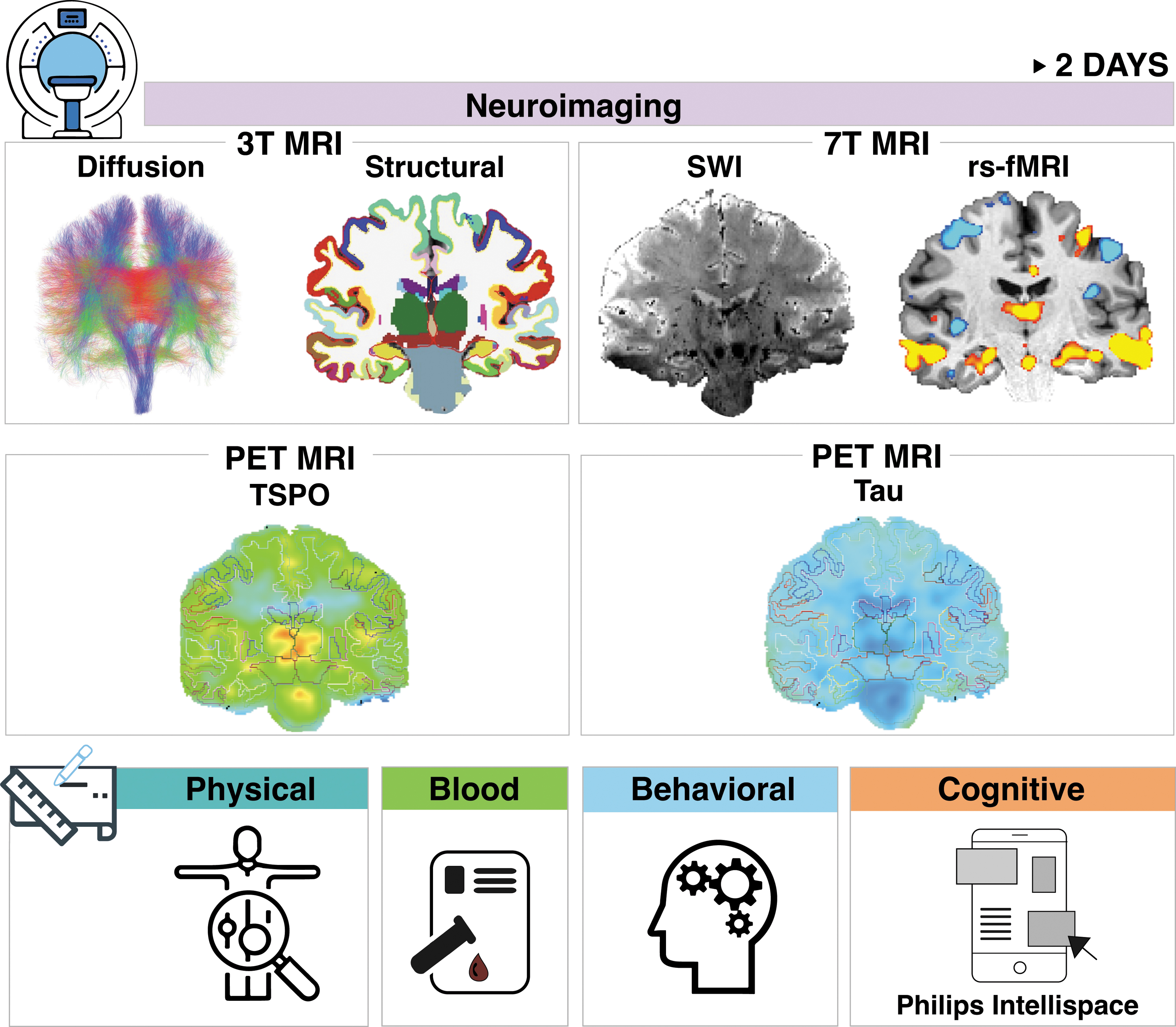
Overview of ReBlast Pilot Study procedures. The 2-day ReBlast study protocol involves four imaging assessments (top and middle rows) and comprehensive cognitive, behavioral, physical symptom and blood biomarker assessments (bottom row). 3T, 3 Tesla; 7T, 7 Tesla; MRI, magnetic resonance imaging; PET, positron emission tomography; rs-fMRI, resting-state functional MRI; SWI, susceptibility-weighted imaging; TSPO, translocator protein.
State of the science
Blast exposure terminology and quantification
There is ongoing debate about the appropriate nomenclature for describing blast severity and the optimal tools for quantifying blast exposure. Multiple terms have been proposed to characterize blast severity (See Belding et al. 20215 for a comprehensive review), including “low level blast” (LLB) and “high level blast” (HLB). Currently, there is no validated definition of blast severity, and thus no objective way to differentiate LLB from HLB. Indeed, the recently developed GBEV, 28 which we use in ReBlast as the primary measure of blast exposure, includes both LLB exposure (e.g., small arms) and HLB exposure (e.g., large improvised explosive devices). Further, some munitions commonly classified as generating a LLB because they are “outgoing munitions” (e.g., Carl-Gustaf recoilless rifle) 9 produce shockwaves that exceed 4 pounds per square inch, the proposed threshold above which a blast may be defined as HLB. 29 This inconsistency in nomenclature reinforces the need for future studies to link objective measures of blast exposure (e.g., blast gauges) to subjective self-reports. It is also important to consider that once SOF personnel experience combat, an inclusion criterion for this study, their exposure profiles reflect a broad range of blast intensities. Thus, given that SOF personnel with combat experience are exposed to blast severities that are variable and cannot be objectively classified, we use the general term “RBE” when describing blast exposure in the ReBlast study.
Neurocognitive biomarkers
Studies of the effects of RBE on neurocognitive function, which have been largely conducted in conventional forces and breacher populations, report mixed findings. 6,9 Some studies revealed negative effects of RBE on neurocognitive function (e.g., memory, processing speed, attention). 30 –36 Other studies reported no independent effects of RBE, 24,37 -39 demonstrating the challenge of dissociating neurocognitive performance from co-occurring post-traumatic stress disorder (PTSD), depression, and non-blast mild TBI in a blast-exposed population. These discrepancies may reflect heterogeneity in outcome measures, domains assessed, study design, participant characteristics (e.g., acute versus chronic exposure), and statistical approach. 6,9 Overall, the association of RBE with domain-specific neurocognitive function in humans remains poorly understood. This knowledge gap is especially marked in U.S. SOF, as there are currently no published studies investigating the impact of RBE on neurocognitive function in this high-risk group.
Neurobehavioral and physical biomarkers
PTSD and post-concussive symptoms, such as headaches, anxiety, and insomnia, have been reported in breachers and service members with combat-related mild TBI. 19,21,40,41 Preliminary evidence also suggests that breachers are at higher risk of developing tinnitus than are non-breacher military service members. 42 Among those who receive inpatient rehabilitation for TBI, SOF personnel report more neurobehavioral symptoms than conventional forces, 43 which can lead to suicide-related thoughts and behaviors. 44,45 It is therefore critical to develop objective biomarkers and determine levels of RBE at which clinically important distress begins to emerge.
Neuroimaging biomarkers
Multiple advanced MRI 11,13,26,46 -49 and PET 50 techniques have shown promise in detecting blast-induced brain injury in military personnel. Structural MRI, particularly diffusion MRI (dMRI), detects microstructural changes in regions of white matter that are susceptible to blasts because of their proximity to openings in the skull. 11,26,48 Further, functional MRI (fMRI) reveals disruptions of brain networks related to blasts. 26,47 Tau PET reveals deposition of tau protein in the brains of civilians 51 and military personnel with a history of blast injury. 50 Additionally, TSPO PET identifies microglial activation, 52 a pathophysiological process that may contribute to the astroglial scarring observed postmortem in brain tissue of military personnel with a history of blast exposure. 53 However, there is currently no validated in vivo imaging test to detect astroglial scarring. 53 Moreover, there have been no comprehensive, multi-modal neuroimaging studies of RBE in SOF personnel, which are needed to identify clinically relevant biomarkers.
Blood biomarkers
Plasma proteomic and metabolomic studies have revealed multiple potential biomarkers of neuronal and glial injury in individuals with blast and blunt head trauma. 54 Biomarkers of axonal injury include neurofilament light protein (NfL) 13,55 -57 and tau, 56,57 while biomarkers of glial injury include glial fibrillary acidic protein (GFAP) and S100 calcium-binding protein B (S-100B), 56 –61 which can be detected in plasma following TBI. 62 -64 Few studies have investigated blood markers related to RBE. 13,33 There is evidence that tau and P-tau isoforms increase with number and severity of TBIs, 54 providing potential chronic biomarkers of the burden of brain injury sustained over a career. Given the well-established association between repeated TBI and neurodegenerative diseases, 8 potential candidates for RBE-induced brain injury can be further informed by the Alzheimer's disease literature, 65 where elevated plasma concentrations of tau, 66 P-tau, and Aβ40/42 67,68 are reported.
Methods
Study design overview
The ReBlast Pilot study is a cross-sectional observational study of active-duty U.S. OEF/OIF/OND-era SOF personnel with RBE. Inclusion and exclusion criteria are presented in Table 1. An overview of study procedures is provided in Table 2.
Inclusion/Exclusion Criteria
This pilot study excludes females to avoid imbalance across the blast exposure groups, given that the majority of SOF are male.
Deployment is defined as being deployed to a region of conflict while serving in the U.S. military.
Endorsement of any CES item ensures that all participants will have experienced combat situations during their military career.
The VA/DoD definition of TBI is used for this study: initial Glasgow Coma Scale score <13, loss of consciousness duration >30 min, post-traumatic amnesia duration >24 h, or abnormal structural brain imaging.
Magnetic resonance imaging (MRI) contraindications included: metal in the body that would make an MRI scan unsafe, pre-existing medical conditions including a likelihood of developing seizures or claustrophobic reactions, inability to lie supine for up to 2 h in the MRI scanner, and >300 pounds due to the MRI table's weight limit. Prior radiation exposure of ≥50 mSv over the past 12 months is considered a contraindication for positron emission tomography (PET) imaging.
SOF, Special Operations Forces; VA, Veterans Affairs; DoD, Department of Defense; CES, Combat Exposure Scale; TBI, traumatic brain injury.
Study Procedures
Study activities can vary between Day 1 or Day 2, depending on scheduling availability for scans and other logistical changes.
Philips Intellispace Cognition iPad-based tests are based on widely used analog neuropsychological assessments, with slight modifications to administration procedures and scoring criteria.
ACT, Auditory Consonant Trigrams; ANAM, Automated Neuropsychological Assessment Metrics; AUDIT-C, Alcohol Use Disorders Test-Consumption; BISQ, Brain Injury Screening Questionnaire; BGLHA, Brown-Goodwin Assessment for Lifetime History of Aggression; BPAQ, Buss-Perry Aggression Questionnaire; CES, Combat Exposure Scale; DAST-10, Drug Abuse Screening Test; DKEFS, Delis-Kaplan Executive Function System; DRRI-CES-SS, Deployment Risk and Resilience Inventory; Combat Exposure Scale, modified for STRONG STAR; FrSBE, Frontal Systems Behavior Scale; GBEV, Generalized Blast Exposure Value; GOSE, Glasgow Outcome Scale-Extended; HIT-6, Headache Impact Test; mBIAS, mild Brain Injury Atypical Symptoms; MOS, Military Occupational Specialty; MRI, magnetic resonance imaging; MSVT, Medical Symptom Validity Test; NIH TBI CDE, National Institutes of Health Traumatic Brain Injury Common Data Elements; NSI, Neurobehavioral Symptom Inventory; PCL-5, Post-Traumatic Stress Disorder Checklist for DSM-5; PET, positron emission tomography; PHQ-9, Patient Health Questionnaire-9; PROMIS, Patient-Reported Outcomes Measurement Information System; PSQI, Pittsburgh Sleep Quality Index; RAVLT, Rey Auditory Verbal Learning Test; SBQ-R, Suicide Behaviors Questionnaire-Revised; STOP-BANG, Snoring history, Tired during the day, Observed stop breathing while sleep, high blood Pressure, BMI more than 35 kg/m2, Age more than 50 years, Neck circumference more than 40 cm and male Gender; TBI-QOL, Traumatic Brain Injury Quality-of-Life; TSPO, translocator protein; WAIS-IV, Wechsler Adult Intelligence Scale, 4th edition; TOPF, Test of Premorbid Functioning; WHODAS 2.0, World Health Organization Disability Assessment Schedule.
Recruitment
Potential study subjects are identified by USSOCOM Surgeon's Office personnel, posting of recruitment flyers on USSOCOM media outlets, and word of mouth. A preliminary Consort Diagram reporting screening and enrollment data from July 2021 through March 2022 is provided in Figure 2. We have screened 40 SOF, consented 15, and completed study activities with 11. One consented participant withdrew consent prior to initiating study activities.
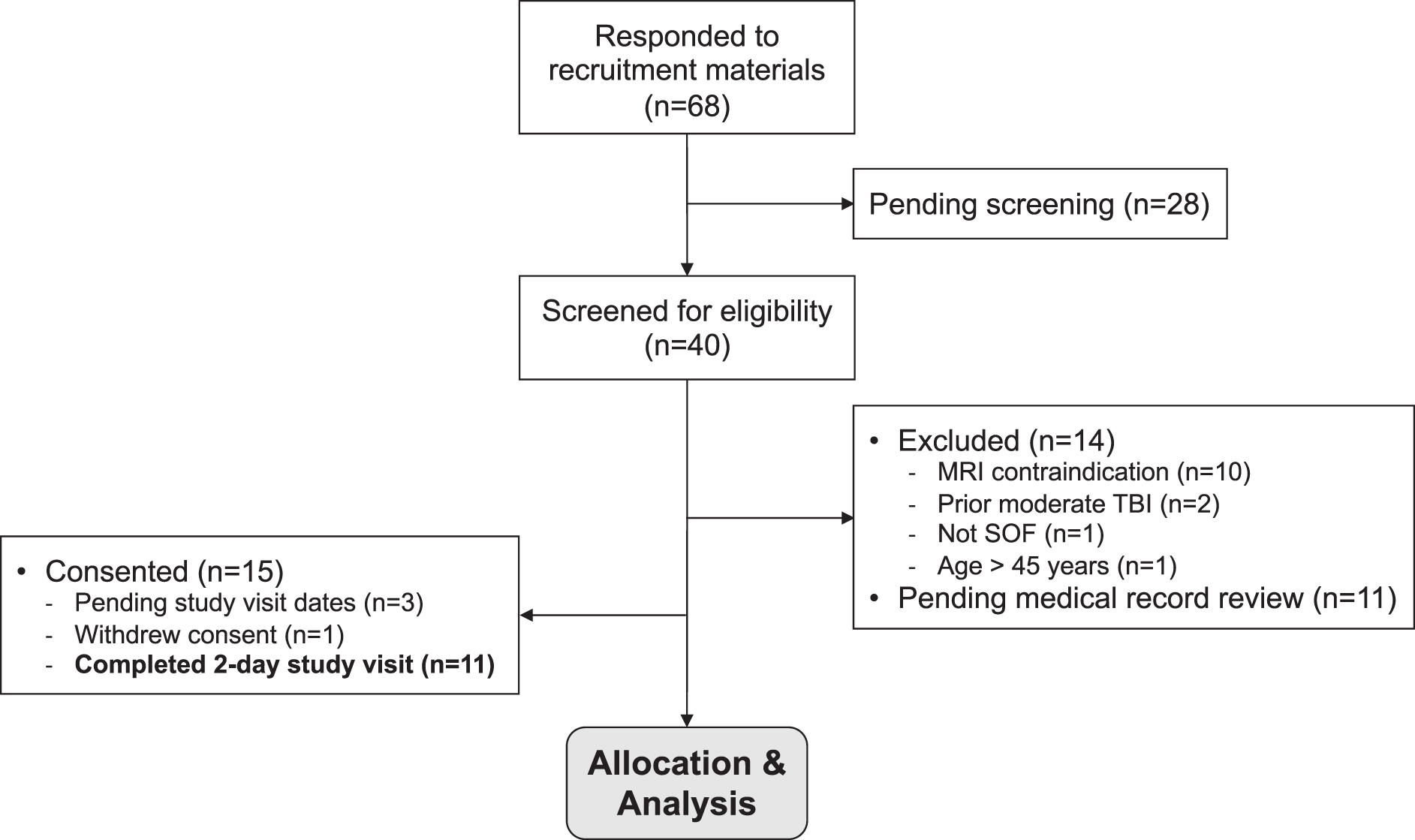
Consort diagram of ReBlast Pilot screening and enrollment—July 2021 through March 2022. MRI, magnetic resonance imaging; SOF, Special Operations Forces; TBI, traumatic brain injury.
Blast exposure
The primary measure of blast exposure is the GBEV, 28 an assessment tool designed to capture units of lifetime blast exposure from weapons and explosives. GBEV asks respondents to self report exposure to five categories of blast: 1) small- and medium-sized arms such as handheld firearms and rifles; 2) large arms, often shoulder-fired, that can be carried on a person; 3) artillery, missile weapon systems, or large arms carried by vehicle, aircraft, or boat; 4) small explosives or grenades; and 5) large explosives or targeted explosives in close range. For each category, respondents provide the number of years, average months per year, days per month, and rounds per day of exposure, as well as how often exposures occurred on 2 or more consecutive days. An automated algorithm calculates the final GBEV, which has a minimum score of zero but no ceiling score.
For the ReBlast Pilot, GBEV is administered via telephone interview by a member of the research team who has expertise in assessing blast exposure. Although GBEV has not been validated, in a sample of 984 service members, 200,000 GBEV units was established as a threshold at which participants were likely to report significant symptoms on the Neurobehavioral Symptom Inventory. 28 Importantly, as described above, GBEV does not distinguish between LLB and HLB exposure.
Brain injury assessment
The Brain Injury Screening Questionnaire (BISQ) 69 Part I is a structured questionnaire that characterizes incidence and severity of lifetime exposure to head trauma. The respondent is asked if they have ever experienced a blow to the head in 19 specific situations (e.g., sports, combat, motor vehicle accidents, etc.). To increase the likelihood of recalling a blow to the head, specific questions are asked for six common contact sports as well as for military training and combat. For every event, participants report if they lost consciousness or had a period of being dazed and confused, and if so, the duration of symptoms. Finally, hospitalizations and emergency department admissions for 13 specific medical events are documented. Table 3 28,70 –101 provides detailed information regarding measures of a participant's lifetime history of brain injury, exposure to wartime stress, and the extent and frequency of exposure to RBE and large weaponry.
Cognitive and Behavioral Assessments
STOP-BANG, Snoring history, Tired during the day, Observed stop breathing while sleep, High blood pressure, BMI more than 35 kg/m2, Age more than 50 years, Neck circumference more than 40 cm and male Gender; PTSD, post-traumatic stress disorder; DSM, Diagnostic and Statistical Manual of Mental Disorders; WHO, World Health Organization, PROMIS, Patient-Reported Outcomes Measurement Information System; REDCap, Research Electronic Data Capture.
Cognitive assessment
The cognitive assessment battery measures executive function, memory, learning, intellectual performance, and fine motor functioning, as detailed in Table 3. Assessments are distributed across the first and second days of the study. Some assessments are administered via a computer (e.g., Automated Neuropsychological Assessment Metric) 102 or iPad (e.g., Philips Intellispace Cognition) 103 to complement traditional tests and obtain granular performance metrics (e.g., reaction time). Pupillometry measurements are obtained to assess the integrity of pupil constriction and dilation—a metric of cognitive processing load tolerance. 104 The Medical Symptom Validity Test 105 and mild Brain Injury Atypical Symptoms scale 106 are administered to assess effort and potential exaggeration of impairment. These measures contextualize the cognitive, behavioral, and physical symptom assessments during statistical analysis.
Neurobehavioral function, physical symptoms, and quality of life
The battery of measures used to assess psychological health, sleep, pain, protective factors, and overall function and life quality are listed in Table 2. Some measures are collected via structured interview or self-report at the in-person visit, while others are collected before arrival online via REDCap, 107 a secure, web-based software platform.
Neuroimaging
Connectome MRI for structural connectivity analysis
Acquisition
We perform a multi-shell dMRI protocol on the 3T Connectome scanner (MAGNETOM CONNECTOM Siemens Healthineers, Erlangen, Germany) 108 -110 using a 64-channel head coil. 111 Complete sequence parameters are provided in Supplementary Table S1 and have been previously reported. 112 –115 Sequence parameters include: 2 mm isotropic voxels; eight linearly spaced gradient strengths in the range G = 30-290 mT/m per diffusion time; 16 b-values ranging from 50 to 17,800 sec/mm2 with either 32 diffusion-encoding directions (for shells with b ≤ 2300 sec/mm2) or 64 directions (for shells with b ≥ 2400 sec/mm2). We also acquire a three-dimensional T1-weighted multi-echo magnetization-prepared rapid gradient echo (MEMPRAGE) sequence for anatomic coregistration. 116,117
Processing and analysis
We correct data for gradient nonlinearity distortions 108,118.119 and susceptibility-induced distortion, head motion, and eddy-current artifacts in the Functional Magnetic Resonance Imaging of the Brain Software Library (FSL). 108,120 Quality control steps include visual inspection of the dMRI volumes before and after pre-processing and automated extraction of motion-related measures (Supplementary Fig. S1). We perform two types of analyses: connectivity-based and region of interest (ROI)–based. For both analyses, we use multi-fiber reconstruction methods 121,122 to extract estimates of fiber orientations in each voxel (Supplementary Fig. S2). We perform connectivity analysis of probabilistic tractography data 110,123 to quantify axonal connections linking canonical networks whose nodes are segmented via FreeSurfer. 124,125 For the ROI-based analysis, 42 white matter bundles are automatically reconstructed using TRActs Constrained by UnderLying Anatomy (TRACULA; Supplementary Fig. S3). 126,127 To quantify changes in white matter microstructure we fit a tensor model and extract measures of fractional anisotropy, mean, radial, and axial diffusivity at each voxel. We also fit a three-compartment model 113,115,128 of intra-axonal restricted diffusion, extra-axonal hindered diffusion, and free diffusion to extract estimates of axonal diameter in regions of white matter that are susceptible to blast injury.
7 Tesla MRI for functional connectivity analysis
Acquisition
We acquire ultra-high spatial resolution blood-oxygen level dependent (BOLD) fMRI data on the 7T scanner, as previously described 129 and as detailed in Supplementary Table 1. We use the Terra 7T platform (Siemens Healthineers, Erlangen, Germany) with the vendor-supplied 32-channel head-only receive coil array and birdcage transmit coil (Nova Medical, Wilmington, MA). Notable BOLD sequence parameters include: whole–brain single-shot simultaneous multi-slice 130 gradient-echo echo-planar imaging at 1.2 mm isotropic voxel size, with repetition time = 2.25 sec and multiband factor 3. We acquire four runs of resting-state fMRI (rs-fMRI), each with 150 measurements. Before the first run and between runs, the participants are reminded to remain awake with their eyes open and asked to confirm that they are comfortable and alert by squeezing a pneumatic ball. We acquire fMRI data alongside structural MRI data, such as T1-weighted and susceptibility-weighted imaging data, to provide an anatomical reference for the fMRI data, as well as standard calibration and auxiliary data such as magnetic field maps (B0 and B1 +) that are used to adjust the system and remove artifacts.
Processing and analysis
We analyze data in the FreeSurfer 124 Functional Analysis Stream (FSFAST) and the CONN toolbox. 131 Preprocessing includes B0 distortion correction, motion correction, slice-timing correction, and temporal detrending. In FSFAST, the fMRI volume is sampled onto the cortical surface created by FreeSurfer and surface-smoothed; the subcortical areas are resampled into the MNI305 and volume-smoothed. We perform seed-based 132 and independent component analyses 133 of functional networks to estimate functional connectivity between the cortical and subcortical nodes of canonical neural networks, such as the default, salience, executive control, and dorsal attention networks. 125
Translocator protein PET-MRI
TSPO genotyping
A venous blood sample is drawn to perform genotyping for the Ala147Thr polymorphism in the TSPO gene.
Acquisition
We scan each subject using the TSPO ligand [ 11 C]PBR2852 with a simultaneous PET-MRI scanner, a unique Siemens BrainPET photodiode-based PET scanner operated in the bore of a 3T whole–body magnetic resonance scanner. 134 We have developed MR-based methods for generating attenuation correction maps. 135 An intravenous bolus injection of [ 11 C]PBR28 (up to 15 mCi) is administered. After an uptake period of approximately 45 min, PET data are acquired for up to 60 min (overlapping with magnetic resonance data acquisition) and stored in list-mode format.
We acquire brain MRI data using the Siemens 3T Trio scanner, equipped with a standard radiofrequency head coil positioned inside the PET insert, similar to standard MRI. During the PET-MRI, we acquire an MEMPRAGE sequence to be used as an anatomical reference, an arterial spin labeling (ASL) sequence to measure regional brain perfusion, and a time-of-flight magnetic resonance angiography sequence to image brain vasculature.
Processing and analysis
Standardized uptake values (SUV) from 60-90 min post-injection are calculated as previously described. 52 We coregister the SUV images to the MEMPRAGE image and then register to the MNI template and normalize to a pseudo-reference region (SUVR). Partial volume correction using region-based voxel-wise (RBV) correction is applied using PETsurfer. 136 Voxel-wise analysis of ligand binding 52 is performed using a combination of software packages, including FreeSurfer and FSL. TSPO genotype is controlled for in all analyses. Individuals with a low-binding genotype, estimated at 10% of the general population, 137 will be excluded from analysis.
Tau PET-MRI
Acquisition
Each subject is scanned using the ligand [18F]MK6240. 138 As with the TSPO PET scan, the tau PET scan utilizes a combined PET-MRI scanner. An intravenous bolus injection of [ 18 F]MK6240 (up to 5 mCi) is administered. After an uptake period of approximately 60 min, PET data are acquired for up to 60 min. We acquire a MEMPRAGE sequence, a T2-weighted SPACE fluid-attenuated inversion recovery (T2 SPACE FLAIR) sequence, and a two inversion-contrast magnetization-prepared rapid acquisition gradient echo (MP2RAGE) sequence (Supplementary Table S1).
Processing and analysis
The tau PET data analysis pipeline is similar to that used for analysis of the TSPO PET data, including voxel-wise group comparisons of ligand binding. 52 SUV from 70-90 min post-injection is calculated as a primary outcome, normalized to a pseudo-reference region, and partial volume corrected.
Integration of multi-modal neuroimaging data
To facilitate precise neuroanatomic correlation of lesions and abnormal signals across modalities, all MRI and PET data are coregistered to the same spatial coordinates (Fig. 3). 139 All MRI data are corrected for gradient nonlinearity distortion 119 to reduce differential distortion between the images from any of the MRI or PET scanners. In addition, a B0 map is computed from images collected with reversed phase-encode blips and used to correct the B0 distortion in the fMRI. 140 Cortical surface and whole-brain segmentation are computed using FreeSurfer tools from the minimally distorted T1-weighted image 124 acquired on the Connectome. All images are registered to this T1-weighted image: MRI data are registered via boundary-based registration, 117 and PET images are registered using normalized mutual information. 141 All coregistered MRI and PET data undergo quality assessment for artifacts or noise, and study investigators manually correct anatomic errors observed on the cortical surfaces or subcortical segmentations.
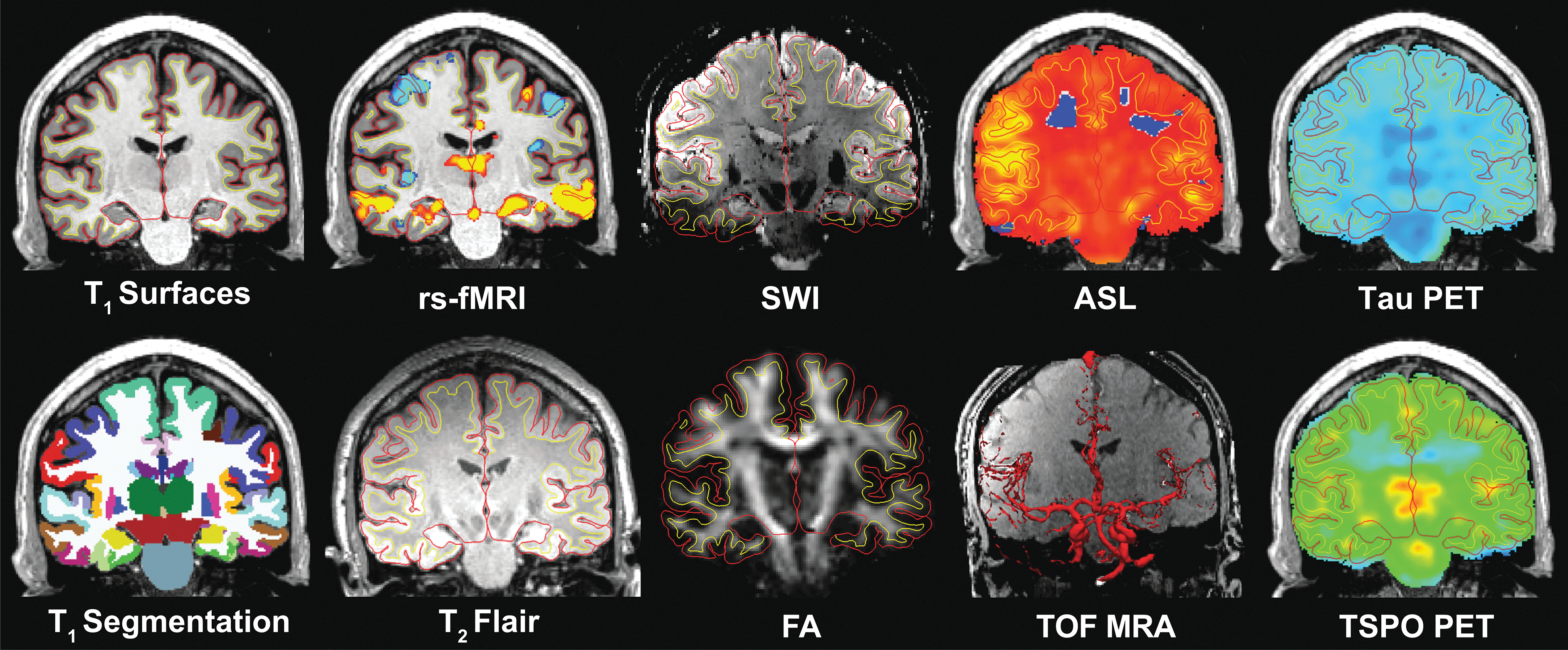
Multi-modal integration of neuroimaging data. MRI and PET data are coregistered and segmented using FreeSurfer tools, allowing precise neuroanatomic correlation of lesions and abnormal signals across modalities. The red line indicates the pial surface, and the yellow indicates the cortical gray-white matter junction. In the T1 segmentation image, cortical and subcortical regions are segmented according to the Desikan-Killiany atlas. 139 FA, fractional anisotropy; ASL, arterial-spin labeled perfusion imaging; MRA, magnetic resonance angiography; PET, positron emission tomography; rs-fMRI, resting-state functional MRI; SWI, susceptibility-weighted imaging; T1, T1-weighted; T2 FLAIR, T2-weighted fluid-attenuated inversion recovery; TOF, time of flight; TSPO, translocator protein.
Blood biomarker proteomic and metabolomic analysis
Acquisition
We collect ∼20 cc of blood, in the fasting state (i.e., no food for at least 6 h prior), for plasma and serum analyses.
Blood biomarker processing and analysis
Proteomic measurements are performed using proximity extension assay technology (Olink Proteomics). Protein markers include NfL, GFAP, as well as other candidate biomarkers available on the platform and new biomarkers that emerge during the study period. 54,64,142 Candidate protein markers will be validated as needed using alternative measurement methods such as enzyme-linked immunosorbent assays, electrochemiluminescence, and/or mass spectrometry. Metabolomics measurements are performed using liquid chromatography-tandem mass spectrometry for targeted detection of candidate metabolites. Raw data values are normalized to interspersed pooled plasma samples using our standard approach. 143 -145
Statistical analysis
The primary aim of the ReBlast Pilot study is to generate preliminary data that will inform the design of a larger, hypothesis-driven investigation. As such, due to feasibility constraints associated with enrolling active-duty SOF personnel for a 2-day study visit that requires travel away from their training bases, the ReBlast Pilot's target enrollment is 30 participants. Upon completion of study enrollment, we will aggregate GBEV scores from all 30 participants. We will use the median GBEV score to allocate participants into two groups: 1) GBEVb (n = 15 below the median GBEV), and 2) GBEVa (n = 15 above the median GBEV). Our primary statistical analyses will aim to answer the question: Does the GBEVa group differ from the GBEVb group with respect to cognitive, behavioral, physical, neuroimaging, and blood biomarkers? We will use Wilcoxon rank-sum tests to assess the null hypothesis that there is no difference in median biomarker values between the GBEVb and GBEVa groups. Of note, this pilot study's sample size of 30 participants is only sufficient to detect large differences between groups (effect sizes >1.2) with 80% power and an unadjusted Type 1 error control of 0.05.
Secondary analyses
We will use Spearman's rank correlation coefficients to test for associations between biomarkers and GBEV scores. Given the exploratory nature of this pilot study, both original and adjusted p values (correcting for the number of biomarkers within each domain) will be calculated. We will also use linear regression to identify biomarkers that significantly differ between the GBEVb and GBEVa groups. Biomarkers will be treated as dependent variables in each model and GBEV as the independent variable, controlling for lifetime history of TBI exposure (from the BISQ) and combat exposure (from the Combat Exposure Scale), within the limits of our sample size. These analyses aim to identify associations between RBE and alterations in cognitive, behavioral, physical, neuroimaging, and blood biomarkers.
Given the inherent limitations of performing a median-split analysis (e.g., exposure to RBE may not differ significantly between the GBEVb and GBEVa groups) and of evaluating each biomarker independently, we will also implement an unsupervised learning approach, such as k-means clustering, to derive clusters of personnel whose biomarker profiles are similar within and across domains. This clustering approach minimizes within-group differences, maximizes between-group differences, and increases our ability to identify combinations of biomarkers that account for complex, heterogeneous phenotypes. In turn, we will evaluate whether these clusters, consisting of participants with similar phenotypes, are associated with GBEV scores (Fig. 4).
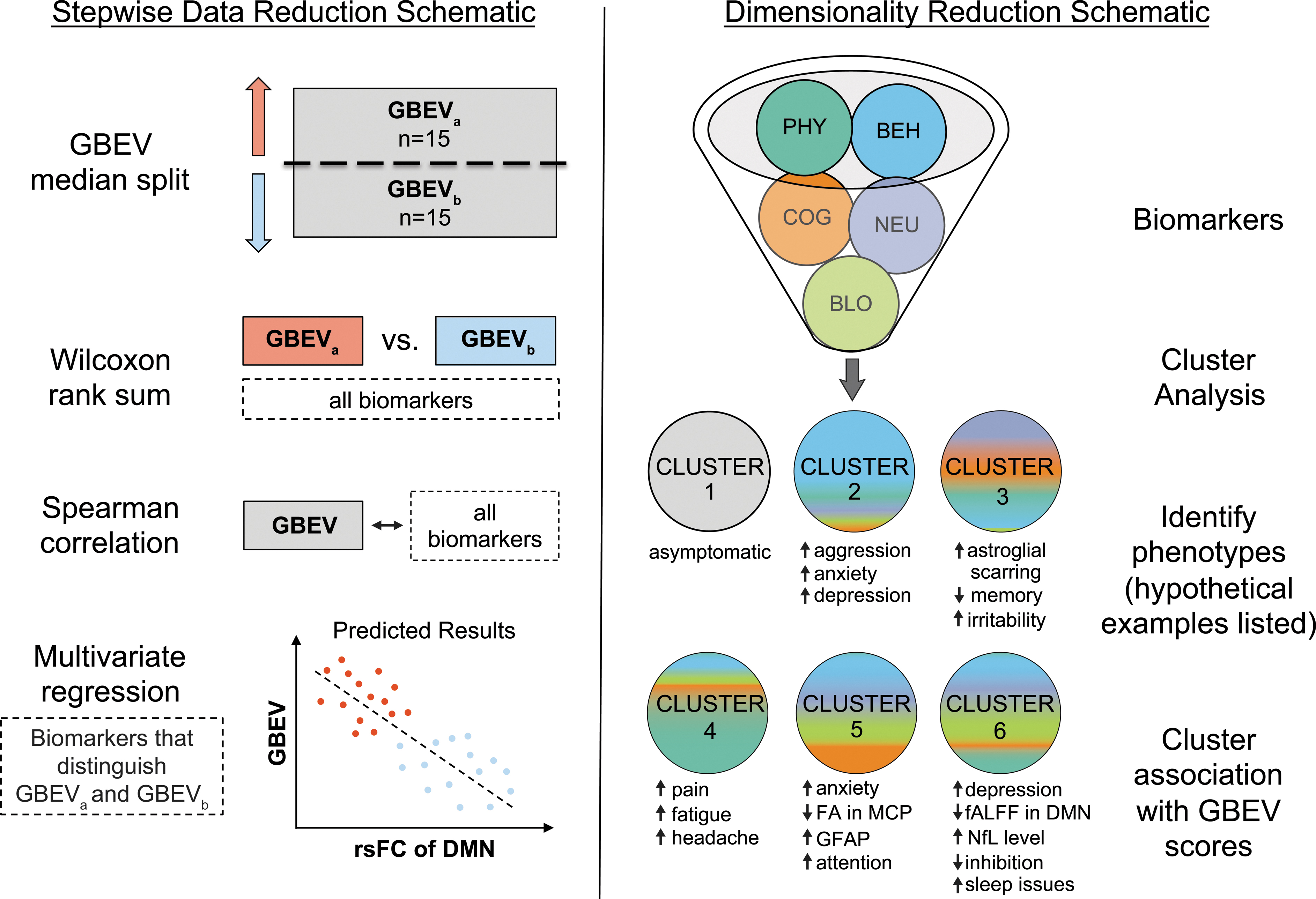
Statistical plan for ReBlast pilot study. This figure depicts the primary statistical analyses proposed in this study: 1) a stepwise method to reduce the data into a meaningful set of biomarkers that distinguish the groups and then, assessing their relationship with GBEV scores (left panel); and 2) an unsupervised learning approach to reduce the data into meaningful clusters (representing groups of individuals with similar phenotypes) and then, evaluating their association with GBEV scores (right panel). Of note, the scatter plot in the left panel and the clusters in the right panel are hypothetical, not actual results. BEH, behavioral; BLO, blood; COG, cognitive; DMN, default mode network; FA, fractional anisotropy; fALFF, fractional amplitude of low-frequency fluctuations; GBEV, Generalized Blast Exposure Value; GBEVa, participant group with GBEV values above the median; GBEVb, participant group with GBEV values below the median; GFAP, glial fibrillary acidic protein; MCP, middle cerebellar peduncle; NEU, neuroimaging; NfL, neurofilament light chain; PHY, physical; rsFC, resting-state functional connectivity.
Limitations
There are four key limitations associated with our analytic approach. First, with a sample size of n = 30, our ability to detect significant differences between the GBEVb and GBEVa groups will be limited in both the primary and secondary analyses, including the k-means clustering approach. Second, a median split of GBEV scores may artificially create two participant groups whose exposure to blast is relatively similar. Our secondary analyses aim to address this concern by using GBEV as a continuous variable when evaluating the relationship between the GBEV and biomarkers. Third, we likely lack statistical power to robustly control for variance in mild TBI and combat exposure. Fourth, our study does not include a control group of participants without RBE, because all SOF personnel who have experienced combat (a study inclusion criterion) undergo training that results in RBE. Non-SOF military personnel may have less RBE but did not go through the SOF selection process and would not have similar training or combat exposures. Thus, non-SOF active-duty military personnel would not be an adequate control group. Some of these limitations can be mitigated in future studies by following SOF personnel longitudinally, as discussed below.
Discussion
The ReBlast Pilot study aims to identify biomarkers that individually, and in combination, detect RBE-related brain injury in SOF personnel. We designed the study to include a wide range of diagnostic modalities, recognizing that they have complementary strengths. The results of this exploratory, hypothesis-generating study will inform biomarker selection and sample size calculations for a subsequent, longitudinal study designed to validate a new diagnostic test for RBE-related brain injury. By identifying the underlying mechanisms, risk and resilience factors, and clinical phenotypes associated with RBE-related brain injury in U.S. SOF personnel, the results of ReBlast will support the development of future therapies aimed at preventing or repairing RBE-related brain injuries. The results of subsequent studies will also inform evidence-based recommendations to modify training experiences and deployment schedules to optimize brain health and performance in SOF warfighters.
ReBlast is one of several studies currently investigating the impact of blast exposure on military service members. 21,25,146 We anticipate that the ReBlast Pilot study will be synergistic with ongoing studies and will provide unique insights from the SOF community. Specifically, SOF warfighters differ from conventional forces both on pre-selection characteristics such as education, physical and mental health 147 and on post-selection experiences such as blast and combat exposures. 23 -25,43 Studying SOF and conventional forces together generates averaged results that do not reflect the distinct demographic, clinical, and exposure characteristics of these cohorts. Further, a long-term goal of ReBlast is to generate preventive measures, diagnostic tools, and interventions that enhance career longevity and improve quality of life. Thus, unlike studies that focus on Veterans, 146 ReBlast enrolls only active-duty service members who may benefit from the findings during their careers and are more likely to accurately report blast and combat exposures because of their temporal proximity. Though enrollment is limited to SOF personnel, ReBlast findings may generalize across military forces and provide new avenues for future research on blast-related brain injury in all forces.
Future directions
Upon completion of the ReBlast Pilot study (target date June 30, 2023), we will determine which candidate biomarkers have the strongest association with RBE. We then plan to launch follow-up studies to validate the diagnostic utility of these biomarkers in a larger cohort of active-duty SOF. Central to this follow-up effort will be longitudinal data acquisition, in which each participant serves as his own control. This study design may elucidate the temporal dynamics of biomarker changes and identify which biomarker changes are attributable to ongoing RBE versus other types of exposures. A longitudinal study design will also allow us to detect when SOF personnel become symptomatic and to identify factors that lead to resilience or recovery.
Another future goal of ReBlast is to validate in vivo neuroimaging biomarkers using ultra high-resolution ex vivo MRI and histopathology. Emerging evidence suggests that ex vivo MRI scans of formalin-fixed whole–brain specimens and tissue sections reveal pathological findings associated with TBI, including astrogliosis 148 and axonal injury. 149 -151 These ex vivo MRI data can be spatially coregistered to both in vivo MRI data 152 and immunohistochemistry data, 148,149,152,153 providing a methodological bridge between gold-standard microscopy and in vivo neuroimaging tests. Thus, ex vivo MRI and histopathological analysis of brain specimens donated by SOF personnel have the potential to enhance the methodological rigor of future studies that aim to validate a diagnostic test of blast-induced brain injury.
Ultimately, the goal of the ReBlast study—both the current pilot and future follow-up studies—is to define a diagnostic battery that will provide actionable data to inform clinical care for SOF personnel in training and in theater. A diagnostic battery is essential for future efforts to prevent and treat blast-induced brain injury in SOF personnel. ReBlast thus has the potential to not only improve battle readiness and career longevity for SOF warfighters, but also optimize long-term brain health and veteran quality of life.
Footnotes
Acknowledgments
We thank Kelsey Radmanesh and Jennifer Michaud for their guidance and support in implementing the study protocol. We thank Claire Modica, PhD, LT, MSC, USN, for consultation regarding study design and application of the Generalized Blast Exposure Value. We thank Mary Tresvalles, Kyle Droppa, and Riana Schleicher for assistance with data acquisition. We thank John E. Kirsch, Jacob C. Calkins, Amy L. Kendall, and Grae E. Arabasz for serving on the ReBlast MRI Safety Committee. We also thank Tobias Kober of Siemens Healthineers for use of the MP2RAGE WIP 944.
The views expressed in this manuscript are entirely those of the authors and do not necessarily reflect the views, policy, or position of the United States Government, Department of Defense, or United States Special Operations Command.
Authors' Contributions
Brian L. Edlow, MD: study concept, design, and oversight; acquisition, analysis, and interpretation of data; drafting/revising the manuscript; critical revision of the manuscript for intellectual content. Yelena G. Bodien, PhD: study concept, design, and oversight; acquisition, analysis, and interpretation of data; drafting/revising the manuscript; critical revision of the manuscript for intellectual content. Timothy Baxter, MSc: study design and oversight; critical revision of the manuscript for intellectual content. Heather G. Belanger, PhD: study concept and design; acquisition, analysis, and interpretation of data; critical revision of the manuscript for intellectual content. Ryan J. Cali, BS: acquisition, analysis, and interpretation of data; critical revision of the manuscript for intellectual content. Katryna B. Deary, NP: study concept and design; acquisition, analysis, and interpretation of data; critical revision of the manuscript for intellectual content. Bruce Fischl, PhD: acquisition, analysis, and interpretation of data; critical revision of the manuscript for intellectual content. Andrea S. Foulkes, ScD: study design; acquisition, analysis, and interpretation of data; critical revision of the manuscript for intellectual content. Natalie Gilmore, PhD: acquisition, analysis, and interpretation of data; drafting/revising the manuscript; critical revision of the manuscript for intellectual content. Douglas N. Greve, PhD: study design; acquisition, analysis, and interpretation of data; critical revision of the manuscript for intellectual content. Jacob M. Hooker, PhD: study concept, design, and oversight; acquisition, analysis, and interpretation of data; critical revision of the manuscript for intellectual content. Susie Y. Huang MD, PhD: study concept, design, and oversight; acquisition, analysis, and interpretation of data; critical revision of the manuscript for intellectual content. Jessica N. Kelemen, BA: study oversight; acquisition, analysis, and interpretation of data; critical revision of the manuscript for intellectual content. W. Taylor Kimberly, MD, PhD: study concept, design, and oversight; acquisition, analysis, and interpretation of data; critical revision of the manuscript for intellectual content. Chiara Maffei, PhD: acquisition, analysis, and interpretation of data; drafting/revising the manuscript; critical revision of the manuscript for intellectual content. Maryam Masood, MS: study oversight; acquisition, analysis, and interpretation of data; critical revision of the manuscript for intellectual content; Daniel P. Perl, MD: study concept and design; critical revision of the manuscript for intellectual content. Jonathan R. Polimeni, PhD: study concept, design, and oversight; acquisition, analysis, and interpretation of data; critical revision of the manuscript for intellectual content. Bruce R. Rosen, MD, PhD: study concept and design; acquisition, analysis, and interpretation of data; critical revision of the manuscript for intellectual content. Samantha L. Tromly, BS: study design and oversight; critical revision of the manuscript for intellectual content. Chieh-En J. Tseng, PhD: acquisition, analysis, and interpretation of data; drafting/revising the manuscript; critical revision of the manuscript for intellectual content. Eveline F. Yao, MD: study concept and design; critical revision of the manuscript for intellectual content. Nicole R. Zürcher, PhD: acquisition, analysis, and interpretation of data; critical revision of the manuscript for intellectual content. Christine L. Mac Donald, PhD: study concept and design; acquisition, analysis, and interpretation of data; drafting/revising the manuscript; critical revision of the manuscript for intellectual content. Kristen Dams-O'Connor, PhD: study concept, design, and oversight; acquisition, analysis, and interpretation of data; drafting/revising the manuscript; critical revision of the manuscript for intellectual content.
Funding Information
Support for this work is provided by the U.S. Department of Defense (USSOCOM Contract No. H9240520D0001).
Author Disclosure Statement
No competing financial interests exist.
Supplementary Material
Supplementary Table S1
Supplementary Figure S1
Supplementary Figure S2
Supplementary Figure S3
References
Supplementary Material
Please find the following supplemental material available below.
For Open Access articles published under a Creative Commons License, all supplemental material carries the same license as the article it is associated with.
For non-Open Access articles published, all supplemental material carries a non-exclusive license, and permission requests for re-use of supplemental material or any part of supplemental material shall be sent directly to the copyright owner as specified in the copyright notice associated with the article.