




Abstract
Purpose:
To determine the comparative efficacy of ganciclovir ophthalmic gel and famciclovir oral tablets in cats with experimentally induced ocular feline herpesvirus-1 (FHV-1) epithelial infection.
Methods:
A randomized, placebo-controlled trial was performed using 16 nonvaccinated, specific pathogen-free cats with experimental FHV-1 infection induced by topical ocular inoculation. Cats received topical ganciclovir 0.15% ophthalmic gel (1 drop 3 times daily, n = 6 cats), oral famciclovir (90 mg/kg twice daily, n = 6), or topical artificial tear gel (1 drop 3 times daily, n = 4) for 14 days. Cats were monitored after inoculation for 30 days. Ophthalmic examinations were performed every 2 days and ocular disease scores calculated. In vivo confocal microscopy was performed, and corneal leukocyte infiltrates quantified. Ocular samples for FHV-1 quantitative polymerase chain reaction (qPCR) and virus isolation assays were collected every 3 days. Hemograms and serum biochemistry panels were performed at intervals.
Results:
Clinical ocular disease scores and corneal leukocyte infiltrates were significantly lower in the ganciclovir and famciclovir groups compared with placebo, but no significant differences were detected between the antiviral treatment groups. Ocular viral loads determined by qPCR were significantly lower in the ganciclovir group compared with the placebo group, but there were no significant differences between the other study groups. Hemograms and biochemistry panels were unremarkable.
Conclusion:
Topical application of ganciclovir gel 3 times daily was well-tolerated and displayed similar efficacy at reducing clinical ocular disease scores and corneal inflammation as twice daily oral famciclovir treatment in cats with experimental ocular FHV-1 infection.
Introduction
Feline herpesvirus-1 (FHV-1) is a common etiology of ocular surface infection in domestic cats.1,2 Ocular disease resulting from naturally acquired and experimentally induced FHV-1 infection is clinically similar to that observed with herpes simplex virus (HSV) infection in humans and can be associated with numerous, diverse clinical presentations including blepharitis, conjunctivitis, ulcerative keratitis, and nonulcerative keratitis. 3 Despite the high prevalence of FHV-1 ocular disease in cats, there are few evidence-based antiviral treatment options available for ocular FHV-1 infections. 4
Prior controlled studies that evaluated antiviral medications in experimental models of ocular FHV-1 infection are limited.5–9 At present, the only antiviral treatment options that were described as both safe and effective in cats with experimental ocular FHV-1 infection are oral raltegravir, oral famciclovir, and topical cidofovir therapy.5–7 There are also no prior studies comparing the relative efficacy of 2 or more antiviral treatments in cats with experimentally induced FHV-1 ocular disease.
Cats with ocular FHV-1 infection are a well-established, host-adapted, large animal model of HSV ocular disease and they are frequently used for investigations of therapeutic strategies and viral pathophysiology.10,11 Ganciclovir is a synthetic nucleoside analog of guanosine that displays a broad-spectrum of in vitro activity against many diverse herpesviruses.12,13 Prior studies have determined that ganciclovir is a potent inhibitor of FHV-1 replication in vitro.14,15 Ganciclovir is relatively nontoxic as its active metabolite, ganciclovir triphosphate, accumulates only in virus-infected host cells. 16 Ganciclovir is commercially available as an 0.15% aqueous ophthalmic gel under a variety of trade names. The gel formulation of ganciclovir extends ocular surface contact time, enhances ocular tissue penetration, and increases ocular tissue concentrations. 17
Topical ocular application of ganciclovir 0.15% aqueous ophthalmic gel is an effective treatment for both ocular HSV infection in humans and canine herpesvirus-1 infection in domestic dogs.18,19 A recent study determined that ganciclovir 0.15% aqueous ophthalmic gel was well-tolerated and nontoxic when applied to a small group of laboratory cats without ocular disease 3 times daily for 1 week, but the efficacy of ganciclovir has not been evaluated in cats with ocular FHV-1 infections. 20
The purpose of this study was to evaluate the efficacy of ganciclovir 0.15% ophthalmic gel versus placebo in cats with experimentally induced ocular FHV-1 infection. In addition, the efficacy of the ganciclovir gel was compared with famciclovir oral tablets. Famciclovir is approved by the USA FDA for the treatment of herpes labialis, genital herpes, and acute herpes zoster in humans. 21 Systemic famciclovir treatment was previously demonstrated to be a highly effective treatment for experimentally induced and naturally acquired ocular FHV-1 infections in cats.6,21,22
Methods
Animals
All protocols were approved by the Animal Care and Use Committee of Cornell University (Protocol No. 2019-0073, approved 7/18/2019) and were conducted in accordance with the Association for Research in Vision and Ophthalmology Statement for the Use of Animals in Ophthalmic and Vision Research. Sixteen 3-month-old, nonvaccinated, specific pathogen-free (FHV-1, feline leukemia virus, and feline immunodeficiency virus seronegative) domestic shorthair laboratory cats were used in the study. Eight sexually intact male and 8 sexually intact female cats were included.
Cats were single-housed in an isolation housing facility and direct contact among cats was prevented for the duration of the active study. Strict bioisolation was maintained in the housing facility for all personnel in contact with the cats, including the use of surgical isolation gowns, gloves, face masks, and boot covers, throughout the study. Cats were acclimated to the housing facilities for 4 weeks before the beginning of the study. All cats were adopted by private households after the completion of the study.
Study groups and induction of primary FHV-1 infection
A randomized, 30-day placebo-controlled study was performed. Baseline study data were collected 1 day before initiation of the trial (study day 0). FHV-1 ocular infection was then induced using the ocular drop method, as previously described. 5 In brief, a 0.1 mL solution containing 106 plaque-forming units (PFU) of the FHV-1 strain FH2CS 23 was topically applied into the inferior conjunctival fornix of each eye and the eyelids were manually held closed for 1 min. Cats were randomly assigned to one of the 3 study groups (block randomization with even numbers of males and females in each group).
The ganciclovir group (n = 6 cats) received 1 drop of ganciclovir 0.15% ophthalmic gel (Zirgan®; Bausch & Lomb, Inc., Tampa, FL) in both eyes 3 times daily at 08:00, 14:00, and 20:00 hours. The famciclovir group (n = 6 cats) received 90 mg/kg of an FDA-approved famciclovir tablet (Camber Pharmaceuticals, Inc., Piscataway, NJ) twice daily at 08:00 and 20:00 hours using a pet pill dispenser. The famciclovir dose and frequency selected were based upon current feline recommendations. 4 The placebo group (n = 4 cats) received 1 drop of artificial tear gel (Optixcare®; CLCMEDICA, Ontario, Canada) in both eyes 3 times daily at 08:00, 14:00, and 20:00 hours. All treatments were administered for 14 days beginning 24 h postviral inoculation on study day 1.
Clinical examination
Complete physical examination and ophthalmic examination, including Schirmer I tear tests, slit-lamp biomicroscopy (Kowa SL-17; Kowa Co, Tokyo, Japan) before and after application of fluorescein stain (FUL-GLO® fluorescein sodium strips USP; Moore Medical LLC, Farmington, CT), applanation tonometry (Tono-Pen® XL; Reichert, Inc., Depew, NY), and indirect ophthalmoscopy (Heine Omega® 600; Heine Optotechnik, Herrsching, Germany) were performed on both eyes of each cat on study day 0 (baseline examinations) and study day 30. Abbreviated ophthalmic examinations were performed on both eyes of each cat at 2-day intervals throughout the 30-day study. These abbreviated ophthalmic examinations included slit-lamp biomicroscopy before and after corneal application of fluorescein stain.
A previously described ocular surface clinical scoring system 24 was used to quantify examination findings, with a minimum daily cumulative clinical score of 0 and a maximum daily cumulative score of 15. Clinical signs of blepharospasm, ocular discharge, conjunctival hyperemia, and chemosis were scored by means of the following classifications: 0 = none, 1 = mild, 2 = moderate, and 3 = severe. Corneal epithelial ulceration was scored by means of the following classifications: 0 = none, 1 = punctate ulcerations, 2 = one or more linear or dendritic ulcerations, and 3 = geographic ulcerations.
When cats had multiple classifications of corneal ulcerations present during an individual examination, they were assigned the highest ulcer score present for that day. A single cumulative ocular surface disease score was calculated for each cat on each examination day. A mean ocular surface clinical disease score for each group at each time point was calculated.
The respiratory rate of each cat was recorded at 2-day intervals throughout the 30-day study period. The respiratory rate was the number of thoracic excursions observed during a 1-min period and was calculated by visual observation of the undisturbed cat through the cage door before the ophthalmic examination was performed. Blood samples were collected from each cat by peripheral venipuncture on study day 0 (baseline samples) and study day 30 for complete blood count and serum biochemistry panel analyses.
FHV-1 virus isolation and real-time polymerase chain reaction
After initial clinical ocular disease scoring, but before fluorescein stain application, conjunctival swab samples were collected from both eyes of each cat. Samples were collected for FHV-1 virus isolation and real-time polymerase chain reaction (PCR) assays by gently brushing a sterile polyester-tipped swab across the superior and inferior conjunctival fornices. Conjunctival swab samples were collected at 3-day intervals throughout the duration of the study, starting on study day 0. Swabs were immediately placed in sterile tubes on ice containing virus transport media composed of 10% neonatal calf serum and 3 × antibiotic/antimycotic solution in phosphate-buffered saline. Tubes were incubated on ice for 4 h and then vortexed for 15 s.
Viral titers were determined as previously described. 25 In brief, 6 sequential 10-fold dilutions of virus were added to monolayers of Crandell Rees Feline Kidney (CRFK) cells in a 12-well plate for 2 h. Inoculum media were then removed and replaced with media containing 1.88% carboxylmethylcellulose. Plates were fixed after 48 h and used to calculate plaque-forming units per mL (PFU/mL).
To assess viral genome copies by real-time PCR, DNA was isolated from 200 μL of virus transport media using the Qiagen DNeasy Blood and Tissue (Qiagen, Valencia, CA) according to the manufacturer's protocol, and diluted to 15 ng/μL. Viral DNA was quantified using a previously described plasmid standard and primers targeting the US7 gene of FHV-1. 26 Feline genome equivalents in virus transport medium were quantified using a previously described plasmid standard and primers targeting feline albumin (fAlb). 5
Real-time PCR was performed using an QuantStudio3 Real-Time PCR System (Thermo Fisher Scientific, Inc., Waltham, MA) using SYBR green reagents (Thermo Fisher Scientific, Inc.). The standard curves were used to interpolate the viral and host genome equivalents, and results were expressed as viral genome copies per 10 copies of fAlb.
Confocal microscopy
In vivo confocal microscopic examinations of the cornea were performed with a Heidelberg Retina Tomograph II and Rostock Cornea Module (Heidelberg Engineering, Heidelberg, Germany) using a 63 × objective (Carl Zeiss Meditec AG, Jena, Germany). Confocal microscopic examinations were performed on both eyes of each cat on study day 0 and then again on study day 10.
Examination of both eyes was performed after the application of a single drop of topical anesthetic (proparacaine hydrochloride 0.5% ophthalmic solution, USP; Akorn, Lake Forest, IL). Several drops of contact gel (GenTeal® tear gel; Novartis Pharmaceuticals Corp., East Hanover, NJ) were applied to the front of the microscope lens and ocular surface. A sterile, single-use polymethyl methacrylate cap (TomoCap®; Heidelberg Engineering) mounted on the microscope lens was positioned perpendicular to, and in slight contact with, the corneal surface. The polymethyl methacrylate caps were changed between each eye and cat examined.
After examination, digitized corneal images were analyzed for pathology. Images of a standardized anatomic location in the cornea were acquired and used for leukocyte infiltrate scoring. Images of the basal corneal epithelium in the axial cornea were collected and leukocytes were quantified (cells/mm2 of tissue) in 3 noncontiguous corneal images from each eye using semi-automated cell counting software (Rostock Cornea Module Software Version 1.3.3, Heidelberg Engineering).
Statistical analysis
Regression analysis was used to evaluate the association between the clinical ocular disease scores and the study groups (ie, placebo, famciclovir, and ganciclovir) over time and within each study day. The dependent outcome was the clinical ocular disease scores, and the reference group was the placebo group. The significance of the association was evaluated by the significance of the respective regression coefficient. The appropriate transformation of time was considered before the analysis and the proper transformation of the variable time that increased the fit of the regression model was performed. Post hoc analysis was performed using Tukey's test.
Similar analyses were performed for viral loads, as detected by both qPCR and virus isolation individually, corneal leukocyte infiltrate counts determined with in vivo confocal microscopy, and respiratory rates. The analysis was performed using statistical software (SPSS Statistics, version 27; IBM Corp., White Plains, NY). All tests applied were 2-tailed and significance was set at α (P-value) <0.05 for all analyses.
Results
Baseline examinations
No significant abnormalities were detected in any of the study cats during baseline physical and ophthalmic examinations. Schirmer I tear test results and intraocular pressure measurements were within normal ranges in both eyes of each cat. Results of complete blood counts and serum biochemical analyses were unremarkable for each cat. In vivo confocal microscopy examination of the cornea was unremarkable in all cats with no leukocytes detected in any cornea (Fig. 1). FHV-1 was not detected by either virus isolation or qPCR analysis of ocular samples in any of the cats.

Representative in vivo corneal confocal photomicrographs of cats with experimentally induced ocular FHV-1 infection treated with topical ophthalmic ganciclovir, oral famciclovir, or topical artificial tear gel for 14 days. Ocular FHV-1 infection was induced on study day 0 and study medications were administered for 14 days starting on study day 1. In vivo confocal microscopy was performed on study day 0
Clinical ophthalmic examinations and respiratory rates
All cats developed clinical signs consistent with ocular FHV-1 infection by study day 4 (Fig. 2). Ocular disease was characterized in all cats by bilateral intermittent blepharospasm, mucopurulent ocular discharge, conjunctival hyperemia, chemosis, and corneal epithelial ulceration (Fig. 3). All cats from all study groups developed punctate and dendritic superficial corneal ulcerations during the study. Geographic superficial corneal ulcers were only detected in a single study cat from the placebo group.
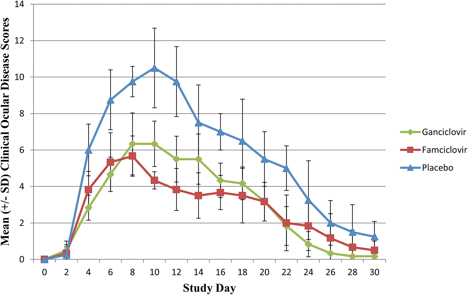
Mean ± SD clinical ocular disease scores for cats with experimentally induced ocular FHV-1 infection treated with topical ophthalmic ganciclovir (green line), oral famciclovir (red line), or topical artificial tear gel (blue line) for 14 days. Ocular FHV-1 infection was induced on study day 0 and study medications were administered for 14 days starting on study day 1 (administered on study days 1–15). Clinical ocular disease scores were calculated every 2 days for 30 days. The ocular surface clinical scoring system used to quantify examination findings had a minimum daily clinical score of 0 and a maximum daily score of 15.

Representative clinical photographs of 3 cats with experimentally induced ocular FHV-1 infection treated with topical ophthalmic ganciclovir, oral famciclovir, or topical artificial tear gel for 14 days. Ocular FHV-1 infection was induced on study day 0 and study medications were administered for 14 days starting on study day 1.
Clinical ocular disease scores rose more rapidly and remained elevated for a longer duration in the placebo group than either the ganciclovir or famciclovir groups (Figs. 2 and 3). The maximum mean (± SD) clinical ocular disease scores and the study day when they were recorded were 10.5 (± 2.2) in the placebo group on day 10, 6.3 (± 1.3) in the ganciclovir group on both days 8 and 10, and 5.7 (± 1.1) in the famciclovir group on day 8. Clinical ocular disease scores slowly declined after the maximal values and were at or near the baseline scores by study day 30 in all cats.
Clinical ocular disease scores were significantly (P ≤ 0.001) lower over time in the ganciclovir and famciclovir study groups versus the placebo group. Clinical scores initially declined more rapidly in cats treated with famciclovir versus ganciclovir, but the differences between these study groups were not statistically significant (P = 0.4) and the mean clinical scores in the ganciclovir group were consistently lower than the famciclovir group after study day 20.
The mean (± SD) respiratory rate over the entire study period after viral inoculation was 33.3 (± 4.7) breaths per minute in the placebo group, 33.1 (± 5.1) breaths per minute in the ganciclovir group, and 34.4 (± 5.2) breaths per minutes in the famciclovir group. There were no significant differences in mean respiratory rate between any of the study groups over time.
Virus isolation
Ocular viral shedding was detected in all study cats by virus isolation on study day 3 (Fig. 4). The mean (± SD) duration of viral shedding as detected by virus isolation was 15 (± 0.1) days in the placebo group, 15 (± 2.4) days in the ganciclovir group, and 17.5 (± 4.4) days in the famciclovir group. The mean (± SD) daily viral load over the entire course of viral shedding was 24,475 (± 7,854) PFU/mL in the placebo group, 22,166 (± 10,410) PFU/mL in the ganciclovir group, and 17,041 (± 9,860) PFU/mL in the famciclovir group.

Mean ± SD ocular viral load (PFU/mL) determined by virus isolation for cats with experimentally induced ocular FHV-1 infection treated with topical ophthalmic ganciclovir (green line), oral famciclovir (red line), or topical artificial tear gel (blue line) for 14 days. Ocular FHV-1 infection was induced on study day 0 and study medications were administered for 14 days starting on study day 1 (administered on study days 1–15). Samples for determination of ocular viral load were collected every 3 days for 30 days.
Ocular viral shedding was detected by virus isolation in the placebo group until study day 15, the ganciclovir group until study day 18, and the famciclovir group until study day 24. There were no statistically significant differences detected in ocular viral shedding duration or viral load between any of the study groups as determined by virus isolation.
FHV-1 real-time PCR
Ocular viral shedding was detected in all study cats by qPCR on study day 3 (Fig. 5). The mean (± SD) duration of viral shedding as detected by qPCR was 20 (± 5) days in the placebo group, 22 (± 7) days in the ganciclovir group, and 25 (± 4) days in the famciclovir group. Ocular viral shedding was detected by qPCR in at least one cat until day 27 for the placebo group, day 30 for the ganciclovir group, and day 27 for the famciclovir group. The mean (± SD) daily viral load over the entire course of viral shedding was 1,504 (± 355) FHV-1 copies/5 cells in the placebo group, 1,126 (± 325) FHV-1 copies/5 cells in the ganciclovir group, and 1,308 (± 590) FHV-1 copies/5 cells in the famciclovir group.

Mean ± SD ocular viral load (copies FHV-1/5 cells) determined by FHV-1 qPCR for cats with experimentally induced ocular FHV-1 infection treated with topical ophthalmic ganciclovir (green line), oral famciclovir (red line), or topical artificial tear gel (blue line) for 14 days. Ocular FHV-1 infection was induced on study day 0 and study medications were administered for 14 days starting on study day 1 (administered on study days 1–15). Samples for determination of ocular viral load were collected every 3 days for 30 days.
Ocular viral load detected by qPCR was consistently higher in the famciclovir groups relative to both the placebo and ganciclovir groups after study day 15. Ocular viral load measured by qPCR was significantly (P = 0.01) lower in the ganciclovir group relative to the placebo group. No significant differences were detected in ocular viral load between the ganciclovir and famciclovir groups, or between the famciclovir and placebo groups. There were no statistically significant differences detected in ocular viral shedding duration between any of the study groups as determined by qPCR.
In vivo confocal microscopy
Corneal epithelial leukocytes were detected by in vivo confocal microscopy in all cats on study day 10 (Fig. 1). The mean (± SD) in vivo confocal microscopy keratitis scores from study day 10 were 404 (± 216) leukocytes/mm2 in the right eye (OD) and 442 (± 205) leukocytes/mm2 in the left eye (OS) for the placebo group, 65 (± 37) leukocytes/mm2 OD and 54 (± 32) leukocytes/mm2 OS for the ganciclovir group, and 132 (± 101) leukocytes/mm2 OD and 94 (± 73) leukocytes/mm2 OS for the famciclovir group. Leukocyte infiltrate counts were significantly lower in both eyes of the ganciclovir and famciclovir groups versus placebo (P ≤ 0.001), but no significant (P ≥ 0.3) differences were detected between the ganciclovir and famciclovir groups.
General diagnostic assays and examinations
All hemogram and serum biochemistry panel results performed on study day 30 were unremarkable. No evidence of adverse reactions to the study medications, including ocular irritation after ganciclovir administration, was observed in any study cat at any time during the study. No significant abnormalities were detected in any of the study cats during physical and ophthalmic examinations performed on study day 30, except for mild residual conjunctivitis in a few cats from each study group. Results of Schirmer I tear tests and intraocular pressure measurements were within normal ranges for all cats.
Discussion
In this study using an experimental model of ocular FHV-1 epithelial infection, topical application of ganciclovir 0.15% ophthalmic gel 3 times daily was well-tolerated and reduced clinical disease scores, corneal leukocyte infiltrates detected by in vivo confocal microscopy, and ocular viral loads measured with qPCR compared with a placebo artificial tear ophthalmic gel. When compared with oral famciclovir treatment, topical application of ganciclovir ophthalmic gel resulted in a similar reduction in clinical ocular disease scores, corneal leukocyte infiltrates, ocular viral loads (as measured by both virus isolation and qPCR), and a similar duration of ocular viral shedding.
Earlier clinical reports of topical antiviral therapy for FHV-1 infections in cats often describe a relatively high daily frequency of medication administration. In part, this is because all ophthalmic antiviral medications have a virustatic mechanism of action. The topical antiviral agents idoxuridine 0.1% solution, trifluridine 1% solution, vidarabine 3% ointment, and acyclovir 0.5% ointment are generally recommended to be administered every 4–6 h in cats with FHV-1 ocular disease based primarily upon anecdotal clinical reports.27,28 In human patients with ocular HSV infection and dogs with ocular canine herpesvirus-1 infection, ganciclovir 0.15% aqueous ophthalmic gel is typically administered 5 times daily initially.17,19
The behavior and temperament of many domestic cats with FHV-1 infections can present unique challenges to the frequent administration of topical ophthalmic medications. Theoretically, the stress associated with frequent medication administration may exacerbate herpetic infections in cats.29,30 In addition, clients many be unable to locate, restrain, and administer a topical ophthalmic medication at a high frequency in some cats safely and reliably. It is for these reasons that a subjectively more practical 3 times daily ganciclovir administration frequency was evaluated in this study. The pragmatic study design that was selected proved successful by both clinical and virological outcome measures, but it is currently unknown if administration of ganciclovir at a higher frequency could prove more efficacious in cats with FHV-1 infection.
In vitro studies evaluating the ability of various antivirals to inhibit FHV-1 replication have repeatedly demonstrated ganciclovir to be among the most potent drugs tested against FHV-1. Reported IC50 (number) values for ganciclovir in these studies ranged from 5.2 to 12.5 μM, values much lower than most other drugs evaluated in the studies including foscarnet, acyclovir, adefovir, penciclovir, and cidofovir.14,15
Ganciclovir triphosphate, the active metabolite of ganciclovir, blocks viral replication by 2 independent mechanisms. It is incorporated into viral strand primary DNA where it induces DNA strand breaks and viral DNA chain termination. 31 Ganciclovir triphosphate also competes with deoxyguanosine triphosphate for binding to viral DNA polymerase resulting in the interruption of new viral DNA synthesis. 32 The combination of a high level of in vitro activity against FHV-1 and the extended ocular surface contact time provided by the gel formulation likely contributed to the efficacy of the ganciclovir 0.15% aqueous ophthalmic gel evaluated in this in vivo study.
Famciclovir therapy has been demonstrated under a variety of circumstances to be a highly effective medical treatment for ocular FHV-1 infection in cats. A previous placebo-controlled study evaluated cats with experimental primary ocular FHV-1 infection treated with 90 mg/kg famciclovir administered 3 times daily for 3 weeks. 6 Famciclovir was determined in the study to be an effective treatment based upon clinical, virological, and histological outcome measures. 6 Several studies have evaluated the effects of twice daily famciclovir therapy administered from 1 to 3 weeks in cats with naturally acquired upper respiratory tract disease with mixed and inconsistent results.33–35 This study is the first to demonstrate efficacy for experimentally induced FHV-1 ocular disease with twice daily famciclovir administration.
Although overall clinically effective, the mean clinical ocular disease score in the cats treated with famciclovir slightly increased immediately after the conclusion of the 14-day treatment course of famciclovir. The clinical scores then continued to decline at a slower rate for the next several days after famciclovir was discontinued compared with what was observed in the few days before cessation of famciclovir. In addition, ocular viral loads slightly increased immediately after famciclovir was discontinued. This suggests that, under the evaluated experimental conditions, 3 weeks of famciclovir therapy might be preferable to a 2-week course for cats with primary FHV-1 infections. The optimal duration of famciclovir therapy is currently unknown, may be different for primary and recurrent FHV-1 infections, could vary between different clinical scenarios, and requires additional investigation.
Numerous clinical studies have compared the relative efficacy of different topical antivirals to result in the resolution of active HSV epithelial keratitis in human patients. 36 Relatively fewer clinical studies have compared oral antiviral therapy with topical antiviral therapy in humans with HSV keratitis.37,38 A comparison of 5 times daily oral and topical acyclovir treatment found no significant different between treatment groups in the number of patients healed after 14 days (97% vs. 89% for topical and oral treatment, respectively). 38 Similarly, the combination of treatment with both oral and topical antiviral versus treatment with a topical antiviral alone has been evaluated relatively infrequently.36,39,40
In one study comparing topical trifluridine alone or in combination with oral acyclovir, a similar time until corneal ulcer healing was reported in the 2 groups. 39 In contrast to HSV infection, the paucity of controlled clinical and experimental studies directly comparing the efficacy of different antiviral medications for the treatment of ocular FHV-1 infections in cats often results in an antiviral selection based upon other factors, including availability, formulation, and dosing frequency.
Evaluation of the efficacy and potential toxicity of novel antiviral drugs, and the objective comparison of existing treatment options, in animal models of ocular HSV infection is an integral part of the development of clinical treatment strategies. Studies directly comparing different antiviral agents in animal models of ocular HSV infection have generally utilized rabbits or mice with experimentally induced infections.41–43 These animal models have the advantage of permitting a high number of animals to be used to increase the statistical power of the study. In addition, murine models of recurrent ocular HSV infection are described that may more accurately represent the most common clinical scenarios where antiviral therapies are administered in humans. 44
The feline model of ocular herpesvirus infection provides several advantages compared with these more traditional animal systems. These benefits include a larger anatomic ocular surface facilitating detailed clinical examination and sample collections. In addition, experimental FHV-1 infection represents a natural host-adapted pathogen model that is distinct from murine or rabbit models of HSV infection. 10 Feline models of recurrent ocular infection are described, but a primary experimental infection model was selected for the present study as clinical ocular lesions are more reliably induced with this system.5,30
Limitations of this study include the relatively small number of cats included. Inclusion of a higher number of cats might have permitted the statistical detection of additional differences between the study groups for the evaluated parameters. Although not statistically significant, ocular viral loads were higher in both antiviral groups compared with the placebo group after study day 21. This surprising finding might reflect the small number of cats included in the placebo group and individual variation in viral shedding between cats. With the experimental infection model used in this study, the antiviral treatments were initiated 24 h after viral inoculation. Early initiation of antiviral treatment is performed in this model for humane reasons to mitigate the development of more severe clinical ocular disease; however, this may not reflect some clinical scenarios where antiviral therapy is started later in the infection course.5,6
The duration of clinical ocular disease and ocular viral shedding detected in this study was typical for this experimental feline model utilizing direct ocular inoculation to induce primary infection and both are longer than what is typically observed with naturally acquired infection in cats and humans.5–9 Interpretation of the ocular viral loads measured by qPCR is limited by the fact that positive results do not always represent actively replicating virus, but can result from viral remnants previously inhibited by the antiviral drugs. 45 It is for this reason that virus isolation was also performed in this study.
Conclusions
Topical application of ganciclovir ophthalmic gel 3 times daily was well-tolerated and displayed similar efficacy at reducing clinical disease scores and tissue inflammation as twice daily oral famciclovir treatment in cats with experimental ocular FHV-1 epithelial infection.
Footnotes
Acknowledgment
Preliminary results of this study were presented as an abstract at the American College of Veterinary Ophthalmologists Conference, Indianapolis, IN, USA, September 29–October 2, 2021.
Authors' Contribution
All authors meet the criteria for authorship.
Author Disclosure Statement
No competing financial interests exist.
Funding Information
No funding was received for this article.