
Abstract
Hemophilia A, a bleeding disorder, affects 1:5,000 males and is caused by a deficiency of human blood coagulation factor VIII (hFVIII). Studies in mice and macaques identified
Introduction
Hemophilia A is a common hereditary bleeding disorder affecting 1:5,000 males and is characterized by a deficiency of human blood coagulation factor VIII (hFVIII). 1,2 Depending on the level of hFVIII deficiency, disease severity can range from mild to severe. 1 Protein replacement therapy via intravenous (IV) infusion of recombinant hFVIII can effectively maintain a mild-to-moderate phenotype, but the requisite frequency is burdensome to patients. 3 Adeno-associated virus (AAV) vector-based gene therapy for the treatment of hemophilia A may represent an alternative treatment approach, which only requires a single administration.
The major limitation for an AAV-based gene therapy approach for hemophilia A is the size of the hFVIII coding sequence. The native hFVIII protein is a large, multidomain glycoprotein with complementary DNA (cDNA) that exceeds the packaging capacity for recombinant AAV (>7 kb). 4,5 We have extensively engineered the promoter and enhancer in the expression cassette 6 to complement the reduction in size of the hFVIII cDNA performed by others (also referred to as F8). 7 The protein replacement therapy drug—ReFacto® (Pfizer, New York, NY)—was successfully designed to mimic the smallest active form of hFVIII by replacing the B domain with a 14-amino acid SQ linker. 8 Subsequent codon optimization of this B-domain-deleted (BDD) hFVIII-SQ (hFVIIIco-SQ) resulted in efficient packaging and increased expression from lentiviral 7,9 and AAV 6,10 vectors.
We have extensively evaluated AAVrh10 vectors that are designed to drive liver-specific expression of the codon-optimized BDD hFVIII gene within the size constraints of AAV.
6
Within a total genome size of <5,250 bp, we generated 42 enhancer/promoter combinations of 3 shortened liver-specific promoters and up to 3 liver-specific enhancer sequences. After evaluating hFVIII activity and immunogenicity of the transgene in a mouse model of hemophilia A (FVIII knockout [KO] mice), we performed an additional capsid-specific immunogenicity evaluation.
6
Based on our analyses, we selected AAVrh10 and AAVhu37 capsids and the
Upon systemically administering 1.2 × 10¹³ genome copies (GCs)/kg of
Materials and Methods
AAV vector production
AAV vector for the pilot dose-ranging study was produced by the Penn Vector Core at the University of Pennsylvania, as described previously.
12
In brief, plasmids expressing hFVIIIco-SQ from
Mice
We obtained FVIII KO mice (B6;129S-F8tm1Kaz /J) from The Jackson Laboratory (Bar Harbor, ME). A colony was maintained at the University of Pennsylvania under specific pathogen-free conditions; the mice used for the pilot dose-ranging study were derived from this colony. All animal procedures and protocols were approved by the Institutional Animal Care and Use Committee of the University of Pennsylvania. Each cohort included 10 animals. Before the study, we determined five animals per cohort to be the minimal number to enable statistical analysis of study outcome; we included five additional animals per time point to ensure that enough study animals would be available for meaningful analysis in the occurrence of unexpected deaths, antibody generation to the hFVIII transgene, or other unanticipated events.
Pilot dose-ranging study
Male FVIII KO mice aged 6–12 weeks received an IV injection of 1.5 × 10¹⁰, 1.5 × 10¹¹, 5 × 10¹¹, 1.5 × 10¹², 5 × 10¹², or 1.5 × 10¹³ GC/kg of
MED study
We obtained mice from Jackson Laboratories and housed five animals per cage in disposable microisolator mouse caging with corn cob bedding. Nestlets were provided for enrichment (Innovive, San Diego, CA). We provided certified irradiated Laboratory Rodent Diet 5002 (LabDiet, St. Louis, MO) ad libitum. All interventions were performed during the light cycle. Mice were not fasted before blood collection.
In this study, we used male FVIII KO mice (n = 100) aged 8–14 weeks, weighing 18.7–28.8 g in body weight. Before dosing, mice were first allocated to groups. Mice in the same cage belonged to the same group (mixing male mice from different cages into a new cage would cause fighting and possible death as these are hemophilic mice). Each group was randomly assigned to one of the dosing groups using an online program (Research Randomizer,
FVIII KO mice received an IV injection of 3 × 10¹¹
, 1 × 10¹², 3 × 10¹², or 1 × 10¹³ GC/kg of
Plasma was collected on days 7, 14, 28, and 56 via retro-orbital bleeds into sodium citrate collection tubes. Mice were necropsied on days 28 and 56. As the expressed transgene is human FVIII, most mice would likely have mounted an immune response to the transgene by day 56, neutralizing the activity of any further expressed transgene. Therefore, continuing the study past day 56 was not expected to yield additional pharmacological data.
Clinical chemistries
We collected blood at the time of necropsy by cardiac puncture in labeled serum gel separator brown-top tubes. After allowing the blood to clot for at least 30 min at room temperature, we centrifuged the samples at 3,500 g for 5 min at room temperature. The serum was separated and then shipped to Antech GLP (Morrisville, NC) for analysis of alanine aminotransferase (ALT), aspartate aminotransferase (AST), alkaline phosphatase, gamma glutamyl transferase, total bilirubin, direct bilirubin, and total protein.
hFVIII activity
We measured hFVIII activity in plasma using the Chromogenix Coatest® SP4 kit, according to the manufacturer's protocol (DiaPharma, West Chester, OH). 6 In brief, this kit works by combining mouse plasma with unknown hFVIII levels with calcium, phospholipids, and factors IXa and X. The rate of activation of factor X to Xa is dependent on the levels of hFVIII in the plasma sample. A standard curve was generated using known concentrations of BDD-hFVIII-SQ (Xyntha; Wyeth Pharmaceuticals, Inc., Dallas, TX).
Anti-hFVIII immunoglobulin G
We detected immunoglobulin G (IgG) antibodies against hFVIII in mouse plasma at the time of necropsy via enzyme-linked immunosorbent assay (ELISA) as described previously. 6 Plasma samples were diluted 1/100 or more and values that were fivefold over background levels (naive mouse samples) were considered positive. The data were reported as anti-hFVIII IgG titers. Negative values are denoted as a titer of 1/50 to enable them to be visualized.
Histopathology
We harvested tissues at necropsy for comprehensive histopathological examination. These tissues included the injection site, right testis, brain, liver, right kidney, lung, heart, and spleen. Tissues were fixed using 10% neutral buffered formalin, paraffin embedded, sectioned, and stained for histopathology using H&E stain. An experienced, board-certified veterinary pathologist evaluated liver sections in a blinded manner using scoring criteria. Histopathology slides for other tissues were evaluated and peer-reviewed for the highest vector dose group and the vehicle control group. If any findings were reported in the highest dose group, the next lower dose group was evaluated, and so on.
Vector biodistribution
At the time of necropsy, the liver was collected for biodistribution analysis, frozen on dry ice, and stored at less than or equal to −60°C. We extracted DNA and RNA from liver samples and performed quantitative polymerase chain reactions (qPCRs), as described previously. 6 Using qPCR targeting a vector-specific sequence, we assayed DNA and RNA samples for vector GC and vector-derived hFVIII transgene levels, respectively. Vector GC assay results were reported as GC per microgram of DNA (GC/μg). We then calculated the vector GC per diploid genome, assuming that one microgram of DNA contains ∼2 × 10⁵ diploid genomes. 13
Statistical analyses
We calculated and reported the group average and standard error of the mean for the following: ALT, AST, total bilirubin, total protein, hFVIII activity, vector GCs, and hFVIII RNA transcript levels. We compared groups administered with vector and the vehicle control using a Wilcoxon rank-sum test at each time point for ALT, AST, total protein, total bilirubin, hFVIII activity, vector GCs, and hFVIII RNA transcript levels (nonparametric evaluation as the data appeared to be nonnormally distributed). We compared vector-administered groups to each other using a two-sample Wilcoxon rank-sum test at each time point for hFVIII activity, vector GCs, and hFVIII RNA transcript levels. We used linear mixed-effect modeling to compare the overall change between the vector- and vehicle control-administered groups across all time points. No multiple testing adjustment was performed. A p-value of <0.05 was considered significant.
Results
Pilot dose-ranging study in FVIII KO mice
Based on the results of our previous studies,
6,11
we selected the clinical candidate vector,
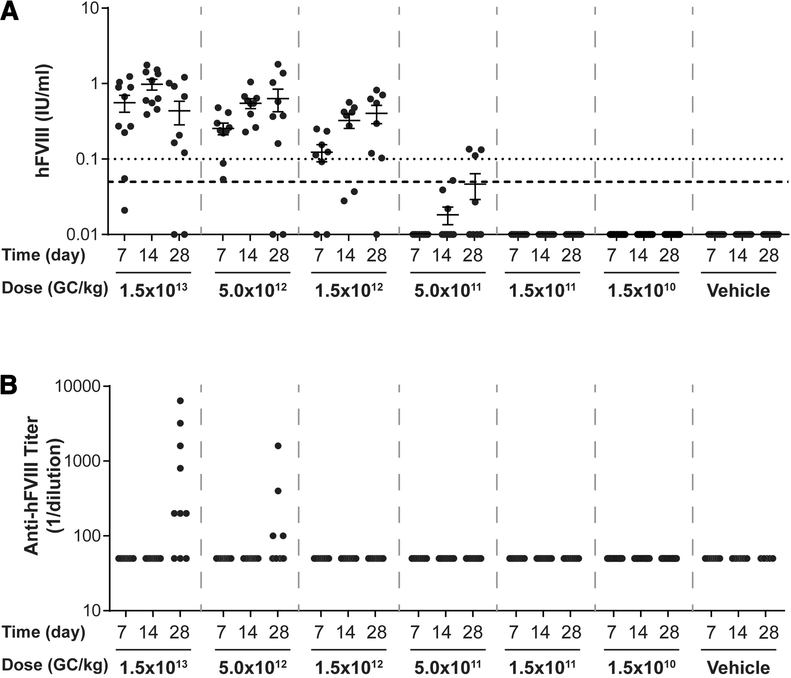
Plasma hFVIII activity levels and anti-hFVIII IgG titers in pilot dose-ranging study. Male FVIII KO mice (n = 10/group) were injected IV with 1.5 × 10¹³ GC/kg, 5.0 × 10¹² GC/kg, 1.5 × 10¹² GC/kg, 5.0 × 10¹¹ GC/kg, 1.5 × 10¹¹ GC/kg, or 1.5 × 10¹⁰ GC/kg of
We injected IV male FVIII KO mice at 6–12 weeks of age with
MED study rationale and design
Male FVIII mice aged 8–14 weeks received an IV tail vein injection of vehicle control or
Clinical findings
During the study, two mice from the vehicle control group were euthanized for humane reasons (on days 15 and 24). In both cases, we performed a full necropsy and collected tissues for analysis. Histopathology findings pointed to evidence of blood loss in conjunction with the clinical signs, although no direct evidence of hemorrhage or blood loss was observed. During the in-life phase of the study, we recorded clinical observations for 18 out of the 100 mice enrolled in this study that did not affect the outcome. An additional eight mice required supportive care.
Dose-dependent increase in hFVIII activity
We analyzed plasma hFVIII activity levels throughout the in-life phase of the study. As expected, hFVIII activity levels displayed a dose-dependent increase following IV administration of increasing vector doses (Fig. 2). hFVIII activity increased over the duration of the study from day 7 until the necropsy time point, unless anti-hFVIII IgG antibodies developed (Fig. 3).
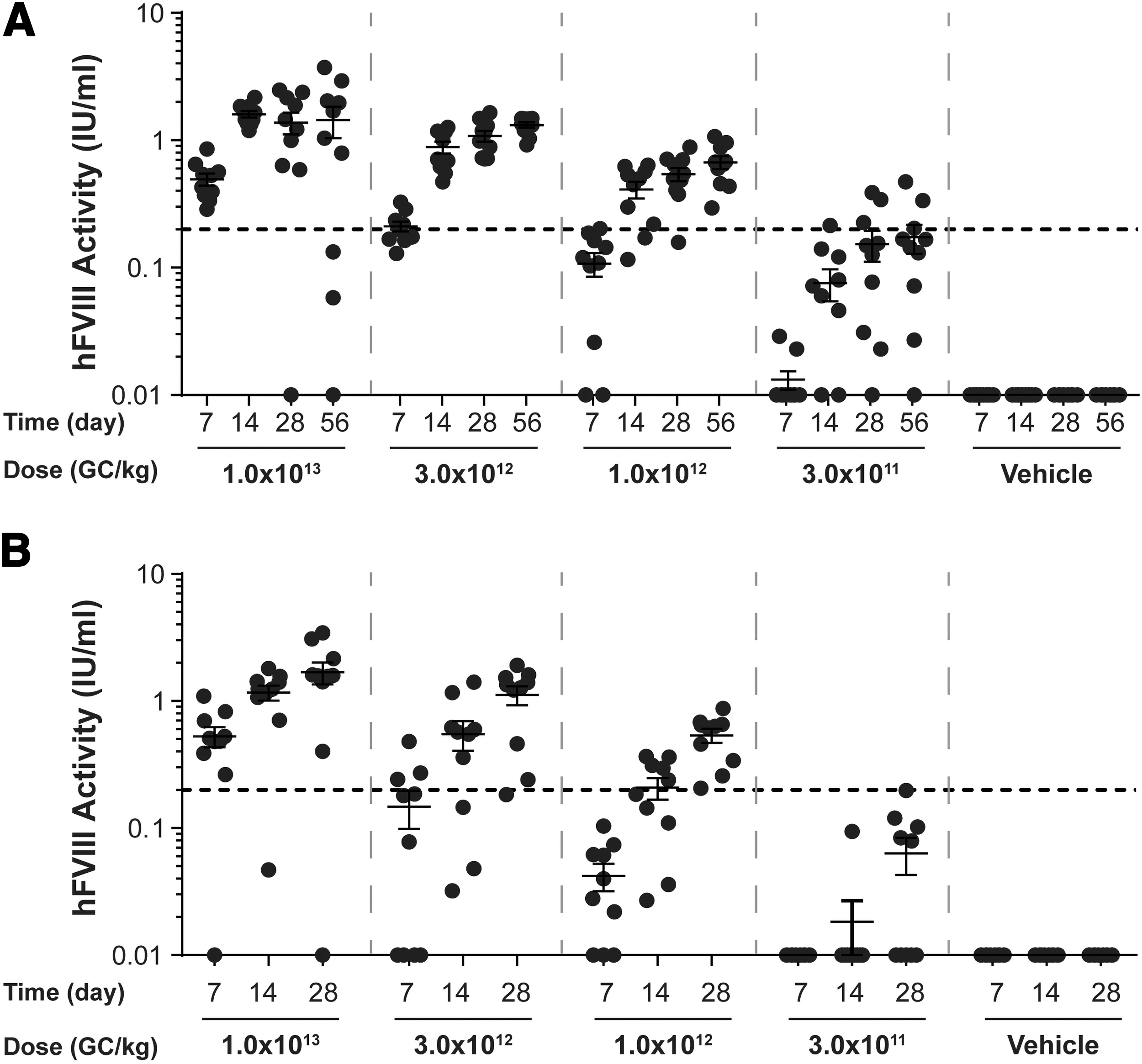
Plasma hFVIII activity levels in vector-administered FVIII KO mice. Male FVIII KO mice (n = 10/group) were injected IV with 1 × 10¹³ GC/kg, 3 × 10¹² GC/kg, 1 × 10¹² GC/kg, or 3 × 10¹¹ GC/kg of
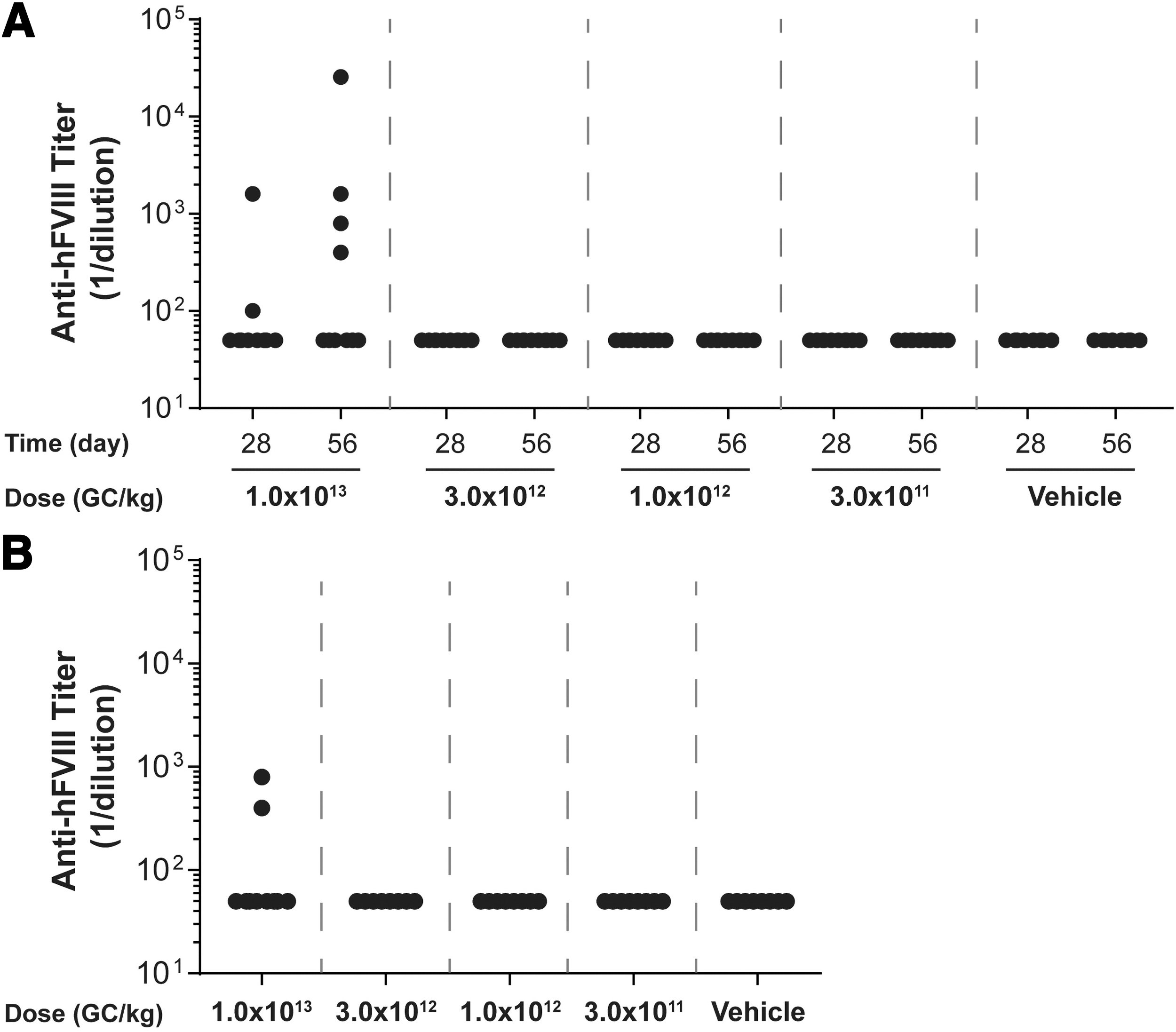
Plasma anti-hFVIII IgG titers in vector-administered FVIII KO mice. Male FVIII KO mice (n = 10/group) were injected IV with 1 × 10¹³ GC/kg, 3 × 10¹² GC/kg, 1 × 10¹² GC/kg, or 3 × 10¹¹ GC/kg of
For mice necropsied on day 56 postvector administration, the average peak activity level in the high-dose group (1 × 10¹³ GC/kg) was 1.438 IU/mL (equivalent to 143.8% of normal FVIII levels) (Fig. 2A). By day 28 postvector administration, anti-hFVIII IgG antibodies had developed in 2 out of the 10 mice administered the high dose, with an additional two mice in this group developing anti-hFVIII IgG antibodies by day 56 (Fig. 3A). All mice with detectable anti-hFVIII IgG antibodies exhibited a reduction in their individual hFVIII activity levels (Figs. 2 and 3).
For mice necropsied on day 28 postvector administration, the average peak activity level in the high-dose group (1 × 10¹³ GC/kg) was 1.684 IU/mL (Fig. 2B). Similar to mice necropsied at day 56 (Fig. 3A), anti-hFVIII IgG antibodies had developed in two mice in the high-dose group by day 28 postvector administration, which resulted in a decline in their individual hFVIII activity levels (Fig. 3B).
At the lowest dose evaluated in this study (3 × 10¹¹ GC/kg), the average peak activity level was 0.173 IU/mL at day 56 (Fig. 2A). As we detected hFVIII activity at all doses of
No correlation between vector dose and elevations in serum ALT or AST levels
Serum chemistry panels were performed on samples collected at necropsy by Antech GLP (Fig. 4). We evaluated the blood chemistry results for statistical differences (p < 0.05) in mice administered vector compared to vehicle controls on days 28 and 56 postvector administration. To compare ALT and AST levels in vector- and vehicle control-administered mice, we applied the Wilcoxon rank-sum test. For mice necropsied at day 28, we observed no significant differences in ALT or AST levels in mice administered any vector dose compared to those administered with the vehicle control (Fig. 4A, C). For mice necropsied at day 56, we observed a significant reduction in ALT levels following administration of 3 × 10¹¹ GC/kg of the vector compared to the vehicle-treated cohort (Fig. 4B), but observed no significant differences in AST levels (Fig. 4D).
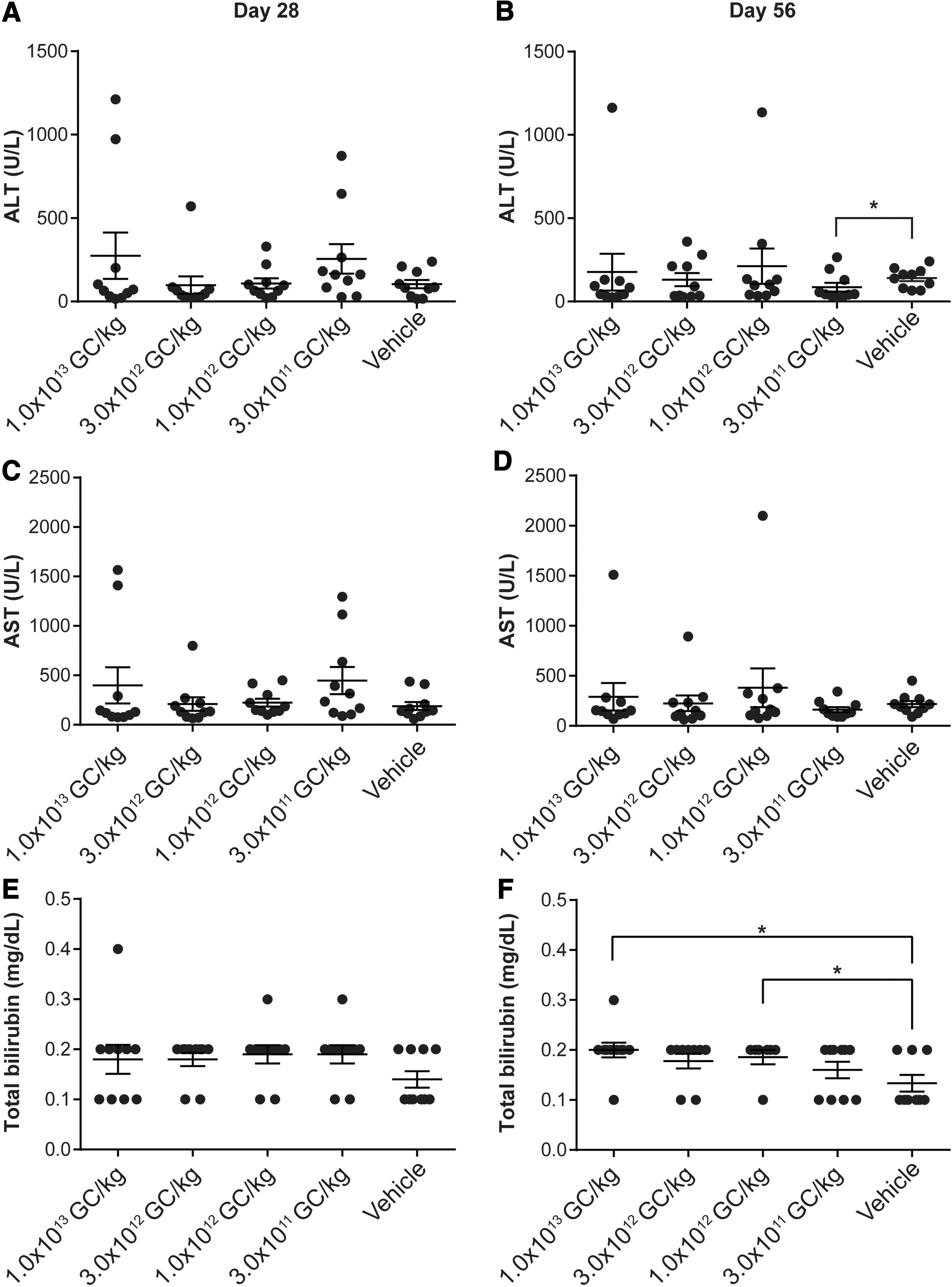
ALT, AST, and total bilirubin levels in vector-administered FVIII KO mice. Male FVIII KO mice (n = 10/group) were injected IV with 1 × 10¹³ GC/kg, 3 × 10¹² GC/kg, 1 × 10¹² GC/kg, or 3 × 10¹¹ GC/kg of
We compared serum total bilirubin levels following administration of vector or vehicle control. Mice necropsied on day 28 displayed no significant differences (Fig. 4E). Mice necropsied at day 56 that were administered 1 × 10¹² GC/kg or 1 × 10¹³ GC/kg of vector exhibited a significant elevation in total bilirubin levels compared to the vehicle control-administered group (Fig. 4F).
In addition, comparing serum total protein levels revealed significant differences in mice administered with 3 × 10¹² GC/kg on day 28 (Supplementary Fig. S1A) or 3 × 10¹¹ GC/kg of vector at day 56 (Supplementary Fig. S1B).
Histopathological findings
We harvested tissues from all animals at the time of necropsy, stained them with H&E, and performed a full histopathological analysis. An experienced board-certified veterinary pathologist evaluated the liver sections in a blinded manner using predetermined scoring criteria. Histopathology slides for other tissues were evaluated and peer-reviewed for the highest vector dose group and the vehicle control group. No vector-related microscopic findings were observed (Table 1).
Summary of histopathology findings for vector-administered FVIII knockout mice
Male FVIII KO mice were injected IV with 1 × 10¹³ GC/kg, 3 × 10¹² GC/kg, 1 × 10¹² GC/kg, or 3 × 10¹¹ GC/kg of
AAV, adeno-associated virus; EME, extramedullary erythropoiesis; GCs, genome copies; IV, intravenous; KO, knockout.
Most of the microscopic findings were observed in vehicle-administered animals and were considered potentially secondary to blood loss. However, no macroscopic or microscopic evidence of hemorrhage was observed (Table 1). These findings included centrilobular hepatocellular necrosis (ischemia), extramedullary erythropoiesis in the spleen and liver, epicardial fibrosis with pigment-laden macrophages (consistent with hemosiderin), pigment-laden macrophages with perivascular distribution in the heart, and pigmented (hemoglobin, presumptive) granular casts within renal tubules. Acute alveolar hemorrhage occurred in the lungs of some mice (1/10 mice administered with 1 × 10¹³ GC/kg necropsied at both day 28 and 56, 1/10 mice administered with vehicle control necropsied at day 28). With no histologic evidence of chronicity (e.g., hemosiderophages), we suspected that this finding was perimortem alveolar hemorrhage (potentially secondary to cardiac puncture). The observation of renal interstitial mononuclear cell infiltrates associated with minimal tubule basophilia was considered incidental. Other microscopic findings were incidental, background, or secondary to IV administration, and included myocardial/epicardial mineralization, pulmonary interstitial infiltrates, focal pulmonary foreign body granuloma (hair shaft), focal inflammation in the liver, mononuclear cell infiltrates within the kidney, and a squamous cyst in the brain. The mineralization observed in the heart of a vehicle-administered mouse and two mice from the high-dose group may represent mineralized thrombi, especially given the proximity of the lesions to blood vessels. We found a single acute nonocclusive fibrin thrombus in the lung of one mouse from the high-dose group. The hematopoietic infiltrates in the liver in one vehicle-administered animal were associated with minimal single hepatocellular necrosis.
Liver vector GC and transgene RNA analysis
At the time of necropsy, we collected the liver for biodistribution analysis. We detected a dose-dependent increase in both vector GC and hFVIII RNA levels in the liver (Fig. 5). Using a two-sample Wilcoxon rank-sum test, we compared each vector-administered group for mice necropsied on day 28 or 56 (Fig. 5A, B). We observed significant differences between doses in vector GCs for all vector-administered groups, except for mice administered 1 × 10¹³ GC/kg and 3 × 10¹² GC/kg and necropsied at day 28 (Fig. 5A).
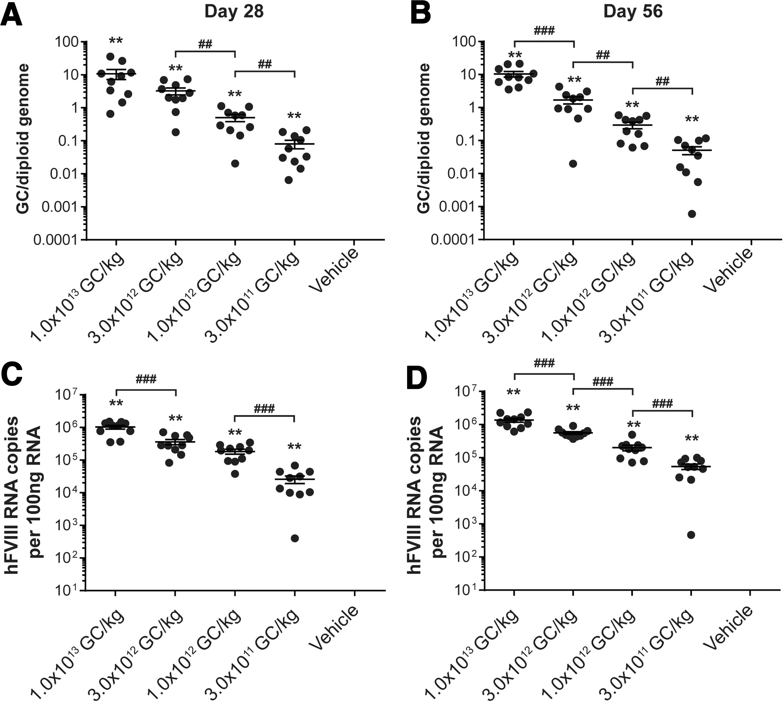
Vector GC and hFVIII RNA transcript levels in livers from vector-administered FVIII KO mice. Male FVIII KO mice (n = 10/group) were injected IV with 1 × 10¹³ GC/kg, 3 × 10¹² GC/kg, 1 × 10¹² GC/kg, or 3 × 10¹¹ GC/kg of
We performed the same comparisons for hFVIII RNA transcript levels between each vector-administered group (two-sample Wilcoxon rank-sum test, Fig. 5C, D). For mice necropsied on day 28, all vector-administered groups displayed significant differences, except for the comparison between mice administered 3 × 10¹² GC/kg and 1 × 10¹² GC/kg (Fig. 5C). For mice necropsied on day 56, all vector-administered groups displayed significant differences in hFVIII RNA transcript levels (Fig. 5D).
Discussion
In this study, we determined the MED of our clinical candidate vector, AAVhu37.E03.TTR.hFVIIIco-SQ.PA75, in a hemophilia A mouse model. We elected to conduct the experiments in FVIII KO mice (rather than in wild-type C57BL/6 mice) for two reasons. First, using this strain of mice enabled us to evaluate efficacy in parallel with additional safety measurements. Second, we aimed to evaluate potential vector-associated safety signs in the setting of any pathology associated with the defect in FVIII and the associated severe hemophilia and its sequelae. While we did not expect the model to exhibit liver pathology, we were concerned that coagulation deficiencies could influence the response of the host liver to vector.
Following IV administration of AAVhu37.E03.TTR.hFVIIIco-SQ.PA75, hFVIII activity increased over the duration of the study from days 7 to 56, unless anti-hFVIII IgG antibodies developed in individual mice. We detected activity levels of >1.4 IU/mL (equivalent to 140% of normal) at the time of necropsy. It is well established that FVIII levels directly correlate with clinical efficacy. 2 Indeed, hemophilia A patients are classified into different severity levels depending on the percentage of normal hFVIII; mild (5–40% of normal, 0.05–0.40 IU/mL), moderate (1–5% of normal, 0.01–0.05 IU/mL), and severe (<1%, 0.01 IU/mL). Given this, our results strongly suggest that our gene therapy product would demonstrate clinical efficacy in hemophilia A patients.
While some FVIII KO mice did develop anti-hFVIII IgG antibodies, the relevance of this to the clinical application of this gene therapy approach is unknown as this vector expressed a human protein in mice. Also, we did not determine the region of the hFVIII protein to which the antibodies were binding. Other investigators have used immunodeficient FVIII KO mouse models (either Rag2 −/− or CD4−/−) to evaluate expression and activity in the absence of antibody generation. 14 –17
We found no significant differences in liver transaminases in mice administered any vector dose compared to mice administered vehicle control (except for one group administered 3 × 10¹¹ GC/kg). There was a significant elevation in total bilirubin levels following administration of 1 × 10¹² GC/kg or 1 × 10¹³ GC/kg of vector at day 56 compared to the vehicle-treated group.
Importantly, we did not observe any gross or histological vector-related pathology findings. The majority of the microscopic findings were in mice administered the vehicle control and were considered to point to evidence of blood loss in conjunction with clinical signs, although no direct evidence of hemorrhage or blood loss was observed.
As we observed no dose-limiting vector-related safety measurements, the maximally tolerated dose was greater than or equal to the highest dose tested, which was 1 × 10¹³ GC/kg. Moreover, conducting this study in a hemophilia A animal model allowed us to estimate the MED. We detected hFVIII activity at all doses of the test article administered; hFVIII activity levels were significantly elevated for test article cohorts administered more than 3 × 10¹¹ GC/kg at all time points. Therefore, the MED is equal to 3 × 10¹¹ GC/kg.
Direct comparison of the MED for
Due to the low MED for this treatment approach for hemophilia A, combined with expression data following systemic administration of the same vector in nonhuman primates,
11
we believe that this AAVhu37-based gene therapy approach has therapeutic potential in humans. In macaques, the hFVIII activity levels following IV administration of 1.2 × 10¹³ GC/kg would be sufficient to ameliorate a severe hemophilia A phenotype,
11
thereby reducing or potentially removing the need for recombinant hFVIII infusions. Moreover, a single-administration gene therapy could significantly reduce the cost of treating hemophilia A, particularly in comparison to current IV factor VIII replacement therapy.
18
Based on this promising preclinical evidence and with an ongoing clinical trial for evaluation (
Footnotes
Authors' Contributions
J.A.G.—conceptualization, investigation, methodology, supervision, writing—original, writing—review and edits; M.K.S.—investigation; J.M.L.N—investigation; T.G.—investigation; E.A.C.—validation; E.L.B.—formal analysis, investigation, writing—original, writing—review and edits; N.S.—conceptualization, writing—review and edits; L.M.K.—conceptualization, methodology, writing—review and edits; S.W.—conceptualization, methodology, writing—review and edits; J.M.W.—conceptualization, funding acquisition, methodology, supervision, writing—review and edits.
Acknowledgments
We thank the Penn Vector Core for providing the AAV vector for the pilot dose-ranging MED study, and the Program in Comparative Medicine, Histology Core, Quality Assurance Unit, and Regulatory Affairs and Compliance unit within the Gene Therapy Program. We thank Christine Draper, Peter Hewins, Deirdre McMenamin, and Regina L. Munden for invaluable technical assistance and support, as well as Mingyao Li, and Yan Che for help with statistical analysis. We thank Nathan Denton for assistance with article preparation and graphics.
Author Disclosure
The authors declare having potential competing financial interests. J.M.W. is a paid advisor to and holds equity in Scout Bio and Passage Bio; he also has sponsored research agreements with Amicus Therapeutics, Biogen, Elaaj Bio, FA212, Janssen, Passage Bio, Regeneron, and Scout Bio, which are licensees of Penn technology. J.M.W. and J.A.G. are inventors on patents that have been licensed to various biopharmaceutical companies and for which they may receive payments. N.S. is an employee and stockholder of Bayer Aktiengesellschaft. For the remaining authors, no competing financial interests exist.
Funding Information
This research was supported by Ultragenyx Gene Therapy (formerly Dimension Therapeutics).
Supplementary Material
Supplementary Figure S1
References
Supplementary Material
Please find the following supplemental material available below.
For Open Access articles published under a Creative Commons License, all supplemental material carries the same license as the article it is associated with.
For non-Open Access articles published, all supplemental material carries a non-exclusive license, and permission requests for re-use of supplemental material or any part of supplemental material shall be sent directly to the copyright owner as specified in the copyright notice associated with the article.