
Abstract
We conducted a comprehensive, multiphase laboratory evaluation of the Plague BioThreat Alert® (BTA) test, a lateral flow immunoassay (LFA), for the rapid detection of Yersinia pestis. The study was conducted in 7 phases at 2 sites to assess the performance of the LFA. The limit of detection (LOD) was determined using both a virulent and avirulent strain of Y. pestis, CO99-3015 (105 CFU/ml) and A1122 (104 CFU/ml), respectively. In the other phases, 18 Y. pestis strains, 20 phylogenetic near-neighbor strains, 61 environmental background microorganisms, 26 white powders, and a pooled aerosol sample were also tested. A total of 1,110 LFA test results were obtained, and their analysis indicates that this LFA had a sensitivity of 97.65% and specificity of 96.57%. These performance data are important for accurate interpretation of qualitative results arising from testing suspicious white powders and aerosol samples in the field. Any positive specimen in this assay is considered presumptive positive and should be referred to the Centers for Disease Control and Prevention Laboratory Response Network for additional testing, confirmation, and characterization for an appropriate public health response.
The authors conducted a comprehensive, multiphase laboratory evaluation of the Plague Bio Threat Alert® (BTA) test, a lateral flow immunoassay (LFA), for the rapid detection of Yersinia pestis. A total of 1,110 LFA test results were obtained, and their analysis indicates that this LFA had a sensitivity of 97.65% and specificity of 96.57%. These performance data are important for accurate interpretation of qualitative results arising from testing suspicious white powders and aerosol samples in the field.
Yersinia pestis is the causative agent of plague. It is a Gram-negative, nonmotile, non–spore forming coccobacillus, which is urease and indole negative.1-7 As a facultative anaerobe belonging to the Yersiniaceae, Y. pestis evolved from Yersinia pseudotuberculosis between 2,600 and 28,000 years ago, after the acquisition of 2 Y. pestis–specific plasmids: pMT1 and pPCP1.8,9 Y. pestis grows at 28°C, which is the normal body temperature for fleas, and at 37°C, the normal body temperature for humans.1,4,6,7 In the laboratory, the bacterium can grow on standard microbiologic media. 4 Observable growth is present at 24 to 48 hours, although colonies are smaller than those seen with other bacteria.4,7 In the wild, Y. pestis infects rodents, including rats, squirrels, and prairie dogs.1,4,10,11 Infection usually results from being bitten with a Y. pestis–infected vector such as the Oriental rat flea (Xenopsylla cheopis).1,3,4,10,12-15 Less commonly, infection can occur through handling infected animals or from inhaling infective respiratory droplets or aerosols. 3
All 3 pathogenic species of Yersinia (Y. pestis, Y. pseudotuberculosis, and Yersinia enterocolitica) carry the 70 kbp pCD1 plasmid, which encodes for a type III secretion system and Yersinia outer membrane proteins (Yops) that enable these bacteria to evade the host immune system. The pCD1 plasmid is not found in nonpathogenic Yersinia species.9,13,16-18 Virulent strains of Y. pestis also possess 2 additional plasmids: the 90 kbp pMT1 plasmid, alternatively known as pFra, which encodes the F1 capsular protein, and murine toxin. 19 The F1 capsule enables Y. pestis to resist engulfment by neutrophils and macrophages.1,12 The F1 gene is temperature regulated and expressed at ≥33°C. The 9.5 kbp pPCP1 plasmid, also known as pPla or pPst, encodes for plasminogen-activating factor as well as pestin and coagulase.10,19 In the laboratory, the plasmids can be lost during storage or on subculture.19,20
There are 3 clinical forms of plague: bubonic, septicemic, and pneumonic. The most common form is bubonic plague, acquired through the bite of an infected flea, which accounts for approximately 80% to 95% of all cases worldwide. Infected humans develop regional lymph node swelling and fever; bubonic plague is not transmissible from person to person and has a mortality rate of approximately 50% if untreated. Following entry into the body, Y. pestis is phagocytized by both neutrophils and macrophages; however, the bacteria survive and replicate in macrophages. Following infiltration of the lymph nodes, bacteria may enter the bloodstream, resulting in septicemic plague, which can also be caused directly via wound exposure.4-6,14,15,21-23
Pneumonic plague is far more dangerous but is very rare. Secondary pneumonic plague may result from the dissemination of bacteria to the lungs in cases of bubonic or septicemic plague, leading to severe bronchopneumonia, chest pain, dyspnea, cough, and hemoptysis. Primary pneumonic plague results from the direct inhalation of airborne droplets or aerosols of Y. pestis and is the clinical form most likely to occur following an aerosol release in a bioterrorism attack. Pneumonic plague is transmissible from person to person via airborne droplets. Infected individuals experience a 2- to 4-day incubation period, followed by rapid onset of chills, fever, general malaise, increased heart and respiratory rates, elevated body temperature, and a cough that becomes bloody as the disease progresses. During the terminal stage of the disease, patients experience hemorrhagic necrosis, acute respiratory failure, sepsis, and circulatory collapse. Diagnosis is based on immunostaining, PCR, and culture. In the absence of prompt treatment with antibiotics, pneumonic plague has a mortality rate that approaches 100%. Recommended treatment involves a 10- to 14-day regimen with an antibiotic such as streptomycin, gentamicin, ciprofloxacin, levofloxacin, or doxycycline.
First responders often encounter unknown and suspicious white powders in the field, and it is important to quickly evaluate them for the possible presence and identification of biological threat agents. The results of these evaluations can inform decisions to initiate public safety actions, including area evacuation, facility closure to prevent additional exposures, decontamination and initiation of medical countermeasures for potentially exposed individuals, collection of samples for law enforcement and public health purposes, sample transfer to a reference laboratory for immediate testing, and containment of contamination to prevent secondary dissemination or reaerosolization. In order to support first responders with the appropriate tools to carry out their mission, there is a need to understand performance of rapid assays for screening suspicious white powders.
The purpose of the present study was to evaluate the sensitivity, specificity, limit of detection (LOD), reproducibility, and limitations of the Plague BioThreat Alert test (Tetracore, Inc., Rockville, MD) as pertains to its use in the field to screen for the presence of Y. pestis. The goal of this study was to provide an understanding of assay performance, including the likelihood of false-negative results (assay is negative, but the target analyte is present at a concentration at or above the LOD), false-positive results (assay is positive, but the target analyte is not present in the sample), the limit of detection, and reproducibility, so that appropriate and effective decisions can be made by first responders to support public safety actions while avoiding unnecessary fear, panic, and costly civil disruptions.
This study was designed and executed through an interagency collaboration with participation from subject matter experts from the Department of Homeland Security (DHS), the Department of Health and Human Services (HHS), the Department of Justice (DOJ), the US Department of Agriculture (USDA), and the US Secret Service (USSS).
Materials and Methods
Biosafety Considerations
Strains used in this study were handled with appropriate biosafety conditions in accordance with the 5th edition of Biosafety in Microbiological and Biomedical Laboratories (BMBL) 24 and the Federal Select Agent Regulations.
Plague BTA Test and BioThreat Alert Reader MX
The Plague BTA test, which employs antibodies to detect the presence of the F1 capsular antigen, BioThreat Alert Reader MX (BTA Reader), and BTA Sample Buffer (BTA buffer) were obtained from Tetracore, Inc. (Rockville, MD). The performance of the Plague LFA and BTA Reader was evaluated at 2 test sites: (1) the Centers for Disease Control and Prevention (CDC), Fort Collins, CO, and (2) Omni Array Biotechnology Corporation, Rockville, MD. Samples for phases 1, 2, and 7 were prepared and tested at CDC, and samples for phases 3 to 6 were prepared and stored at 4°C at Omni Array until use, then analyzed by personnel from DHS Science and Technology (S&T) and FDA Center for Food Safety and Applied Nutrition (CFSAN), according to a standard protocol provided by the manufacturer.
Plague BTA LFA results were read both visually and with a BTA Reader, according to the directions provided by the manufacturer—that is, between 15 and 30 minutes after adding the sample (0.150 mL) to the BTA LFA. Samples with readings of <200 were considered negative, and test strips that failed to develop a control line were noted and discarded. Positive or negative determinations were based on the reader result. BTA buffer was used as a negative control. Y. pestis strain A1122 (avirulent, select agent exempt strain) was used as a positive control at a concentration 2 logs above LOD (107 to 108) at both test sites.
Culture Preparation
All bacterial isolates used in phases 1 and 2 were grown and prepared at CDC. Isolates were inoculated onto 6% sheep blood agar (SBA) and incubated at 35°C for 24 hours. Isolates were subcultured to confirm purity and then used to prepare cell suspensions. Suspensions were prepared in 2 mL of 0.85% sterile saline and lightly vortexed. Stock suspensions were adjusted to an OD600 absorbance of 1.0 (5.5 x 10⁹ CFU/mL) using a Microscan turbidity meter (Dade Behring, Inc., Deerfield, IL). The CFU/mL for a Y. pestis cell suspension with an OD600 of 1.0 was determined by colony counts, and this absorbance was subsequently used for preparing for cell suspensions of assigned concentrations.
Phase 1: Linear Dynamic Range and Repeatability Study
The linear dynamic range of the Plague LFA was determined using both the virulent (CO99-3015) and avirulent (A1122) strains of Y. pestis. Bacterial suspensions for testing were prepared by diluting the stock suspension 10-fold dilution in BTA buffer to achieve concentrations of 108-109 CFU/mL, 107-108 CFU/mL, 106-107 CFU/mL, 105-106 CFU/mL, 104-105 CFU/mL, 103-104 CFU/mL, and 102-103 CFU/mL. Each dilution was quantified by plating 0.1 mL dilutions in triplicate onto SBA agar plates and counting colonies after 48 hours' incubation at 35°C. For testing, each suspension was lightly vortexed and immediately tested by adding 0.150 mL to the sample well of a test strip. Each concentration was tested 5 times, and the results read both visually and with 1 of 2 BTA Readers. The lowest concentration that yielded positive results in 5 out of 5 tests was further evaluated with an additional 120 test strips (repeatability study), with results read both visually and with 1 of 2 BTA Readers.
Phase 2: Inclusivity Panel
In order to determine whether the Plague LFA assay could detect diverse strains of Y. pestis, a total of 18 Y. pestis strains (Table 1) were grown at 35°C for 24 hours. Bacterial suspensions were prepared and diluted in BTA buffer to a final concentration of 107 to 108 CFU/mL (2 logs above the LOD) and vortexed, and a 0.150-mL volume was added to the sample port of 5 Plague BTA test strips. Results were read both visually and with 1 of 2 BTA Readers.
Y. pestis inclusivity strains
Phase 3: Near Neighbor Panel
Suspensions of 20 phylogenetic near neighbors of Y. pestis (Table 2) were prepared and diluted in BTA buffer to a concentration of 108 to 109 CFU/mL (3 logs above the LOD). After vortexing, a 0.150-mL volume of each suspension was added to the sample wells of 5 test strips. Results were read both visually and with 2 BTA Readers.
Y. pestis Near Neighbor Panel
Phase 4: Environmental Background Panel
The 61 diverse environmental background organisms that were selected based on the recommendations of a panel of subject matter experts are listed in Table 3. 25 Each organism was inoculated onto optimal solid medium and incubated under appropriate conditions for 24 to 48 hours. A single, isolated colony was selected and inoculated onto a second plate and incubated for 1 to 6 days, depending on the organism and its growth conditions. Plates were then sealed with parafilm, coded, and stored at 4°C until use. For testing, several colonies were removed and suspended in 4 mL BTA buffer to a final concentration of 109 to 1010 CFU/mL (4 logs above LOD) and 0.150 mL added to the sample wells of 5 Plague LFAs.
Environmental background panel
Phase 5a: White Powder Panel
A stakeholder panel consisting of representatives from state public health laboratories, CDC, DOD, EPA, the FBI, and the commercial sector identified 26 white powders (Table 4) that were commonly encountered by first responders and CDC Laboratory Response Network (LRN) reference laboratories.25,26 These powders were evaluated for their ability to affect the performance of the assay. Ten milligrams (10 mg) of each powder was suspended in 1.00 mL of BTA buffer (final concentration 10 mg/mL) and vortexed for 10 seconds. The suspension was allowed to settle for at least 5 minutes before 0.150 mL aliquots of the supernatant was removed and added to the sample wells of 5 Plague LFAs.
White Powder Panel
Phase 5b: White Powder Spiked with Y. pestis strain A1122
The 26 white powders tested in Phase 5a were spiked with Y. pestis strain A1122 and tested to determine if the white powders affected detection of Y. pestis by the LFA. Ten milligrams of each white powder were suspended in 0.900 mL of BTA buffer, and 0.100 mL of a suspension of Y. pestis strain A1122 was added to a final concentration of 106 to 107 CFU/mL (1 log above the LOD). Each tube was vortexed for 10 seconds. The suspensions were allowed to settle for at least 5 minutes; then 0.150 mL aliquots of the supernatant were removed and added to the sample wells of 5 LFAs to understand the degree, if any, to which the powder inhibited detection of Y. pestis.
Phase 6a: Environmental Filter Extract
A pooled aerosol filter extract (from 30 different filters), which contained 6 μg protein/μL, was prepared as previously described 25 and shipped to the test site. Operators added 1.00 mL BTA buffer to 1.00 mL extract. After mixing for 10 seconds, the suspension was allowed to settle for at least 5 minutes, and 0.150 mL aliquots of supernatant (450 μg total protein) were added to the sample wells of 5 Plague LFAs.
Phase 6b: Environmental Filter Extract Spiked with Y. pestis
The pooled aerosol filter extract was spiked with Y. pestis strain A1122, at a final concentration of 106 to 107 CFU/mL, and retested. After mixing for 10 seconds, the suspension was allowed to settle for at least 5 minutes, followed by removal of 0.150 mL aliquots of the supernatant for testing. The spiked pooled filter extract was tested a total of 5 times.
Phase 7: Temperature Dependent Expression of F1 Capsular Antigen
Y. pestis strains C099-3015 (Orientalis), Nepal 516 (Antiqua), and PyH1R3 (Mediaevalis), Pestoides A (Pestoides) were grown to test detection of temperature-dependent expression of Y. pestis F1 capsular antigen. Strains were inoculated onto 6% SBA and incubated either at 25°C or 35°C for 24 hours. Plates were incubated at 35°C for 24 hours followed by 25°C for an additional 4 days prior to testing. Cell suspensions were prepared in 2 mL of 0.85% sterile saline, lightly vortexed for homogenization, and adjusted to an absorbance of 1.0 (5.5 x 10⁹ CFU/mL) as previously described. Suspensions for testing were prepared by diluting 10-fold in BTA buffer and lightly vortexing immediately prior to testing. A concentration of 1 above the LOD was tested in 20 replicates at each temperature for each strain.
Statistical Analysis
Titration curves, Receiver Operator Characteristic Curves (ROC) based on BTA Reader values were made using GraphPad Prism version 7.04 for Windows (GraphPad Software, La Jolla, CA, www.graphpad.com). Plague BTA test values were used for generating the interactive dot plots of LFA sensitivity and specificity calculations, and assay performance evaluation using MedCalc Statistical Software version 18.11.3 (MedCalc Software bvba, Ostend, Belgium; https://www.medcalc.org, 2019).
Results
A multiphase study was conducted to evaluate and assess the performance of the Plague BTA LFA; 0.15 mL of BTA buffer was used as negative control, and 0.15 mL of Y. pestis strain A1122 at a concentration of 106-107 CFU/mL was used as a positive control in each phase of the study. A total number of 1,182 LFA tests were performed in this study, and details about positive and negative controls tested and number of samples tested in each phase of the evaluation are shown in Table 5.
Details of the number of samples tested, including the positive and negative controls by plague BTA LFA test in each of the 7 phases
In the Phase 1 range finding and reproducibility study, a total of 185 tests were performed: 35 tests for range-finding of strain CO99-3015, 30 tests for range-finding of strain A1122, and 120 tests for reproducibility LOD testing of strain CO99-3015, along with positive and negative controls. All samples tested at a concentration
Data from Phase 1 range-finding study, 35 tests (5 replicates for each of 7 different concentrations) of Yp strain CO99-3015 and 30 tests (6 replicates of 5 different concentrations) of Yp strain A1122 were used in the Probit regression analysis for determining the LOD of Plague BTA LFA visual results. LOD was calculated as the concentration that corresponds to a probability of 0.95, which is equivalent to the estimated LOD within 95% confidence intervals. 27 Probit analysis curves are shown in Figure 1 for estimating the LOD for Y. pestis strains CO99-3015 and A1122. The calculated LOD based on Phase 1 data and Probit analysis for Y. pestis C099-3015 was 2.3 x 10⁵ CFU/mL (3.4 x 10⁴ CFU/assay), and for Y. pestis A1122, it was 4.4 x 10⁴ CFU/mL (6.6 x 10³ CFU/assay).
Probit regressions for the Y. pestis strains C099-3015 and A1122 strains are shown as 2 different lines in the scatter plot. The curves are drawn using the calculated probability of detection as a function of spore concentration. Limit of detection of the Yp LFA test was estimated by finding the Y. pestis strain concentration with a probability of detection at 0.95. For Y. pestis C099-3015, the LOD is 2.3 x 10⁵ CFU/mL, and for Y. pestis A1122 the LOD is 4.4 x 10⁴ CFU/mL.
BTA Reader values were plotted against various concentrations of Y. pestis for determining the limit of detection as shown in Figure 2. The curves show that 105-106 CFU/mL uniformly gave positive results above the BTA Reader cut-off value of 200 and determined as the LOD.
The titration curves depict BTA reader value with respect to the concentration of Y. pestis strains C099-3015 and A1122. The curves were generated using the average of at least 5 replicates, and the error bars are the standard deviations. The cut-off value of 200 is shown as a dashed line. For both strains, the first test concentration that is above the cut-off value is 105 CFU/mL.
The Plague LFA assay was further tested in Phase 1 for repeatability by 2 operators, using a final concentration of 105-106 for a total of 120 tests using Y. pestis strain CO99-3015, of which 120 of 120 tests gave expected positive results (both by visual observation and by BTA Reader call).
In Phase 2, an inclusivity panel of 18 Y. pestis strains was tested 5 times each by 1 of 2 different operators (Table 1). The inclusivity panel consisted of 17 F1 capsular antigen positive strains and one F1 capsular antigen minus strain, Angola O.PE3. The 17 F1 capsular antigen positive Y. pestis strains yielded 85 positive visual results and 84 of 85 BTA Reader positives. The 1 BTA Reader negative plague LFA was re-read on a second reader and was positive. The 1 F1 capsular antigen minus strain yielded 5 negative visual results and 5 negative BTA Reader results.
In Phase 3, a near neighbor panel of 20 Yersinia (non-pestis) strains (Table 2) was tested by 5 different operators at a concentration of 108 to 109 CFU/mL (3 logs above the LOD). All tests yielded negative results both visually and by BTA Reader. In Phase 4, an environmental panel of 61 strains (Table 3) yielded negative test results for 58 of 61 (95%) strains by visual observation and using BTA Reader. Positive plague LFA results were observed with Brevundimonas diminuta, Myroides odoratus, and Staphylococcus aureus. In Phase 5, the white powders did not affect the performance of the LFA as evidenced by the presence of the positive control line in 130 of 130 tested samples and the absence of any false-positive result in the test line. When the powders were spiked with Y. pestis strain A1122, positive results were observed with 125 of 130 tests. Inhibition (5 of 130 tests) of detection of Y. pestis strain A1122 was observed with only chalk dust and drywall dust. In Phase 6, the environmental filter extract did not inhibit the performance of the LFA, as evidenced by the presence of a positive control line in 5 of 5 tested samples without false-positive result. Filter extract spiked with Y. pestis strain A1122 yielded positive results in all 5 replicates.
The Plague LFA assay was also evaluated in Phase 7 for its ability to detect temperature-dependent expression of Y. pestis F1 capsular antigen. Four Y. pestis strains representing each of the biovars C099-3015 (Orientalis), Nepal 516 (Antiqua), PyH1R3 (Mediaevalis), and Pestoides A (Pestoides), were tested 20 times each for the 2 growth conditions (a total of 160 tests were performed). Strains grown at 25°C resulted in 80 of 80 visual negative results, and 73 of 80 negative BTA reads. The 7 positive LFA strips were re-read on a second BTA reader and were negative. Strains grown at 35°C for 24 hours followed by 25°C for 4 days resulted in 100% positive results both visually and by the BTA Reader. The effect of growth temperature on detection was statistically significant (P < 0.0001) (Figure 3).
Bar diagram that summarizes the testing performed on Y. pestis strains Yp, C099-3015 (Orientalis); Yp, Nepal 516 (Antiqua); Yp, PyH1R3 (Mediaevalis); Yp, Pestoides A (Pestoides) were grown either at 25°C or at 35°C, or 24 hours followed by 25°C for 4 additional days. It provides a visual representation of the BTA Reader values in 2 different growth temperatures. The number of tests performed per sample are displayed at the top of each cluster. The cutoff value of 200 is shown as a solid line. Any data points that were above the cut-off value are positive, while any data points below the cut-off value are negative.
Excluding the 33 positive controls and 39 negative controls performed during the evaluation, the data from the 1,110 tests performed during Phases 1 through 7 were used to calculate sensitivity and specificity. Sensitivity, specificity, and accuracy are basic measures of performance for a diagnostic/detection test. Together, they describe how well the test can determine whether the Y. pestis F1 antigen (the analyte) is present or absent in the tested sample. Data from visual reads of the plague BTA LFA are displayed in a 2 x 2 contingency table format (Table 6). Test results fall in 1 of the 4 categories: true positive (TP, Y. pestis antigen present, and test positive); false positive (FP, Y. pestis antigen not present but test positive); false negative (FN, Y. pestis antigen present but test negative), and true negative (Y. pestis antigen absent and test negative). Of the 1,110 tests performed, 460 were true positive, 625 were true negative, 15 were false positive, and 10 were false negative.
2 x 2 Contingency table to assess the accuracy of Y. pestis LFA by visual read
Sensitivity is defined as the proportion of true positives that are correctly identified by the test:
Specificity is defined as the proportion of true negatives that are correctly identified by the test and is calculated as:
Accuracy is defined as the proportion of the total number of true-positive and true-negative tests that are correctly identified by the test and is calculated as:
Data from only 1,110 Yp BTA LFA test results are used for calculations. Data from testing of 72 samples that were used as positive and negative controls were excluded in the diagnostic test sensitivity and specificity determination. Table 6 shows the 2 x 2 contingency table and statistical analysis results for the resulting sensitivity (97.868%), specificity (97.660%), and accuracy (97.748%) of this assay.
To further evaluate the assay, the BTA Reader values were used to generate a Receiver Operating Characteristic (ROC) curve. For Phases 1, 2, and 7, the BTA Reader values used were from the rerun on the second reader. Even though the reader values are not quantitative, the values can be used to further evaluate the accuracy of a detection test to discriminate the test positive samples from those that are test negative using ROC analysis. The sensitivity and specificity were calculated for every possible cut-off point selected to discriminate between the positive and negative populations. This curve was created by plotting the true-positive rate as a function of the false-negative rate for every possible cut-off point. Figure 4 shows the ROC curve for the Plague LFA, having the area under the curve 0.99, thus indicating that this test is highly specific and sensitive. The sensitivity (97.65%) and specificity (96.57%) calculated from the ROC curve are based on a BTA cut-off value of 200. These values are lower than the calculated sensitivity (97.868%) and specificity (97.660%) shown using visual plague BTA LFA results (Table 6). The Youden index J is the maximum vertical distance between the ROC curve and the line of equality. The cutoff value that responds to the Youden index J can give the optimal combination of sensitivity and specificity, if the disease prevalence is 50%. The Youden index J calculated from this ROC curve was 0.9483, and the calculated best sensitivity of 97.01% and specificity of 97.82% at a cut-off of BTA Reader value >238. When reader value is available, different cut-offs can be set to calculate the sensitivity and specificity of the assay.
Receiver operator characteristic (ROC) curve provides a graphic representation of the sensitivity and specificity of the visual test results of Yp LFA test. Each point on the curve is a possible cut-off value, and its place on the curve is determined by its specificity and sensitivity. The calculated area under the curve (AUR) was 0.99, thus indicating that the assay is accurate and reliable.
In addition, data required for ROC analysis can also be depicted as an interactive dot plot (Figure 5) for measuring the sensitivity and specificity. In this plot, the reader values are shown on the Y axis, and different cut-off values can be used to estimate the sensitivity and specificity at that value. In this analysis, a threshold reader value of 200 also gave a sensitivity of 97.65% and specificity of 96.57%.
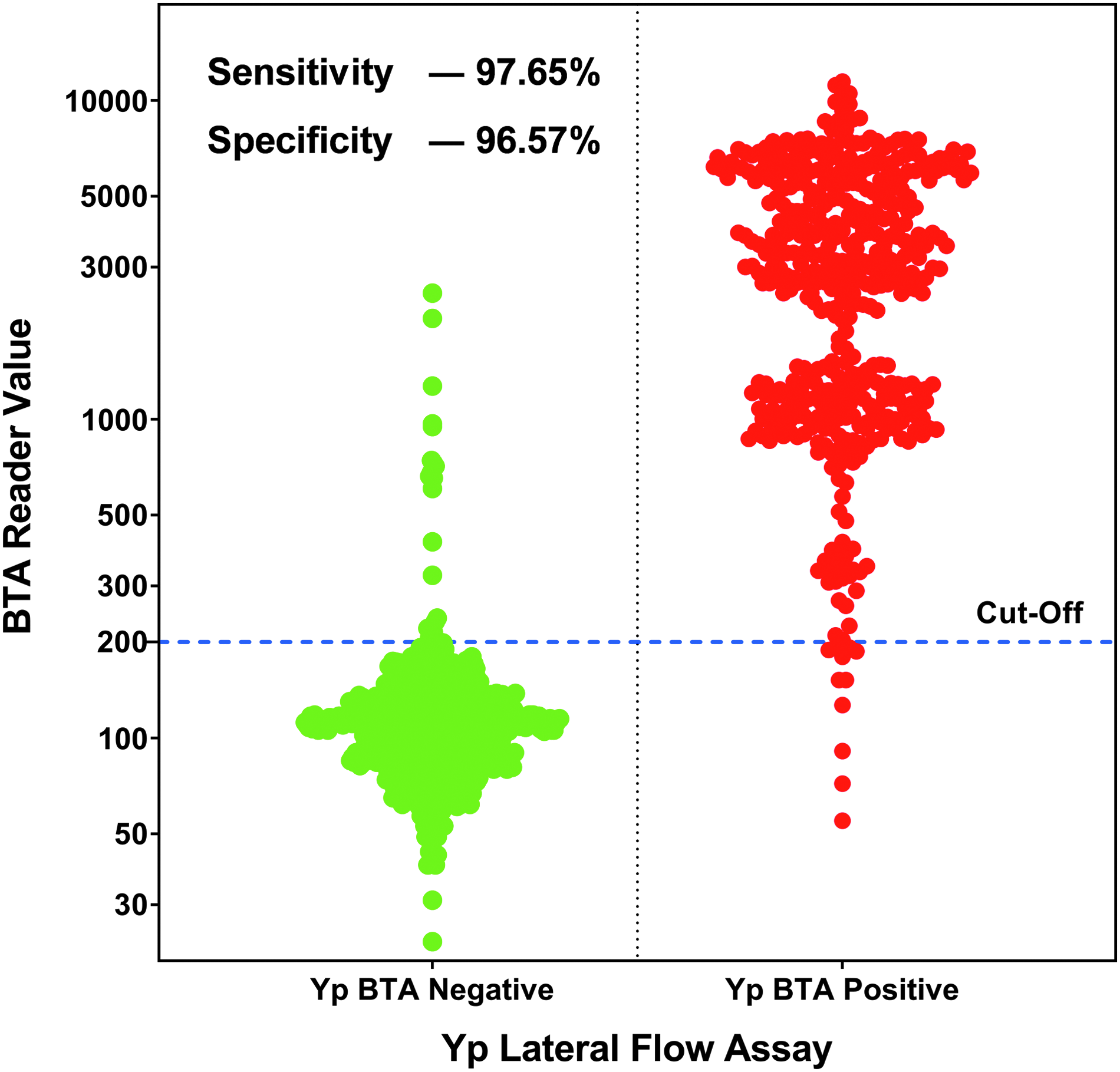
A dot density diagram that shows all 1,110 tests performed grouped as designated positive and designated negative by the BTA Reader. The cut-value of 200 is shown as a solid line. The calculated assay sensitivity is 97.65%, and the specificity is 96.57%. Any data points in the designated negative group that were above the cut-off value are false positive, while any data points in the designated positive group that were below the cut-off value are false negative.
Discussion
In addition to natural outbreaks (ie, the 3 major pandemics), Y. pestis has been employed and/or developed as a biological weapon. During World War II, Japan's infamous Unit 731 purportedly dropped plague-infected fleas over Manchuria. 4 Both the United States and the former Soviet Union investigated the use of Y. pestis as a weapon, including methods of aerosolization allowing direct dispersal of the bacteria.4,7,14
An effective public health response to Y. pestis involves several facets, including use of medical countermeasures (vaccines, antimicrobials) and methods for surveillance and early diagnosis. Because of the high mortality rates associated with untreated pneumonic plague, rapid identification of a deliberate release is critical so that medical countermeasures can be rapidly deployed and potentially exposed individuals can be treated. Currently, many technologies exist for the detection and identification of Y. pestis. Polymerase chain reaction (PCR) assays and platforms, including the RAPID and SmartCycler, have demonstrated sensitivities of 50 fg DNA (∼10 genomic equivalents, GE) for single-probe assays, 28 while the FilmArray detected Y. pestis strains at 250 to 25,000 GE. 29 Yang et al 30 investigated the use of 2 suspension arrays for the detection of Y. pestis, one of which could identify only to the genus level. The species-specific assay had a detection limit of 50 fg DNA. Fluorescence in situ hybridization has also been investigated but is impractical because of its lengthy assay time (∼8 hours to results). 32 Most antibody-based assays target the F1 capsular protein.33-36
LFA assays were first commercially introduced for pregnancy testing in 1988. 37 LFA assays require minimum sample and no specialized equipment 38 and could be used by first responders and law enforcement officers to test suspicious materials in field settings. An F1-based lateral flow that used monoclonal antibodies to F1 has been developed and used in Madagascar to test bubo aspirates from suspected plague patients. 32 F1 antigen is temperature regulated and not expressed at temperatures <33°C; as an alternative, a dipstick employing antibodies against the Pla protein is not limited by variations in growth temperature, although it is present in Y. pseudotuberculosis and Y. enterocolitica, and recent studies have demonstrated it is also found in other bacteria and therefore not a specific to Y. pestis. 33 Anti-LPS antibodies have also been evaluated and may hold promise for species-specific detection. 34
Other BTA LFA assays have previously been evaluated for the detection of biothreat agents, including orthopoxviruses, 39 ricin, 40 abrin, 41 and Bacillus anthracis. 25 Limited evaluations have also been conducted with assays for the detection of Francisella tularensis (unpublished data), botulinum neurotoxins, 42 and staphylococcal enterotoxins. 43 The Plague BTA LFA test uses a combination of a polyclonal capture antibody and monoclonal detect antibody to selectively capture and detect the presence of F1 antigen in aqueous samples. The purpose of the current study was to evaluate the performance of the Plague LFA assay in order to understand its sensitivity, specificity, reproducibility, and limitations for potential use in the field. Using the BTA Reader and the manufacturer's recommended cutoff of 200, we estimated the LOD of the Plague LFA to be approximately 105 to 106 CFU/mL. This LOD is lower than reported in an earlier study, in which Zasada et al demonstrated an LOD of 107 CFU/mL for Y. pestis using the Plague LFA assay. The difference in LOD may be because in the previous study, Y. pestis organisms were inactivated by heating at 60°C for 22 hours prior to testing with the Plague BTA Test Strip. Limitations of the Plague BTA LFA include the relatively high LOD compared to real-time PCR methods and the inability to detect Y. pestis grown at 25°C.
To evaluate the sensitivity and specificity of the Plague BTA LFA assay, 18 Y. pestis strains belonging to 4 different biovars and of different geographic origins were used. Seventeen of these strains yielded positive results visually when tested at a concentration 2 logs above the LOD. The Angola O.PE3 strain is an F1 capsular antigen minus strain and, as expected, was negative by this assay. The Plague LFA was also tested against 20 Yersinia near neighbors as well as 61 other organisms commonly encountered in the environment. Positive results were observed with environmental background strains; B. diminuta, M. odoratus (the F1 capsule antibody used in the Tetracore Plague LFA targets an antigenic epitope that is also present in M. odoratus and B. diminuta where there is an overlap in the F1 capsule antigen protein sequence from positions 103 to 119), and S. aureus. Antibodies used in the Plague BTA LFA are purified on a protein A column, and this protein is found in the cell walls of some organisms (eg, S. aureus).
In this study, we evaluated the ability of this assay to detect Y. pestis in the presence of commonly encountered powders and extracts taken from environmental filters. The Plague LFA yielded positive results in 24 of 26 powders spiked with Y. pestis A1122 at a final concentration of 106-107 CFU/mL. Inconsistent results were observed with chalk dust and drywall dust, both of which consist of relatively large particles that may inhibit the flow of fluid across the test strip and that may also act as a filter to prevent the antigen from interacting with the antibody, resulting in a false-negative result. Pooled environmental filter extracts alone yielded negative results and did not prevent the assay from detecting Y. pestis at a final concentration of 106-107 CFU/mL.
Variability in readings between BTA Readers was encountered, which yielded either false-positive or false-negative readings. In the case of false-positive results, the reader typically produced values near the cut-off. These findings highlight the importance of these assays being performed by trained and experienced users with an understanding of the limitations of sample testing and result interpretation.
In conclusion, the results presented here demonstrate a sensitivity (97.87%), specificity (97.66%), accuracy (97.75%), and limit of detection (104-105 CFU/ml) for the Plague BTA LFA. These performance data are important for accurate interpretation of qualitative results arising from testing suspicious white powders and aerosol samples in the field. It should be noted that specificity of this test has not been evaluated for other environmental specimens such as soil or water. It is not approved for human clinical diagnostic use. Highly suspicious samples should be tested by other methods in a reference laboratory. If only limited or very sparse material is available, it should be collected and submitted to a CDC LRN laboratory for testing. In addition, this assay is based on detection of Y. pestis F1 capsule, an antigen expressed only at ≥33°C. Any positive specimen in this assay is considered presumptive positive and should be referred to the CDC LRN for additional testing, confirmation, and characterization for an appropriate public health response.