
Abstract
Sampling of prostate tissue (n = 97) was performed in conjunction with planned radical prostatectomies, in collaboration with Biobank1®. The tissue used in this study was collected during the period 2003–2016, quickly frozen, and kept at −80°C until assayed in 2018. RNA extraction was performed with two different protocols (miRNeasy and mirVana™), and RNA quality was determined by measuring the RNA Integrity Number (RIN). The level of isoprostanes is widely recognized as a specific indicator of lipid peroxidation both in vitro and in vivo. The level of 8-isoprostane was measured because it is the main oxidation product of arachidonic acid, the most abundant phospholipid fatty acid. The level of 8-isoprostane was measured using enzyme immunoassay. There was no statistically significant difference in yield between the samples isolated with the mirVana protocol compared to the miRNeasy protocol. Average RIN was 2.8 units higher with the mirVana extraction protocol compared to the miRNeasy protocol (p < 0.001). For miRNeasy extractions, RINs were 7.1 for prostatectomies in 2005–2007 and 6.2 for those in 2018 (p < 0.001). For mirVana extractions, the difference in RIN score between the two groups regarding years of collection was not statistically significant. There was no significant increase in the levels of 8-isoprostane between the 2005–2007 samples and the 2018. The conclusion is that there is no oxidation of phospholipids with increasing storage time up to 15 years.
Introduction
The concept of material quality in biobanking
Biological material of high quality is essential for biomedical research aiming at a better understanding of disease mechanisms, as well as at the identification of clinically useful biomarkers for disease and the discovery of new drug targets. Accordingly, vast resources are invested worldwide in the establishment of biobanks, providing collection and storage of biological specimens for use in future studies. Thus, biobanking is becoming an ever more important discipline to serve the growing demand in terms of quality, as well as quantity. Many of the collections, especially in the case of rare diseases, must continue over several years to achieve sufficiently large sample sizes for the planned investigations.
One of the biggest challenges in biobanking is to ensure that samples can be stored for long periods without undue negative impact on their integrity, that is, that there is no uncontrolled alteration of their physical or chemical composition. The consequence of such alterations may be that the result of, say, a biochemical analysis reflects an effect of the storage rather than reflecting the physiological conditions in the sample. Ideally, the material should be preserved in such a way that the original cell structure and composition are faithfully conserved, even for many years. Depending on the material and its intended use, several methods have been used for storage of biological samples, for example, dry state storage, cooling, and freezing, eventually reducing the exposure to atmospheric oxygen using vacuum packaging, or by keeping the sample under the cover of nitrogen or other inert gas. Chemical fixation and/or paraffin embedment can also conserve some of the constituents of the sample, however at the expense of modification or loss of other components.
In Norway, a set of national guidelines was developed and made available in the document “Best practice for Norwegian biobanks,” recommending systematic control of sample quality and exact documentation of all procedures being used during collection, preservation, and storage, including the registration of any deviation from the procedure. 1 At the international level, BBMRI-ERIC strongly advocates that all European biobanks adhere to a set of international standards, technical specifications, and accreditation processes. 2 These standards and specifications describe procedures for the processing of biological material for research. Hopefully, this practice can be standardized both nationally and internationally as a measure to ensure consistently good quality.
In recent years, much effort has gone into the search for correlations between genetic aberrations and propensity for various diseases. However, there is a growing awareness that important questions of risk factors and disease mechanisms cannot be reduced to mere genomics. Thus, the list of relevant “-omics” has been ever lengthening, and the number of chemical classes which may be of interest for biobank research has increased accordingly. This development adds to the challenges in biobanking, since different substances have differing vulnerabilities and may have different storage requirements. Moreover, an organ is an integrated system of different tissues, which again are more or less heterogeneous collections of different cell types. Disease related changes are unevenly distributed among various cell types, and cell- and tissue physiology vary over time, all of which add to the complexity. When it comes to defining sample quality, no parameter fits all types of biological material or all kinds of research. Sample quality must rather be understood as fitness for a particular purpose.
Vulnerable biomarkers
Several studies have investigated the effects of long-term storage on the chemical integrity of DNA, RNA, proteins, and some other chemical compounds.3–6 DNA is known to be relatively stable, capable of withstanding exposure to harsh environments without detrimental effects. RNA, on the other hand, is found to suffer ever-increasing fragmentation with storage time in formalin fixed, paraffin embedded samples, thereby reducing their usefulness for gene expression analysis. 7 Thus, rapid freezing followed by low temperature storage is at present the preferred method for maintaining good quality RNA. Moreover, the ubiquitous presence of RNases represents a challenge for the handling of specimens before freezing. In addition to the effect of chemical fixation, studies have identified tissue fixation time, specimen size, duration of ischemia, and tissue storage conditions as important factors influencing RNA quality. 7
Currently, lipid profiling is emerging as a potential source of useful biomarkers. A variety of signaling molecules originate from cell membrane phospholipids, so phospholipids and their derivatives may potentially function as indicators of biological processes. Diverse pathological conditions are characterized by oxidative stress, leading to increased levels of oxidized lipids in biological fluids and tissues.8–13 Primary products of oxidation are lipid hydroperoxides. 14 Further decomposition of hydroperoxides yields a range of secondary oxidation products, such as malondialdehyde, 4-hydroxynonenal, and isoprostanes, of which the latter are considered the most specific markers of oxidative stress. 15 In particular, 8-isoprostane (8-isoPGF2α) has been proposed as a gold standard for assessing lipid peroxidation 16 and as a general indicator of biochemical integrity of biobank samples, as far as their lipid composition is concerned.16–20 Unsaturated carbon bonds are also susceptible to auto-oxidation on contact with atmospheric oxygen. The deterioration of lipid components in foodstuff, known as rancidity, has been known since ancient times and still constitutes a huge challenge for storage of meat products. 21
The utterly complex mechanisms behind lipid oxidation are the subject of extensive study in food science, but the phenomenon has received much less attention in the biobank setting. In contrast to the situation with nucleic acids, remarkably little is known about optimal preservation of lipids in biobank samples. Blood plasma was reported to show extensive auto-oxidation when kept at −20°C, whereas only limited effect could be seen after 6 months at −80°C. However, we are not aware of any systematic investigation of lipid preservation in tissue specimens. Phospholipids as found in cell membranes are the lipids most vulnerable to oxidation, and especially unsaturated fatty acyl chains. In fact, the oxidation susceptibility increases exponentially with the degree of unsaturation. 22 Thus, we feared that isoprostane levels might tend to increase upon prolonged storage in the biobank, thereby rendering the samples less useful for future research.
Impact of long-term storage on prostate tissue quality
In men over 50 years of age, prostate cancer is the most frequent malignancy, and one in eight men will be diagnosed with this condition during their lifetime. 23 However, there is a wide variation in biological behavior between individual cases, and current diagnostic tools leave a lot to be desired when it comes to distinguishing between indolent and aggressive tumors. Moreover, very little is known about the causes and the cellular mechanisms behind the development of the disease. Thus, there is a pressing need to gain more insight into the underlying processes and to identify biomarkers which characterize the different clinical variants. A huge effort is going on worldwide to achieve these goals, seeking potential biomarkers within both the transcriptome and the lipidome of actual patients. One of the challenges in biobanking is to establish procedures which ensure that the stored material can be used for a wide variety of purposes and that the highest possible quality is maintained over the lifetime of the samples, which can extend to many years. For many years, we have been systematically collecting fresh-frozen tissue material from total prostatectomies and storing it at ultralow temperature. We have previously documented that our material provides a high yield of good quality RNA in the short time perspective. The aim of the present study was to examine whether several years of storage in ultralow-temperature freezers would have a detrimental effect on the integrity of RNA and/or phospholipids.24,25
Materials and Methods
Collection of tissue samples from radical prostatectomies
At St. Olavs Hospital, Trondheim, Norway, we have collected systematically one central slice of thickness 2 mm from radical prostatectomy specimens since 2003. The surgery is performed in patients who have organ-localized cancer and at least 10 years of life expectancy. Currently, the surgery is performed with a robot-assisted laparoscopic technique, but until 2010 it was done as an open laparotomy. A slice is cut perpendicularly to the urethra, within 10–15 minutes after the specimen has been removed from the body, thereafter rapidly frozen between two metal plates precooled with liquid nitrogen, and transferred to a mechanical freezer at −80°C. 3 The material is exposed to atmospheric O2 during sample preparation, and further the material was stored in sealed zip bags, in a box in a −80°C freezer. The freezers used in this study are only for long-term storage, so the doors in the freezer are not frequently opened. There are also separate doors to each shelf inside the freezer.
There are temperature sensors on the freezer with an alarm connected to security central at St. Olavs Hospital. No alarms were recorded during the period.
The remaining two halves of the specimen are fixed in formalin and further processed for routine histopathological examination. The Regional Committee of Medical Research Ethics approved the collection of the material and its subsequent use in research, based on the patients' written consent.
For the present project, a total of 97 frozen samples were selected, as detailed in Table 1. There was only one frozen prostate slice per patient, so the number of patients is the same as the number of prostate slices.
In Total 97 Tissue Slices from Prostate Were Used in This Study, Some for RNA Isolation and Measurement of RNA Integrity Number and Some for Lipid Extraction and Analysis of Oxidation of Phospholipids
All the cylinders were about 30 mg.
Tissue sampling and preparation for extraction of total RNA
For extraction of RNA, tumor-free areas in the frozen tissue slices were identified by adjusting micrographs of the histological slides taken from each side of the frozen slice to the photograph of the slice itself. Tissue cylinders were harvested using a high-speed drill unit with specially made bore bits of diameter 3 mm, as depicted in Figure 1. The slice was kept frozen during the entire procedure, being kept lying flat on an aluminum block continuously cooled by contact with liquid nitrogen. 26
Both protocols give results that are acceptable for what is defined as pure RNA.
The collected cylinders were immediately transferred to a test tube, immersed in liquid nitrogen, and further stored at −80°C until analysis. Two cylinders were taken from the same prostate slice, of which RNA from one cylinder was isolated with the miRNeasy Mini Kit and RNA from the other cylinder was isolated with the mirVana™ miRNA Isolation Kit.
An experienced pathologist confirmed the morphology of some of the tissue cylinders (n = 32), by microscopy of hematoxylin, eosin and safran (HES) stained cryosections.
Extraction and isolation of total RNA with two different protocols: miRNeasy Mini Kit (Qiagen) and mirVana miRNA Isolation Kit (Thermo Fisher)
Total RNA was extracted from samples collected during 2005–2007 (n = 27) and in 2016 (n = 38).
The miRNeasy Mini Kit and QIAcube procedure include semiautomatic isolation of total RNA using Phase Lock Gel tubes, PLG Heavy 733-2478 (Quantabio). The miRNeasy Mini Kit combines phenol/guanidine-based lysis of samples and silica membrane-based purification of total RNA. QIAzol Lysis Reagent is a monophasic solution of phenol and guanidine thiocyanate, designed to facilitate lysis of tissues, to inhibit RNases and to remove most of the cellular DNA and proteins from the lysate by organic extraction. The extractions were carried out over a period of 2 months. First RNA was extracted, and then the RNA was measured collectively.
Frozen tissue cylinders, a total of about 30 mg per sample, were transferred to tubes prefilled with 700 μL QIAzol Lysis Reagent (Precelly Ceramic Kit) homogenization. The tubes were placed in Precellys 24 and homogenized at 6000 rpm for 20 seconds. Thereafter, the tubes were cooled on ice for 5 minutes before a new homogenization cycle. The samples were then left at room temperature for 5 minutes. After addition of 140 μL chloroform, the samples were vortexed for 15 seconds and further incubated at room temperature for 2–3 minutes. The lysate was then transferred to a PLG Heavy tube and centrifuged at 12,000 g, 4°C, for 15 minutes. The upper aqueous phase was transferred to a sample tube and loaded in the QIAcube for purification of total RNA using spin columns. The purified total RNA (30 μL) was immediately transferred to an ultra freezer at −80°C, for temporary storage until use in downstream analysis.
The mirVana miRNA Isolation Kit is used for rapid isolation of total RNA, including small RNAs, from tissue and cells using a fiberglass filter-based method. Frozen tissue cylinders (30–50 mg) were suspended in 300–500 μL of Lysis/Binding buffer (10 volumes per tissue mass) and thoroughly disrupted using a rotor-stator homogenizer. After addition of 1/10th volume of Micro RNA Homogenate Additive, the solution was vortexed and further incubated for 10 minutes on ice. A volume of Acid-Phenol: Chloroform corresponding to the original lysate volume was then added. The solution was vortexed for 30–60 seconds. To separate the aqueous phase and the organic phase, the solution was centrifuged twice at 13,000 g for 5 minutes. The top aqueous phase was transferred to a new RNase-free tube, and the volume was noted. Ethanol 100% (1.25 volumes) was added and mixed thoroughly. The sample was transferred to a filter column and centrifuged (10,000 g, 15 seconds). The liquid was discarded, and the collection tube was saved for use in the washing procedure. miRNA Wash Solution 1 (700 μL) was added and centrifuged (10,000 g, 5–10 seconds). The liquid was discarded, and the collection tube was saved for reuse. The filter was washed twice with 500 μL Wash Solution 2/3 and centrifuged (10,000 g, 5–10 seconds). The liquid was discarded, and the collection tube was saved for reuse. After the second wash, the filter was put back in the same collection tube and centrifuged (10,000 g, 1 minute) to remove excess liquid. The filter column was transferred to a new collection tube, to which was added 100 μL of elution buffer preheated to 95°C. The tube was centrifuged (10,000 g, 20–30 seconds) to elute the RNA. The purified total RNA (100 μL) was immediately transferred to an ultra freezer at −80°C for use in downstream analysis.
Measurement of RNA concentration, purity, and integrity analysis
The concentration and purity of total RNA in each extract were determined using a NanoDrop Spectrophotometer. The RNA concentration in the extract was determined in duplicate, using 1.5 μL of the sample. The RNA Integrity Number (RIN) was determined using Agilent 2100 Bioanalyzer with Agilent RNA 6000 Nano Kit, designed for analysis of total RNA. Each RNA chip contains a set of interconnected microchannels used to separate nucleic acid fragments by electrophoresis. RNA fragments are separated by size, and the RIN score is calculated as a number from 1 to 10, where 10 represents completely intact RNA and 1 indicates totally degraded RNA.
Quantitative measurement of 8-isoprostane
The level of 8-isoprostane in the stored tissue specimens (n = 32) was measured using the ELISA Kit and standards from Cayman Chemical Company (Ann Arbor, MI), which also provided the reagents and remedies for affinity purification of the samples. All procedures were performed according to the producer's recommendations. From each frozen tissue slice, one cylinder of diameter 3 mm was extracted, suspended in 500 μL 0.1 M potassium phosphate buffer, containing 1 mM EDTA and 0.005% butylated hydroxytoluene (Sigma-Aldrich), and homogenized using an Omni Tissue Homogenizer (Omni International, Kennesaw, GA). After removal of 15 μL for protein analysis, the remaining sample was expanded by the addition of an equal volume of 15% (w/v) KOH to hydrolyze any esterified isoprostane. The mixture was incubated for 60 minutes at 40°C, thereafter neutralized by the addition of 5 mL of 1 M potassium phosphate buffer, and loaded onto a rinsed affinity column containing Sepharose 4B with covalently bound monoclonal mouse anti-8-isoprostane antibody. The column was thoroughly washed with buffer and ultrapure water, following which the retained lipid was eluted with 95% ethanol. The eluate was stored at −80°C until assayed.
Before analysis of the 8-isoprostane extracts, the diluent was evaporated under a stream of dry N2, and the dried samples were dissolved in 500 μL ELISA buffer (Cayman). If necessary, the samples were kept at 4°C for up to 2 days. The assay was performed in 96-well plates precoated with rabbit-anti-8-isoprostane antiserum, in compliance with the manufacturer's instructions. Each sample was assayed in two dilutions, both distributed in triplicate wells. A serial dilution of the standard was assayed in duplicate, covering the concentration interval from 0.8 to 500 pg/mL. To each sample was added a fixed amount of a tracer consisting of 8-isoprostane covalently bound to acetylcholinesterase. Thus, the amount of tracer bound to the well bottom is inversely proportional to the concentration of free 8-isoprostane in the sample. The plate was incubated for 18 hours at 4°C and then washed five times in a Tecan Columbus automatic plate washer. The amount of bound tracer was then revealed by the addition of a detection reagent containing acetylthiocholine, which releases thiocholine by the action of acetylcholinesterase. Thiocholine reacts with 5,5′-dithio-bis (2-nitrobenzoic acid), also present in the detection reagent, thereby producing 5-thio-2-nitrobenzoic acid, which has a strong yellow color. The absorbance was measured in a 2300 EnSpire Multimode Reader (PerkinElmer) at 412 nm.
The protein concentration in the homogenate was measured with Quick Start™ Bradford Protein Assay Kit 1 (Bio-Rad), which detects the color shift induced by interaction between amino acid residues in proteins and Coomassie Brilliant Blue G-250. Two dilutions of each sample were mixed with the reagent and distributed in triplicate wells in a microplate (Sarstedt). After 5 minutes incubation at room temperature, the absorbance was read at 595 nm in a 2300 EnSpire Multimode Reader (PerkinElmer). The absorbances were converted to protein concentrations by reference to a standard curve obtained by measuring a serial dilution of bovine serum albumin (Bio-Rad) in parallel with the samples. No positive control was used, but a standard curve was generated for each plate. The protein concentration was calculated with an average of the absorbance.
The cylinders used to determine RIN and measure 8-isoprostane were not from the same prostate.
Statistics
The data were tested for normal distribution by the Shapiro–Wilk test and visual expression of histogram and Q-Q plot. None of the data from the analysis done with the mirVana protocol was normally distributed (p < 0.001), even when transformed. For statistical evaluation of group differences, the Wilcoxon–Mann–Whitney test was used, whereas the Wilcoxon signed-rank test was used for paired data. 14
Time trends, with data obtained from analysis of samples with different lengths of storage, were evaluated using the Jonckheere–Terpstra test. 27 The level of statistical significance was set at p = 0.05.
Results
Yield and purity of RNA from prostate tissue samples
Total RNA was extracted from prostate tissue collected during 2005–2007 (n = 26) and in 2016 (n = 38) using miRNeasy Mini Kit (Qiagen) and mirVana miRNA Isolation Kit (Thermo Fisher).
The mean yield of RNA for tissue collected during 2005–2007 was 31.0 μg (SD 11.6) for samples isolated with the mirVana protocol, and for samples isolated with the miRNeasy protocol the mean RNA yield was 25.4 μg (SD 11.2). The mean RNA yield for samples collected in 2016 was 32.0 μg (SD 13.2) for samples isolated with the mirVana protocol, and for samples isolated with the miRNeasy protocol the mean RNA yield was 28.1 μg (SD 11.0).
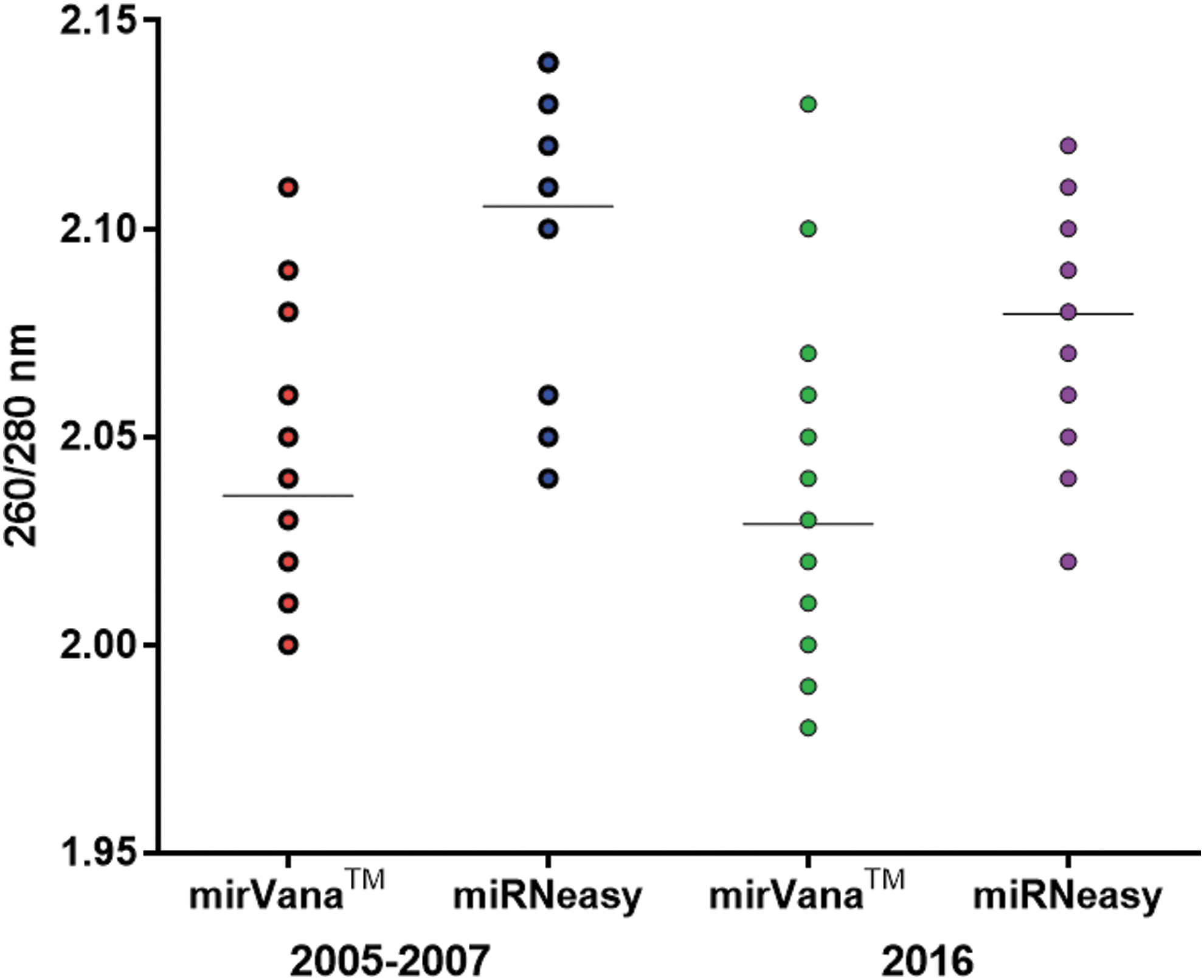
The total yield of RNA from prostate tissue collected during 2005–2007 and in 2016 are on average higher for those samples that are isolated with the mirVana protocol. There is no significant difference in RNA yield between the two protocols, neither between samples collected in the period 2005–2007 (p-value 0.05) nor in 2016 (p-value 0.12). A summary of the RNA yields is presented in Figure 2.
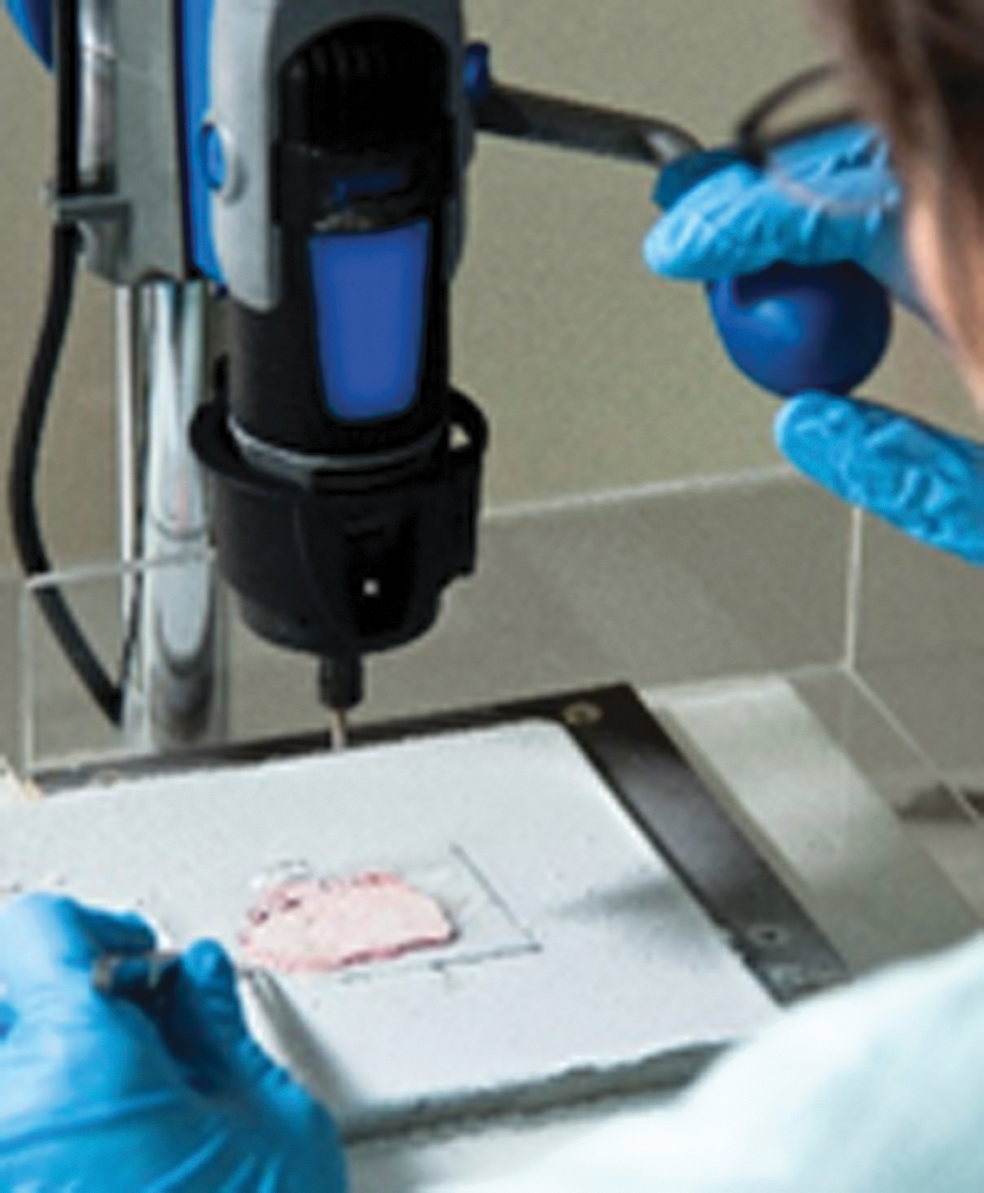
The prostate slice was placed on an aluminum block continuously cooled with liquid nitrogen, and tissue cylinders were collected using a drill. The areas of interest were identified by reference to the histological slides adjacent to the current fresh frozen tissue slice.
The purity of RNA was measured by comparing the absorption at 260 to 280 nm. The ratio of absorbance at 260 and 280 nm may indicate the presence of protein, phenol, or other contaminants.
A 260/280 ratio greater than 1.8 is usually considered as an indicator of high purity RNA. 28 The mean 260/280 ratio of RNA for tissue collected during 2005–2007 was 2.04 for samples isolated with the mirVana protocol, and for samples isolated with the miRNeasy protocol the mean was 2.11.
The mean 260/280 ratio for samples collected in 2016 was 2.03 for samples isolated with the mirVana protocol, and for samples isolated with the miRNeasy protocol the mean was 2.08. The purity analysis is presented in Figure 3.
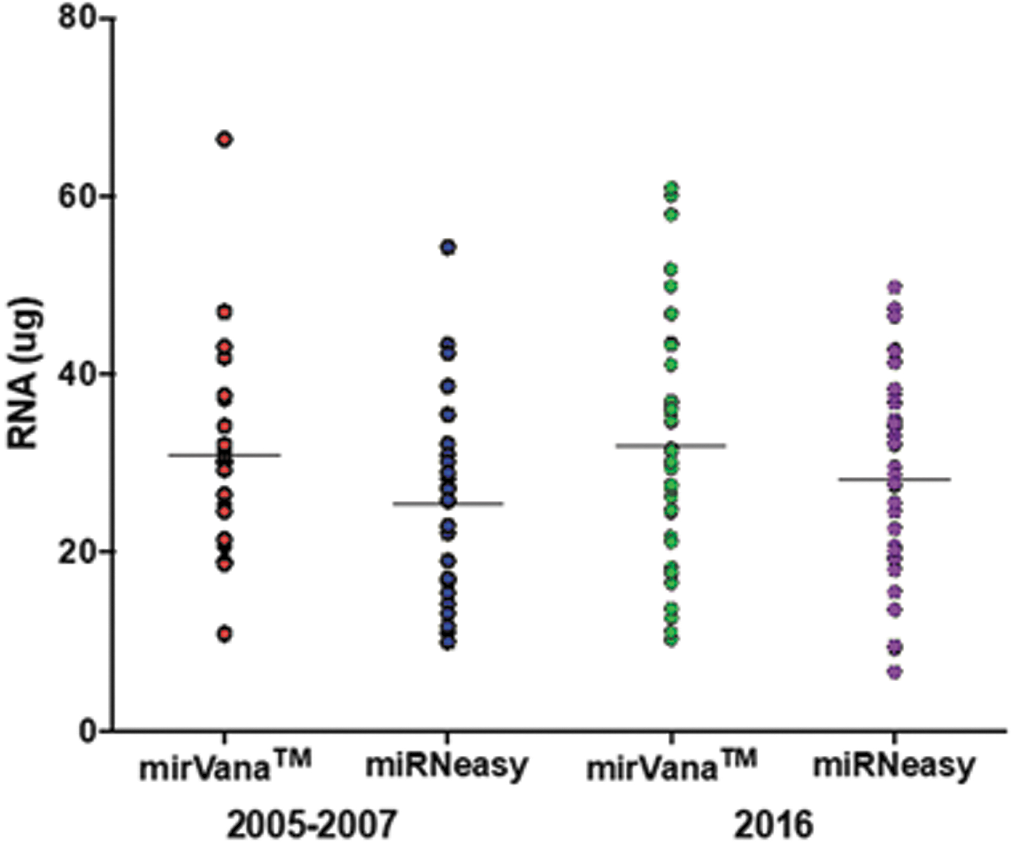
Total amount of RNA extracted from prostate tissue collected during 2005–2007 (n = 26) and in 2016 (n = 38), using two different extraction protocols.
RNA integrity score in prostate tissue after long-term storage in ultralow freezers
The extracted RNA from prostate tissue collected during 2005–2007 (n = 26) had a mean RIN of 9.4 (SD 0.751) for samples isolated with the mirVana protocol, and for samples isolated with the miRNeasy protocol the mean RIN was 6.2 (SD 0.667). Extracted RNA from prostate tissue collected in 2016 (n = 38) showed a mean RIN of 9.7 (SD 0.07) for when isolated with the mirVana protocol, and for samples isolated with the miRNeasy protocol the mean RIN was 7.1 (SD 0.153). Samples isolated with the mirVana protocol showed higher RIN scores than samples isolated with the miRNeasy protocol. The difference in RIN is statistically significant for both groups with regard to protocol (p < 0.001). The variation in RIN between the two groups is illustrated in Figure 4.
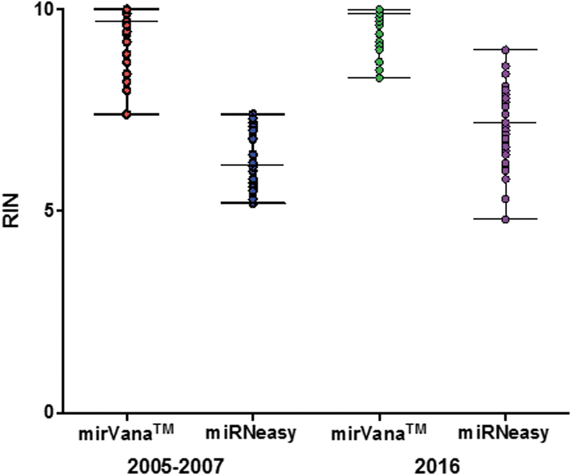
RIN score for samples collected during 2005–2007 (n = 26) and in 2016 (n = 38), using two different extraction protocols. RIN, RNA Integrity Number.
Comparison of samples isolated with the mirVana protocol shows that samples collected during 2005–2007 had a mean RIN score of 9.4 (SD 0.75), and for samples collected in 2016 the RIN was 9.7 (SD 0.46). The difference in RIN score between the two groups with regard to years of collection is not statistically significant (p = 0.083).
Comparison of samples isolated with the miRNeasy protocol shows that samples collected during 2005–2007 had a mean RIN of 6.2 (SD 0.67), and for samples collected in 2016 the RIN was 7.1 (SD 0.95). The difference in RIN score between the two groups regarding years of collection is statistically significant (p < 0.001).
Comparison of RIN in all samples (n = 64) regarding protocol shows that the variation in RIN is statistically significant (p < 0.05), and the mean RIN between the mirVana protocol and the miRNeasy protocol is 2.8 (SD 0.9), which means that the RIN is significantly higher using the mirVana protocol. The comparison is presented in Figure 5.
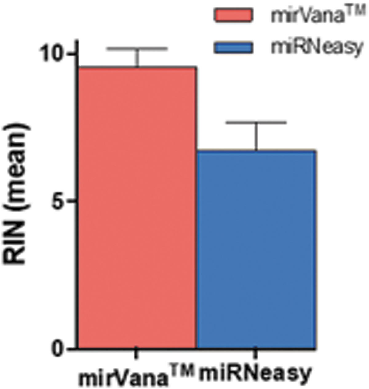
Mean RIN scores in RNA extract from a total of 64 samples, collected during 2005–2007 and in 2016, using two different kits for the RNA extraction. Samples isolated with the mirVana™ protocol had a higher mean RIN (mean RIN = 9.6) than samples isolated with the miRNeasy protocol (mean RIN = 6.7).
8-Isoprostane in prostate tissue after long-term storage in ultralow freezers
The results of the determination of 8-isoprostane are presented in Figure 6. The amount of 8-isoprostane is normalized to the amount of protein in each sample. Most samples show very low values of 8-isoprostane, and there is no significant increase with increasing duration of the storage time. However, for each of the years examined the distribution of the figures shows a heavy tail toward higher values. The causes behind this phenomenon are unclear, but it is most likely due to mechanisms which in individual cases contribute to high oxidative activity, before, during, or after surgery.
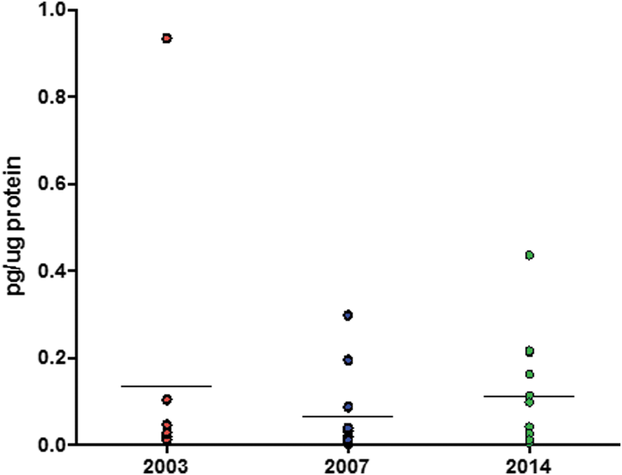
Levels of 8-isoprostane in lipids extracted from homogenates of frozen prostate tissue samples according to the year of collection. The concentrations of 8-isoprostane were normalized by referral to the concentration of protein in the homogenate. The chemical analyses were performed in 2016, thus after 13, 9, and 2 years of storage, respectively. The results show no significant trend with increasing duration of the storage (p = 0.602).
Discussion
There is a need to increase the availability of large numbers of high-quality, well-annotated biological samples. Collections of optimally preserved biological material can provide medical research with valuable molecular information, and the lack of such samples may be what limits research and medical improvements. Research on fresh frozen tissue will hopefully provide the basis for more precise diagnosis, targeted treatment, and a more accurate assessment of the prognosis.
Based on this knowledge, a total of 97 tissue samples from men undergoing radical prostatectomy were collected for use in this study. The conditions for all sampling were standardized and well documented according to guidelines. Fresh frozen prostate tissue was used to examine whether the RNA integrity or phospholipid oxidation was affected during long-term storage in ultralow freezers. RNA integrity and phospholipid oxidation are highly important quality parameters in biobank material for use in research. To study this, RNA yield and RIN score were measured in samples stored for several years compared with samples stored for a short period of time. RNA was isolated with two different protocols. To measure oxidation of phospholipids, the level of 8-isoprostane was measured in extracts from the tissue by enzyme immunoassay (EIA). The results show that storage time, under certain conditions, is not a factor that seems to affect the RNA integrity, but the choice of protocol used for RNA isolation is more important for both RNA yield and RNA integrity. Storage time does not affect the oxidation of phospholipids in fresh frozen prostate tissue.
High yield of RNA in fresh frozen prostate tissue
The mean RNA yield in samples isolated with the mirVana protocol and the miRNeasy protocol is relatively similar, as is the RNA yield in tissues collected in 2005–2007, compared to tissue collected in 2016. The small variance in the RNA yield in the samples can be correlated to the tissue composition. This study is based on noncancerous areas, but if there is a varying proportion of normal glandular tissue or hyperplasia and connective tissue, the RNA yield will differ. 26 Tissue cylinders are taken from the same tissue slice, but it is impossible to have equal cellular composition in each sample, since prostate tissue also is a heterogeneous biological material, so there might be slightly different cellular content in the tissue cylinders.
Significance of RIN for downstream analysis
RIN is often used as a reliable and informative standardized method for evaluating RNA quality in the context of gene expression analyses. Biospecimens contain molecules and enzymes (proteases, lipases, and nucleases) that are strongly affected by temperature, and specific storage conditions are needed to inactivate the molecules and to preserve the cellular composition. Biospecimens have a complex composition, including lipids, water, salt, and proteins, and each component freezes at its own, specific temperature. Various studies report results associated to activity of RNase A. RNase A catalyzes the cleavage of the 3′ end of unpaired C and U residues, and the RNase activity changed at low temperatures.29,30
It is generally recommended that RIN should be >7 in the samples to be used for downstream analyses, like gene expression assays. Sigurgeirsson et al. presented a study which highlights the effect of a decrease in RIN for downstream analyses, more than the actual threshold in RIN (>7 or 8) used for downstream analyses. 31 It turns out that a decrease from RIN 10 to RIN 8 has a more important effect on the results of gene expression analysis than a decrease from RIN 8 to RIN 6. 31 Opitz et al. point out that there are some genes that are directly affected by low RIN. These genes should be excluded from gene expression assays when working with degraded RNA. 32 Reduced gene expression due to low RIN has been shown in other studies as well. In frozen breast cancer samples, some categories are highlighted that are affected by low RIN—deoxyribonuclease activity, collagen, regulation of cell adhesion, cytosolic ribosome, and NADH dehydrogenase activity. 24
There does not seem to be any justification to set a threshold at any specific RIN but rather it is important to be aware of the effects of low RIN, and all samples should preferably be in close range in terms of quality. A study that focuses on this is Mathieson et al., which shows that even samples with very low RIN can be used for downstream analysis, but there are limitations to what can be detected in the sample. It is shown that amplicons of at least 940 bp can be obtained in frozen samples, but not FFPE samples. 25 RNA from FFPE samples will always be degraded. This affects the accuracy and sensitivity of expression measurements based on real-time PCR and in particular differences in the efficiency of cDNA synthesis as the most sensitive step. Reliable measurements can still be made for some genes.33,34
The focus in this study is on long-term storage of prostate tissue at low temperature (−80°C) and based on the results the time and storage do not seem to have impact on the RNA integrity. These findings support the results of previous studies,25,35–38 where the conclusion is that RNA is kept stable during long-term storage. RNA in prostate tissue stored for more than 10 years appears to be more susceptible to degradation during extraction, but this degradation can be ameliorated by protocol optimization. The decline in RIN score is small in its magnitude and does not appear to have an impact on downstream analyses.
There is a significant difference between the samples collected during 2005–2007 and the samples from 2016 regarding the choice of RNA isolation protocol. This can be interpreted as meaning that the storage time affects the RNA quality of the tissue material, when the miRNeasy protocol is used. The main difference between the miRNeasy protocol and the mirVana protocol is the homogenization method. For the miRNeasy protocol, homogenization is done by beads, and for the mirVana protocol the homogenization is done by knife. Homogenization with beads gives varying effect, and if the tissue is not homogenized sufficiently, the RNA in the cells will not be completely released. This allows active RNases in the tissue to degrade RNA as homogenization is insufficient.
All extractions were performed at the same laboratory, by the same personnel, at the same time of the year. All processing done on the sample material is standardized. The cylinders are randomized, and there has been no systematic drilling.
In this study, no gene expression analyses were performed.
8-Isoprostane EIA, a biomarker of oxidative stress
Prostate tissue samples (n = 32), considered to be tumor free, were analyzed with the 8-isoprostane EIA. Retrieval of a sample from the biobank, such as for collection of tissue cylinders, may pose a risk of thawing the sample. Studies have documented how storage/retrieval cycles can affect the sample in question, but also samples stored adjacently to that sample. Abuja et al. showed that room-temperature retrieval, for about 90 seconds, was enough to raise the temperature within a small tissue sample and enough so that solute enriched samples could thaw. 39 If the retrieval of the sample raised the internal temperature to exceed the glass transition temperature it would lead to a recrystallization process, hence facilitating the possibility for oxidation. They also showed that phosphatidylcholines can increase up to 300% in samples with a core temperature of −60°C at retrieval. So, it is recommended that the duration of retrieval at room temperature must be kept at a minimum to avoid thawing of the sample surface.
One might argue that local conditions in the storage box, like the way the bags are stacked in relation to each other, or if it is stacked near the center or the edge, etc., may influence how available the sample is for oxygen from the air. Whenever a sample is taken out of the biobank, all other samples in the same box, and probably adjacently stored boxes, are exposed to the same “near-retrieval” conditions. These “near-retrieval” conditions will involve repeated temperature shifts, which may induce changes in samples that have not been retrieved.
Prostaglandins and fatty acid metabolites have long been known as key factors in many physiological and pathological processes, and there is a general increase in the levels of both free and total 8-iso-PGF2α associated with a variety of conditions and environmental exposures. The collected tissue cylinders were investigated for morphology only on one side and based on the histological sections from the adjacent FFPE-blocks it was thought to be enough to say that the sample was tumor free. Isoprostanes are known to play a role in inflammatory responses, and tumorous tissue could induce an inflammatory state. There are studies that show that 8-isoprostane can be formed not only through oxidative stress but also simultaneously with prostaglandin synthesis that is induced during inflammation. It has been reported that the enzymatic production of isoprostanes, by a cyclooxygenase, was 10-fold higher in prostate cancer tissue than in benign prostatic tissue. 40 So, even though the tissue was thought to be benign prostate tissue it could still contain a small amount of tumorous tissue. This tissue could then influence, and probably explain, the 8-isoprostane levels that stand out in some of the samples or that free 8-isoprostane from tumor tissue could diffuse into nearby benign tissue, thus producing a rise in 8-isoprostane levels measured.
Collections of biological material are the fundamental basis to both diagnostics and clinical research. Results of analyses of biological material can be influenced by different preanalytical conditions like sampling of the material, processing, and storage. This study highlights the stability of RNA and lipids in fresh frozen prostate tissue stored for over 10 years in −80°C.
Footnotes
Author Disclosure Statement
The authors declares that there is no conflict of interest.
Funding Information
The present project received financial contribution from Biobank Norway.