
Abstract
Introduction
Innovation
Gsta4 has by far the highest detoxifying capability of the highly toxic product 4-HNE. Lipid peroxidation is one of the most significant pathophysiological processes in TBI. A naturally occurring genetic variability in Gsta4 is here identified to affect expression and protein levels of the enzyme, which is located in neurons and upregulated in these cells upon injury. A congenic strain with higher expression of Gsta4 displays less nerve cell loss in the hippocampus after TBI, which is the first such congenic strain effect ever to be reported in a TBI model. These findings encourage further studies of the role of polymorphism in human Gsta4 in neurodegenerative diseases and open new perspectives for therapies targeting 4-HNE in TBI.
Indeed, a number of studies have found evidence that polymorphisms in the apolipoprotein E (APOE) gene affect outcome of TBI, with a more unfavorable outcome for individuals carrying the e4 allele of the APOE gene (49). Apart from APOE, a smaller number of association studies have suggested a possible genetic influence on TBI outcome for polymorphisms in the tumor protein 53, interleukin-1β, CACNA1A, dopamine receptor D2, and poly(ADO-ribose) polymerase 1 genes (26). However, all these studies have been conducted with a very limited number of patients, leaving a high risk for false positive findings. From other conditions, we now know that in order to unravel the genetic basis of complex traits, cohorts consisting of many thousand patients are needed to achieve the necessary statistical power to pinpoint genetic influences (36).
Experimental studies conducted in models of TBI are valuable tools for studying the impact of naturally occurring genetic polymorphisms on TBI outcome and thereby revealing possible candidate genes. This approach, by using genetic dissection of complex traits, has been particularly successful in autoimmune diseases such as multiple sclerosis and rheumatoid arthritis, where discovery of important information about underlying genetic regulation has led to increased knowledge of disease pathophysiology and treatment response (15, 31). The impact of genetic heterogeneity has been much less studied in the context of TBI. However, it has been demonstrated that TBI outcome differs across different rodent strains (34, 45), and we recently reproduced this finding by showing considerable differences in TBI outcome in the two inbred strains: dark agouti (DA) and piebald virol glaxo (PVGav1) (2). These two strains have previously been extensively studied in autoimmune models such as experimental allergic encephalomyelitis (EAE), a model of multiple sclerosis (MS), and experimental arthritis, where the DA strain is susceptible while the PVGav1 is resistant (8, 19). We have also demonstrated differences in the response to a standardized peripheral nerve lesion with regard to survival of axotomized nerve cells and local glial activation (8, 19, 44).
In this study, we used microarrays in order to determine genetically regulated pathways between the two strains DA and PVGav1. The most significant differentially regulated pathway was glutathione metabolism. Among the strain-regulated genes in this pathway was glutathione-S-transferase alpha 4 (Gsta4), which plays an important role for detoxification of a toxic product of lipid peroxidation; 4-hydroxy-t-2,3-nonenal (4-HNE). We then used a congenic strain, recently demonstrated to have a phenotype related to axotomy-induced nerve cell loss (42), to investigate if the naturally occurring variation in Gsta4 expression has a significant effect on 4-HNE metabolism and neurodegeneration in experimental TBI with relevance for human TBI.
Results
Differential regulation of the glutathione metabolism pathway between DA and PVGav1
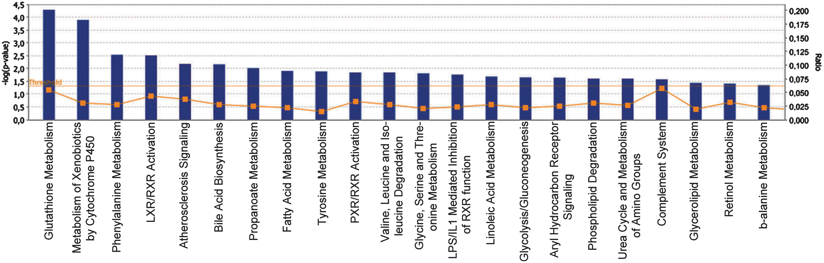
The pericontusional area of 5 DA and 5 PVGav1 rats were analyzed with microarrays in order to provide unbiased information on genetically regulated pathways one day after experimental brain injury. The pericontusional area was collected using a sample corer (Fine Science Tools, Heidelberg, Germany) resulting in a 5×5×6 mm piece of tissue consisting of the contusion core, the pericontusional cortex, the underlying hippocampus, and part of the thalamus. The experimental setup that was used to dissect genetic differences is shown in Figure 1. The molecular pathway showing the largest differences between the two strains was the glutathione metabolism pathway and included the transcripts Gsta1 (5.26E-06; 8.50), Gsta4 (3.94E-04;−1.7), Gstm2 (8.65E-04; −1.50), Gstm4 (2.29E-07; −2.93), and PRDX6 (2.63E-03; −1.56) (Fig. 2). The p value and fold change DA/PVGav1 are shown in the parentheses for each transcript, respectively. All transcripts display higher expression in the PVGav1 strain except from Gsta1. Independent confirmation of differences in transcriptional regulation of Gsta1 and Gsta4 was done with RT-PCR. Gsta1 displayed a similar expression pattern, as in the microarray data, with lower levels in PVGav1 compared to DA (data not shown). RT-PCR for Gsta4 included also the congenic strain R5 and showed that the PVGav1 strain displayed higher Gsta4 levels than the DA strain and similar Gsta4 levels to the R5 congenic (Fig. 3A). Microarray analysis of the 35 genes that are included in the genetic region differing between DA and R5 are shown in Table 1. Only the Gsta4 gene displayed both a significant p value and above threshold fold change in the analysis. Protein quantification with Western blot confirmed that the observed differential Gsta4 expression pattern results in corresponding differences at the protein level (Fig. 3B).


P values and fold change DA/PVGav1 as obtained by microarray analysis are shown. Significant differences p<0.01 or fold change ≥1.4 depicted in
, pseudogene; †, not analyzed in microarrays.
Cellular localization of Gsta4 and 4-HNE
The glutathione metabolism pathway plays a major role in neutralizing oxidative stress products in several conditions (21, 33, 47). Of the different enzymes in this pathway, Gsta4 has the by far most efficient catalytic activity to reduce 4-HNE by conjugating it to glutathione, hence reducing its toxic activity and thereby preventing formation of adducts to other proteins (4). Immunohistological staining of Gsta4 showed labeling mainly in neurons on both the ipsi- and contralateral sides (Fig. 4A and 4B). A semiquantitative assessment of Gsta4 labeling intensity on the ipsi versus the contralateral side suggested an upregulation in neurons after injury in the PVGav1 and R5 strains, but not in DA, which is consistent with previous findings after peripheral nerve injury (42). Immunostaining with two different antibodies recognizing 4-HNE-conjugates showed an essentially similar pattern, with strong staining of blood cell infiltrates in the contusion, a diffuse staining in the pericontusional area, and also a more distinct cellular staining of both glia and neurons. Neuronal cytoplasmic staining of 4-HNE was seen in degenerating cells in the pericontusional cortex and the hippocampal CA1, CA2/CA3, dentate gyrus, and hilus areas at both 1 and 6 days after TBI (Fig. 4C–4F). No discernible qualitative differences in cellular levels of 4-HNE were evident between the different strains.

Analysis of 4-HNE-conjugate levels in cerebrospinal fluid
The detection of 4-HNE-conjugates using ELISA based techniques has been reported previously (7, 33). We therefore attempted to determine CSF 4-HNE-conjugate levels in rat and human cerebrospinal fluid (CSF) after TBI using two different commercial ELISA-kits, of which one gave a positive signal. The analysis was conducted in a clinical material composed of patients with TBI and a control group of patients with other neurological diseases (OND). However, further testing showed that this signal was derived from unspecific binding of the secondary antibody to immunoglobulins (data not shown). In an ELISA set up in the lab using the two different antibodies used for immunohistochemistry, a signal in CSF spiked with 4-HNE down to a concentration of 0.5 μM could be detected. However, no specific signal was present in human CSF as well as rat CSF after TBI or 4-HNE-injection. It is therefore unlikely that 4-HNE-conjugates exist in micromolar concentration in CSF as previously has been reported (38, 39).
The PVGav1 Gsta4 haplotype reduces loss of neurons in hippocampus after TBI
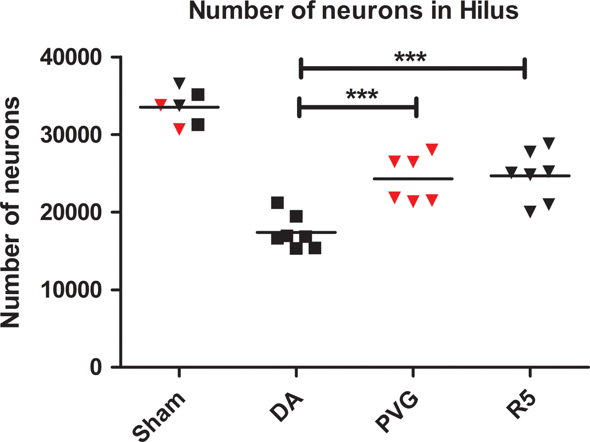
The localization of Gsta4 and 4-HNE in degenerating neurons in the hippocampus prompted us to investigate the effect of the Gsta4 haplotype for neuronal cell loss. Cell numbers in the hilus region were determined with stereology-based cell counts 30 days after TBI in the DA, PVGav1, and R5 strains. All three strains had equal numbers of neurons in sham-operated controls, but lower numbers of neurons after TBI. However, while DA displayed a 47.6% reduction in neuron numbers, the PVGav1 and R5 strain had lost only 24.6% and 29.8% of the neurons, respectively. This corresponds to a more than one-third reduction of neuronal loss in the PVGav1 and R5 strain compared to DA (Fig. 5).
Intraparenchymal injection of 4-HNE induces upregulation of Gsta4 and apoptosis of neurons
The biological cascade that follows TBI includes many toxic molecules apart from 4-HNE. In order to test the effect of 4-HNE more specifically, intraparenchymal injections were performed in DA and R5. Gsta4 labeling was upregulated in the area surrounding the site of injection, revealing a feedback mechanism where neurons upregulate Gsta4 in response to increased 4-HNE levels (Fig. 6A). A blinded semiquantitative assessment of Gsta4 protein staining revealed stronger Gsta4 upregulation in the R5 strain compared to the DA strain (Fig. 6B–6E). In addition, CSF levels of neurofilament light, a marker for axonal injury, were lower in R5 compared to DA, further supporting the in vivo role of Gsta4 to detoxify 4-HNE (Fig. 6F). A strong staining for 4-HNE-conjugates was seen in the area of injection, with both glia and neurons displaying labeling, without discernible qualitative differences between strains (Fig. 6G–6J). Further, TUNEL-staining showed apoptotic cell bodies after injection and co-localization of 4-HNE protein adducts in apoptotic neurons (Fig. 6K–6L).

4-HNE-conjugates are present in neurons in human TBI
A biopsy was taken from a 76-year-old patient who presented with a Glasgow Coma Scale of 9 after head trauma. Radiological evaluation revealed an expansive left contusional hemorrhage and the patient was treated surgically at 15 hours after trauma. Immunolabeling of human pericontusional tissue with antibodies for the neuronal marker NeuN and two different antibodies for 4-HNE, revealed the presence of 4-HNE-conjugates in neurons also after human TBI (Fig. 7).

Discussion
In this study we provide evidence that naturally occurring differences in Gsta4 regulate outcome in experimental TBI. Microarray data was obtained in the two inbred rat strains DA and PVGav1 as a first step to genetically dissect differences in pathway activation after experimental TBI. These strains have previously been extensively studied in autoimmune disease models such as EAE, a model of MS, which has provided both information on genetically regulated pathways as well as specific genes of importance for disease pathogenesis (5, 13, 29, 31, 44). Furthermore, the strains differ both in inflammatory response and neuronal survival in a standardized peripheral nerve injury model (19, 22). Recently, we demonstrated that these two strains also differ in their inflammatory response to experimental TBI, as well as outcome in terms of lesion volume (2). In the present study, we find that the most significantly genetically regulated pathway between DA and PVGav1 after TBI is the glutathione metabolism, with Gsta4 being one of the most differentially regulated transcripts. Glutathione metabolism has long been known to constitute a molecular system that protects tissues by detoxifying toxic molecules produced by oxidative stress and lipid peroxidation (37). In addition to this, oxidative stress and lipid peroxidation is one of the most extensively studied pathogenic processes known to contribute to the secondary damage after TBI. After a primary insult on the brain tissue, free radicals are formed and react with membrane phospholipids in a chain reaction disrupting their function and producing toxic aldehydes such as 4-HNE. 4-HNE reacts with the amino acids lysine, histidine, and cysteine in cellular proteins, which modify their conformation and disrupt important cellular enzymatic functions, making 4-HNE a highly toxic molecule (16). 4-HNE has been shown to be increased early after TBI and antioxidant therapies acting on upstream processes relevant for 4-HNE generation display a neuroprotective effect (11, 14, 18, 40). Once generated by peroxidation of lipids, a physiologic cellular system composed of glutathione (GSH) and a family of enzymes, glutathione-S-transferases, acts as a defense system to limit the deleterious effects of 4-HNE by conjugating it to glutathione, thus, making it unable to react with tissue proteins. Gsta4 has the by far highest catalytic activity for this reaction (4). This prompted us to investigate the role of the here identified naturally occurring genetic variation in Gsta4 expression between the inbred DA and PVGav1 strains for oxidative damage in TBI. This was done by the use of a congenic strain that contains a small genetic fragment including the Gsta4 gene from PVGav1 on DA background, with significantly increased Gsta4 levels. The finding that PVG alleles confer higher expression of Gsta4, which is translated into higher protein levels, while the sequence of the translated protein is identical between the strains (unpublished data) suggests that higher catalytic activity in PVG is a direct consequence of increased protein levels rather than providing higher catalytic activity in the enzyme per se.
We find 4-HNE immunoreactivity both in the pericontusional area and the hippocampus, including neurons, after experimental TBI, suggesting that it may contribute to neuronal cell loss after TBI. Indeed, when 4-HNE is injected directly into the brain, apoptotic nerve cells are present at one day after the injection both in the area of the injection but also in the hippocampus. We have previously performed an unbiased complex trait mapping of motor neuron loss after ventral root avulsion in the DA and PVGav1 strains, where a significant linkage was identified on chromosome 8 (19, 43). Also a whole genome expression QTL mapping in a F2(DAxPVGav1) intercross combined with fine mapping using congenic strains in the ventral root avulsion model identified one single transcript being highly strain regulated; Gsta4 (42). Further, we have found that the DA vs PVGav1 strain difference is equally strong on the protein level (42). We therefore decided to test the smallest of our congenic strain, R5, which contains 35 protein coding genes including Gsta4, in the TBI model. We find that the R5 strain displays a complete reversal of the strain pattern regarding Gsta4 (i.e., the level of expression is similar to the donor PVGav1 strain and significantly higher than the recipient DA strain). Furthermore, the levels of neurofilament light protein, a marker for neurodegeneration (35), were lower in the CSF of the R5 strain compared to DA after injection of 4-HNE, which provides in vivo evidence for a nonredundant importance of naturally occurring genetic variability in Gsta4 for detoxification of 4-HNE. However, the most significant finding is that neuronal cell death, as quantified stereologically in the hilus region, is similar between the congenic R5 and PVGav1 strains and significantly reduced compared to DA. Thus, the difference between the two parental strains in large part can be explained by variation in the R5 fragment, where Gsta4 is the only differentially regulated transcript. This provides strong support for the notion that higher expression of Gsta4 results in a more efficient detoxification of 4-HNE, in turn reducing nerve cell death. This chain of events are supported by findings in previous studies, where it was shown that 4-HNE induces degeneration of a variety of nerve cell populations ranging from hippocampal to motoneurons both in vitro and using experimental in vivo models (3, 17, 25, 27, 48).
Levels of 4-HNE in CSF have been reported previously in the chronic neurodegenerative disorders Alzheimer's disease (AD), Parkinson's disease (PD), and amyotrophic lateral sclerosis (ALS) (38, 39, 41). In the studies involving AD and PD subjects, the CSF concentration is reported to be approximately 0.5 μM in controls and further elevated in patients. The ALS studies show levels of only around 600 pg/ml in controls, corresponding to approximately 5 nM (41). Here we could not detect 4-HNE-conjugates in CSF using ELISA, although the assays had sensitivity down to low micromolar levels. Western blot identified a number of bands in 4-HNE-incubated CSF, but not in unmanipulated samples. Thus, there are presently only a limited number of studies on 4-HNE-conjugate levels in CSF, which have been conducted by two different groups showing very divergent results. Our results indicate that soluble 4-HNE-conjugates are not present at micromolar levels in CSF, which suggests that most HNE-conjugates remain captured in the tissue rather than being dissolved into the CSF. This notion is supported by the finding of immunostaining for 4-HNE-conjugates in contused human brain tissue.
Glutathione-S-transferase polymorphisms have been the subject of prior studies, identifying variability due to ethnic origin, but also to be associated to susceptibility for certain cancers (9). Thus, certain Gsta4 polymorphisms increase the risk to develop melanoma and hepatocellular and lung cancer (1, 28, 32). In CNS diseases there are some indications that a Gstm1 null and a Gstm3 polymorphism increase the risk for PD and AD, respectively (23, 30). The effect of genetic variability in Gsta4 has not been studied, except for a single study in 50 patients with PD. Not surprisingly, given the low number of included patients, the study did not reveal any association (6). In order to determine any possible relevance for neurodegeneration in humans, genotyping of much larger patient materials with available information on clinical follow up data that reflects the rate of neurodegeneration is required.
Taken together, these findings underscore both the importance of genetic diversity for outcome of TBI and also more specifically the role of the Gsta4—4-HNE detoxifying process. The former, if confirmed also for human TBI, could partially explain the failure of antioxidant therapy studies in human TBI. These results encourage studies on any possible genetic influence of polymorphism in Gsta4 for outcome in human TBI as well as on therapeutic approaches that targets the 4-HNE pathway, such as, for example, histidine analogues or hydrazine derivatives that scavenge 4-HNE and rescue cells from 4-HNE toxicity (12, 16, 46).
Materials and Methods
Animals
The DA (RT1av1) strain was originally provided by Professor Hans Hedrich (Medizinische Hochschule, Hannover, Germany), while the MHC congenic PVG-RT1av1 (hereafter called PVGav1) strain was obtained from Harlan UK Ltd (Blackthorn, UK). The DA.PVGav1-Vra1R5 (RNO8: D8Rat24-D8Got132 [82.2–88.6 Mb]) (hereafter called R5) congenic was bred in-house as described previously (43). The genome of the R5 congenic has all genes from the DA strain except 35 genes, including Gsta4, from the PVGav1 strain. All animals were bred in our in-house breeding facility with 12 h light/dark cycles and fed standard rodent chow and water ad libitum.
Experimental brain injury and 4-HNE injections
Experimental traumatic brain contusion was performed in 60 male animals weighing approximately 230–300 grams, at an age of 10–14 weeks, under deep isoflurane anesthesia using the weight drop injury model as described previously (2). In brief, the rats were placed in a stereotactic frame, and a 2 mm craniotomy was drilled 3 mm posterior and 2.3 mm lateral to the bregma. A standardized parietal contusion was made by letting a 24 gram weight fall onto a rod with a flat end diameter of 1.8 mm from a height of 7 cm in contused rats, allowed to compress the tissue a maximum of 3 mm. For 4-HNE injections the same procedure as for TBI was followed except at this time the craniotomy was smaller, with slow injection of 1 μl 64 mM 4-HNE (Cayman Chemical) during 2 min at the same coordinates and at 3 mm depth from the surface. This concentration was chosen since it has been shown to induce axonal and oligodendrocyte damage after intracerebral injection (27). All experiments in this study were approved by the local ethical committee for animal experimentation.
Array hybridization and qRT-PCR and Western blot analysis
Animal sacrifice, tissue preparation and preparation of mRNA was done as previously reported (2). The microarray analysis was performed at the microarray core facility of Karolinska Institutet using Affymetrix Rat gene 1.0 ST Array chips (Affymetrix, Santa Clara, CA) as described before (42). The microarray data is available in MIAME-compliant (minimal information about a microarray experiments) format at the ArrayExpress Database (
For Western blot analysis, protein homogenate from the same region as used for PCR was prepared and analyzed as previously decribed (42). Briefly, Gsta4 was labeled with a rabbit anti-rat Gsta4 antibody (1:2000) and a secondary HRP-conjugated antibody (donkey anti‐rabbit IgG‐HRP conjugated; 1:2000; Amersham NA9340V). ECL solution (Amersham) was applied and membranes exposed to Chemiluminescence film (Amersham HyperfilmTM ECL). Intensity of bands were measured with Gel DocTM XR+/Image LabTM 3.0(BioRad) and normalized against β-actin (Sigma Monoclonal β-Actin antibody Clone AC-15;1: 25 000).
4-HNE and neurofilament light analysis of cerebrospinal fluid after traumatic brain injury
Rat cerebrospinal fluid (CSF) was obtained directly after sacrifice from the cerebellomedullary cistern. The rat was placed in a stereotaxic frame and shaved over the occipital crest. A puncture through the occipital foramen magnum was made and about 100 μl of CSF was slowly drawn. The CSF was stored at −70°C until further processing.
The human CSF samples were obtained from 27 patients with severe TBI while being treated at the intensive care unit (ICU), and from 37 patients with other neurological diseases (OND) that were used as controls. CSF collection used in this study was approved by the regional ethical review board in Stockholm.
To assess the concentration of 4-HNE-protein adducts (μg/ml) in the CSF, we used two different commercial enzyme-linked immunosorbent assay (OxiSelect HNE-His Adduct ELISA, Cell Biolabs, INC, San Diego and HNE ELISA, Cusabio, Hubei Province 430223, P.R.China). These ELISAs were performed according to the manufacturer's instructions. Third, we set up a sandwich ELISA, using mouse monoclonal anti HNE (Abcam) as capture antibody, and rabbit polyclonal anti-HNE (Abcam, 1:6000) as detection antibody. Samples were incubated overnight and a biotinylated anti-rabbit antibody was used together with ABC-kit (Vector labs) for signal enhancement. As standard, control CSF incubated with HNE (Cayman, Burlingame, CA) was used in a dilution series with un-incubated CSF rendering a standard with linear detection in the range of 32–0.5 μM.
Levels of neurofilament light were quantified using NF-Light Neurofilament ELISA RUO kit (Umandiagnostics, Umeå, Sweden) according to the manufacturer's instructions.
Immunohistochemistry and immunofluorescence
Animals were sacrificed with CO2 and perfused first with 100 ml cold PBS containing heparin and after that with 250 ml parafolmaldehyde. The brains were postfixed at the same fixative followed by 15% sucrose and then stored at −70°C. Tissue preparation, immunohistochemistry, and double labeling immunofluorescense was performed as described previously (2). Brain tissue from the pericontusional area was obtained from a 76-year-old female patient who was surgically treated for a left contusional hemorrhage. The biopsy was snap-frozen in liquid nitrogen, stored at −70°C and postfixed in paraformaldehyde. The biopsy used in this study was approved by the regional ethical review board in Stockholm.
The primary antibodies used were rabbit polyclonal anti-HNE Michael adducts (Merck Chemicals Ltd., Nottingham, UK), mouse monoclonal anti HNE-histidine adducts (Abcam), rabbit polyclonal anti-rat Gsta4 (42), mouse monoclonal and rabbit polyclonal anti-NeuN (Millipore, MA). Apoptotic cell death was visualized using TUNEL. The sections were washed in PBS and permeabilized for 5 min in a 2:1 mixture of ethanol and acetic acid at −20°C. The TUNEL reaction was carried out with the "In situ cell death detection kit, fluorescein" (Boehringer Mannheim, Bromma, Sweden) at 37°C for 30 min.
Stereological assessment of neurodegeneration
We determined the number of neurons in the hilus 30 days after experimental TBI using a design based stereological approach. The hippocampi of the side ipsilateral to the injury, of 7 DA, 6 PVGav1, 7 DA.PVGav1R5, and 2 sham animals from each strain were dissected and straightened manually in order to eliminate its natural curvature. They were then embedded in 5% agar and sectioned perpendicularly to the longest axis at 65 μm thickness on a Vibratome 3,000 (Vibratome, St Louis, MO). Every 15th section (ssf=1/15) was chosen from each animal and was stained with thionin with the first section corresponding to a random number. Neuronal cell counting was performed using a light microscope modified for stereology with a microcator, motorized stage, fast digital camera, and the newCAST software (Visiopharm, Hoersholm, Denmark) as reported previously (42). The border of the hilus was outlined at 10× magnification and cell counting was performed using a Zeiss 40× oil objective (FLUAR, NA 1.30). The sampling frame had an area of 4000 μm2 and the step length was 140 μm×140 μm resulting in an area sampling fraction (asf ) equal to 0.20. A z axis distribution was obtained for 2 animals in order to find the range for the dissector height in which the cell nucleoli densities were similar (unpublished data). This range could be defined to be from 6 μm to 28 μm from the top of the section (dissector height; h(dis)=22 μm). Neurons that fulfilled the sampling criteria were counted (ΣQ-) and their total numbers (Ntotal) were estimated for the whole hilus segment using the equation:
Here hsf is the height sampling fraction (hsf=h(dis)/t(Q−), where t(Q−) is the Q-weighted section thickness (10).
Statistical and microarray analyses
All analyses were performed and data illustrated using the software GraphPad Prism 5.0. Data are presented as mean±SD. Partek® Express, version 6.11, copyright 2012 (Partek, St Louis, MO) was used to carry out statistical analyses of microarray data files and Ingenuity Pathway Analysis software (IPA 9.0,
Footnotes
Acknowledgments
This study was supported by the 6th Framework Program of the European Union, NeuroproMiSe, LSHM-CT-2005-018637, EURATools, LSHG-CT-2005019015, and the 7th Framework Program of the European Union, EURATrans, HEALTH-F4-2010-241504, by the Swedish Research Council, the Swedish Brain Foundation and the Swedish Association of Persons with Neurological Disabilities. Centre for Stochastic Geometry and Advanced Bioimaging is supported by Villum Foundation.
Author Disclosure Statement
No competing financial interests exist.