Yersinia pestis is the gram-negative, facultative intracellular bacterium that causes the disease known as plague. Due to the risk for aerosol transmission, a low infectious dose, and the acute and lethal nature of pneumonic plague, research activities with Y. pestis require Biosafety Level 3 (BSL-3) facilities to provide the appropriate safeguards to minimize accidental exposures and environmental release. However, many experimental assays cannot be performed in BSL-3 due to equipment availability, and thus require removal of samples from the BSL-3 laboratory to be completed.
Objectives:
To remove samples from BSL-3 containment and safely handle them at lower containment requires effective inactivation of any viable organisms from the samples prior to removal. While commonly used inactivation methods have been published for various select agents, there is an absence in the literature of a single source providing detailed examples for inactivation methods for Y. pestis. Our objective here is to provide examples of dose-dependent kill curves for commonly used inactivation approaches against Y. pestis.
Methods:
Time- and dose-dependent kill curves using heat, methanol, and formaldehyde inactivation methods, and common nucleic acid extraction procedures.
Results/Conclusions:
We show data demonstrating the complete inactivation of Y. pestis using these methods. While not all-inclusive, this study provides data and examples that can be used by other researchers to develop their own in-house validated inactivation protocols for Y. pestis.
Introduction
Yersinia pestis is the gram-negative, facultative intracellular bacterium that causes the disease known as plague. Historically, there have been three plague pandemics. While the last plague pandemic officially ended in 1945, Y. pestis became endemic in rodent populations throughout the world.1 Within these endemic locations, there is still the potential for spillover events to occur when fleas transfer Y. pestis to humans, highlighted by the 2017 Madagascar and 2022 Democratic Republic of Congo human plague outbreaks.2,3 Mathematical modeling of the impact of climate change on the spread of Y. pestis suggests that the frequency of spillovers is likely to increase—warmer temperatures lead to higher rodent densities and increases in flea populations, which in turn increases the likelihood that humans will come in contact with the infected vectors.4 While Y. pestis is considered a vector-borne disease, there has been recent evidence toward Y. pestis thriving in soil, further increasing the possibility of spillover events.5
Considering these risks, research on plague is still necessary. To ensure laboratorial safety, Y. pestis research is conducted at Biosafety Level 3 (BSL-3) facilities, which provide the appropriate safeguards to minimize accidental exposures and environmental release.
The Federal Select Agent Program (FSAP) was established in 1996 and supervises the possession, use, and transfer of select agents. Select agents are pathogens or toxins determined to have the potential to pose a severe threat to public health and safety. To be considered a select agent, the danger to human health, speed of transmission, and the availability and effectiveness of treatment and/or prevention are all considered. In addition to the acute progression of the plague, Y. pestis also has a history of misuse as a biological weapon.6,7 Because of the rapid nature of plague infection, potential for aerosols and person-to-person transmission, and its risk of deliberate misuse, the FSAP has categorized Y. pestis as a Tier 1 select agent. This designation limits access to government vetted entities and individuals and ensures an increased level of biosecurity to protect the public from accidental or intentional release.
While research with Y. pestis requires work within BSL-3 laboratories to minimize risks to laboratorians and potential release, inactivation of the bacterium (i.e., rendering it nonviable and unable to cause infection) can generate products that can be safely handled at lower containment. However, after the shipment of anthrax spores that were not successfully inactivated, the FSAP has increased safeguards to prevent similar accidents.8 These procedures include the development of standardized inactivation protocols that are validated prior to use, which includes documentation demonstrating that an inactivation protocol successfully inactivates the sample. Important considerations for validation can include kill curves, which identify either minimal concentrations or times required for an inactivation agent to fully inactivate the organism. Furthermore, each validated protocol needs to include an inactivation verification step, which provides proof of successful inactivation each time a protocol is performed prior to removal of the sample from the BSL-3 laboratory.
To aid in developing effective inactivation protocols, the FSAP allows the use of surrogate organisms that can be handled at BSL-2 for the validation of these procedures.8 While commonly used inactivation methods have been published for various select agents, there is an absence in the literature of a single source providing data supporting inactivation methods that can be used for Y. pestis while still providing downstream applications.9,10 Albeit not all-inclusive, our purpose here is to provide the community with examples of several common inactivation approaches used with Y. pestis that can serve as a foundation for their own in-house development of validated inactivation protocols.
Methods
Bacteria
For in vitro studies, we used attenuated derivatives of the Y. pestis KIM biovar missing the high pathogenesis island (pgm) and the large virulence plasmid (pCD1), which is exempt from Select Agent regulation.11 These strains also harbored bioluminescent bioreporters (pLUX or LuxptolC) to monitor bacterial viability as a function of bioluminescence.12,13 These strains represent a surrogate for fully virulent Y. pestis that can be handled safely at BSL-2 to develop and validate inactivation procedures.8,11 For in vivo studies performed within the BSL-3, we used a fully virulent strain of Y. pestis KIM5+ which harbors both the pgm locus and pCD1.14
Bacteria were routinely cultured for 15–18 h at 26°C in Bacto brain–heart infusion (BHI) broth (Cat. No. 237500; BD Biosciences) on a roller drum. Optical densities at 600 nm (OD600) were determined with a spectrophotometer and used as a reference to dilute samples to the desired bacterial concentrations. Final bacterial concentrations were enumerated by serial dilutions of samples and growth on Diffco BHI agar plates (Cat. No. 241830; BD Biosciences) for 2 days at 26°C. Data are reported as colony forming units (CFU) per milliliter.
Heat Inactivation of Y. pestis
Approximately 2.65 × 10⁹ CFU of bacteria were resuspended in 1 mL of phosphate-buffered saline (1 × PBS) in 1.5 mL Eppendorf tubes and placed in a heating block that was preheated to 50°C. For 1 h, 10 μL aliquots were removed every 10 min, and bacterial numbers were enumerated. Enumeration was performed with three technical replicates for each biological replicate at each time point. The limit of detection was determined as 103 CFU. In a separate experiment, samples (n = 3 biological replicates) were incubated for 2 h, pelleted for 1 min at 16,000 g, and resuspended in 100 μL 1 × PBS. The entire 100 μL sample was plated onto BHI agar and incubated for 2 days at 26°C. The limit of detection was determined as 1 CFU. The inactivation was performed four times.
Laemli Buffer and Boiling Inactivation of Y. pestis
Approximately 2 × 10¹⁰ CFU of bacteria were resuspended in 200 μL of 1 × PBS. Samples were either incubated at room temperature in 1 × PBS for 15 min, in 1 × Laemli buffer (6 × Laemli buffer is 5% 2-mercaptoethanol, 2% sodium dodecyl sulfate (SDS), 10% glycerol, 0.012% bromophenol blue, and 0.375 M Tris-HCl) for 5 min, in 1 × PBS in a boiling water bath for 10 min, or in 1 × Laemli Buffer for 5 min at room temperature followed by boiling for 10 min. After each condition, the entire sample was inoculated onto BHI agar and incubated for 2 days at 26°C. The limit of detection was determined as 1 CFU. The inactivation was performed five times.
Paraformaldehyde and Formalin Inactivation of Y. pestis
Approximately 1.95 × 10⁹ CFU of bacteria were resuspended in 1 × PBS. Freshly prepared 4% paraformaldehyde (PFA, Cat. No. P6148; Sigma-Aldrich) was added at final concentrations of 0.5%, 1%, 2%, or 4% (n = 3). Neutral-buffered formalin (NBF, Cat. No. 89370; VWR) was added at final concentrations of 1.25%, 2.5%, 5%, or 10% (n = 3). Final volumes were 1 mL for each condition. As a growth control, 1 mL of 1 × PBS was added to a separate sample. Samples were then mixed by pipetting and incubated at 4°C. Every 15 min for 60 min, 10 μL aliquots were removed to measure bacterial concentration. To confirm starting concentrations, untreated bacteria incubated in 1 mL of 1 × PBS were serially diluted and plated on BHI agar. The limit of detection was determined as 103 CFU. Inactivation was performed three times.
Approximately 1.8 × 10⁹ CFU of bacteria were resuspended in 1 mL of freshly prepared 1% PFA or 2.5% NBF. Samples were then mixed by pipetting and incubated at 4°C. At 30 min, samples were centrifuged for 1 min at 16,000 g, washed once with 1 × PBS, and then resuspended in 100 μL of 1 × PBS. The entire sample was transferred to a BHI agar plate and incubated for 2 days at 26°C. To confirm starting concentrations, untreated bacteria incubated in 1 mL of 1 × PBS were serially diluted and plated on BHI agar. The limit of detection was determined as 1 CFU. The inactivation was performed three times.
Formalin Inactivation of Y. pestis in the Presence of Tissues
Y. pestis was grown at 26°C for 6–8 h, diluted to an OD (600 nm) of 0.05 in Bacto BHI broth (BD Biosciences Cat. No. 237500) with 2.5 mM CaCl2 and then grown at 37°C with aeration for 15–18 h.15 C57BL/6J mice were anesthetized with ketamine/xylazine and administered 20 μL of fully virulent Y. pestis suspended in 1 × Dulbecco's PBS to the left nare as previously described.13,15 At 48 h postinfection, lungs, spleen, and liver were removed by sterile necropsy. Tissues from five mice were cut in half, prior to adding to tissue cassettes, and submerged in 10% NBF for 24 h, 12 mL per tissue. Untreated tissues from four mice were macerated, and bacterial numbers were enumerated by serial dilution and plated on BHI agar. After 24 h, tissues were removed from the 10% NBF and washed with 1 × PBS. The NBF-treated tissues were transferred to 3 mL of BHI and incubated for 2 days at 26°C. Cultures were visually inspected for turbidity as a sign of bacterial growth, and 150 μL (5%) were plated on BHI agar to confirm absence of growth.
Methanol Inactivation of Y. pestis
Approximately 3.2 × 10⁹ CFU of bacteria were resuspended in 1 mL of 1 × PBS+methanol (MeOH, Cat. No. A412; Fisher Chemical) at final concentrations of 0%, 25%, 50%, 75%, and 100% MeOH. Samples were mixed by pipetting and incubated at 4°C. Every 15 min for 60 min, 10 μL aliquots were removed to enumerate bacterial numbers. To confirm starting concentrations, untreated bacteria incubated in 1 × PBS were serially diluted and plated on BHI agar. The limit of detection was determined as 5 × 10³ CFU. The inactivation was performed three times.
Approximately 3.22 × 10⁹ CFU of bacteria were resuspended in 1 mL of 50% MeOH. Samples were mixed by pipetting and incubated at 4°C. At 15, 30, and 60 min, samples were centrifuged for 1 min at 16,000 g, washed once with 1 × PBS, and resuspended in 100 μL of 1 × PBS. The entire sample was transferred to BHI agar and incubated for 2 days at 26°C. To confirm starting concentrations, untreated bacteria incubated in 1 × PBS were serially diluted and plated on BHI agar. The limit of detection was determined as 1 CFU. The inactivation was performed three times.
MeOH Inactivation of Y. pestis in Tissues
Lungs from C57BL/6J mice were removed by sterile necropsy and immediately frozen in a 2 mL tube prefilled with 2.8 mm ceramic beads (Cat. No. 10158-612; VWR). Lungs were thawed and ∼5 × 10⁹ CFU of bacteria in 1 × PBS were added. Additional 1 × PBS was added to fill the remaining air space within the tube. Tissues were homogenized with an Omni Bead Ruptor 4 at speed 5 (5 m/s) for three cycles of 30 s with 1-min pauses in which the lungs were placed on ice to prevent samples from overheating.
Bacterial CFU were enumerated by serial dilution of 100 μL of the homogenized samples to determine the effects of the homogenization process on bacterial survival. Tissue debris was then centrifuged for 10 min at 1,500 g. The supernatants (∼1.5 mL) were then transferred to a fresh Eppendorf tube. From this, 250 μL aliquots were added to MeOH+butylated hydroxy toluene (BHT) (75% +0.1% final concentration, respectively) and incubated at 4°C for 24 h. After incubation, samples were pelleted for 1 min at 16,000 g. MeOH+BHT was removed, and pellets were washed three times with 1 × PBS. Samples were then resuspended in 250 μL of 1 × PBS, and the entire sample was transferred to BHI agar supplemented with irgasan (1 μg/mL) and polymyxin B (12.5 μg/mL) (the Y. pestis strain used is resistant to these antibiotics and allows for differentiation from potential contamination by the host microbiota) and incubated for 2 days at 26°C. The limit of detection was determined as 1 CFU. The inactivation was performed four times.
C57BL/6J mice were anesthetized with ketamine/xylazine and administered 20 μL of fully virulent Y. pestis as previously described.13,15 At 6, 12, 24, 36, and 48 h postinfection, whole lungs were necropsied and homogenized as described above. Bacterial CFU were enumerated from each sample by serial dilution of 100 μL of the homogenized samples to determine the bacterial concentration prior to inactivation. Two hundred microliters aliquots from the fresh Eppendorf tube were added to MeOH+BHT (75% +0.1% final concentration, respectively) and incubated at 4°C for 24 h. After incubation, 50 μL from each sample was transferred to a BHI agar plate supplemented with irgasan (1 μg/mL) and polymyxin B (12.5 μg/mL) and then incubated for 2 days at 26°C. The limit of detection was determined as 50 CFU. The inactivation included five biological replicates.
Y. pestis Inactivation with Promega Wizard Genomic DNA Purification Kit
Approximately 5.7 × 10⁹ CFU of bacteria were collected into 1.5 mL Eppendorf tubes. Bacteria were pelleted for 1 min at 12,000 g and DNA extraction was performed per manufacturer's instructions (Cat. No. A1120; Promega). The entire elution after purification was transferred to BHI agar and incubated for 2 days at 26°C. The limit of detection was determined as 1 CFU. The inactivation was performed three times.
Y. pestis Inactivation with TRIzol and Chloroform
Approximately 5.4 × 10⁹ CFU of bacteria were collected into a 1.5 mL RNase free Eppendorf tube. Bacteria were pelleted for 1 min at 10,000 g and gently resuspended in 1 mL TRIzol reagent (Cat. No. 15596026; Thermo Scientific) and incubated for 5 min at room temperature. Two hundred microliters of chloroform (Cat. No. 513-35-9; Fisher Chemical) was added, followed by 15 s of vigorous shaking and a 3 min incubation at room temperature. Samples were then centrifuged for 15 min at 12,000 g at 4°C. The aqueous phase was transferred to BHI agar and incubated for 2 days at 26°C. The limit of detection was determined as 1 CFU. Inactivation was performed three times.
Statistics
Before statistical analysis, values for samples that were below the limit of detection were converted to the limit of detection and log transformed. All statistical calculations were performed using GraphPad Prism, and the tests used for comparison are reported in the figure legends. When comparing groups to untreated samples, a one-way ANOVA with Dunnett's post hoc test was used. For comparing multiple groups to each other, a one-way ANOVA with Tukey's post hoc test was used. When there were only two groups in the experiment, a two-tailed unpaired T-test was used.
Results
Heat Inactivation of Y. pestis
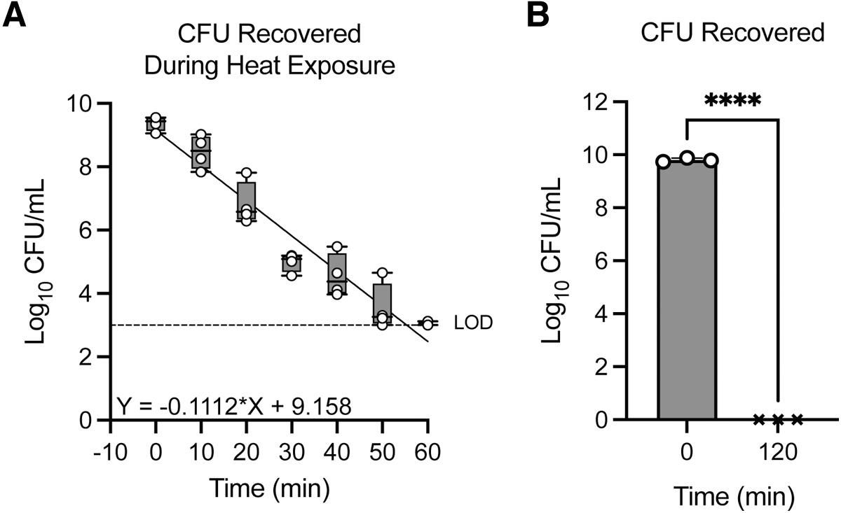
To determine the impact of elevated temperature on the survival of Y. pestis, we examined two conditions, extended incubation at 50°C and boiling bacteria with and without Laemli buffer, a buffer commonly used for protein analyses. To determine the viability of Y. pestis at 50°C, bacteria were incubated at the elevated temperature for 60 min, and recovery of viable bacteria was determined by enumeration. By 10 min, we observed a 10-fold decrease in viability, which continued to decrease over the next 50 min. By 60 min, bacterial viability decreased by >5 orders of magnitude, with one sample below the limit of detection (Figure 1A).
Heat shock at 50°C inactivates Yersinia pestis within 2 h. Y. pestis was incubated at 50°C. (A) Enumeration of viable bacteria at 10 min intervals for 60 min. The limit of detection was 103 CFU, indicated by the dotted line. Each symbol represents an independent biological experiment, and the box plot represents the median of the group ± the range. The equation represents the linear regression analysis of the data used to predict how much time would be required for complete in activation. (B) Enumeration of bacteria from a sample before (0 min) or after incubation at 50°C (120 min). The limit of detection was 1 CFU. Each symbol represents an independent biological experiment, and the bars represent the mean of the group ± the standard deviation. Two-tailed unpaired T-test. ****p ≤ 0.0001. CFU, colony forming units.
Using these data, we calculated that Y. pestis should be completely inactivated after 81 min when incubated at 50°C. Based on this prediction, we incubated a separate group of samples at 50°C for 120 min (a time frame exceeding the minimum inactivation calculated above to ensure complete inactivation of the bacteria). No viable bacteria were recovered from the 120 min samples (Figure 1B; p ≤ 0.0001). Together these data indicate that complete inactivation of Y. pestis can be achieved by incubating the bacteria at 50°C for ≥120 min.
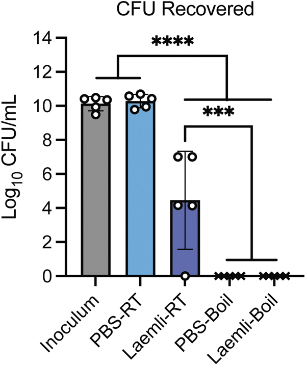
Samples are often prepared for SDS–polyacrylamide gel electrophoresis (SDS-PAGE) analysis by adding Laemli buffer and boiling. To determine if incubation with Laemli buffer inactivated Y. pestis, bacteria were resuspended in the buffer with and without boiling. Incubation of Y. pestis at room temperature (RT) with Laemli buffer significantly reduced bacterial viability compared to samples without Laemli buffer (Figure 2; Laemli-RT vs. PBS-RT, respectively; p ≤ 0.0001). However, boiling the bacteria for 10 min with or without Laemli buffer resulted in no recovery of viable bacteria (Figure 2; PBS-Boil and Laemli-Boil vs. PBS-RT; p ≤ 0.0001). Together, these data demonstrate that boiling samples for 10 min, with or without Laemli buffer, is sufficient to inactive Y. pestis.
Yersinia pestis is inactivated by boiling for 10 min. Y. pestis was incubated at room temperature in 1 × PBS for 15 min (PBS-RT), in 1 × Laemli buffer for 5 min (Laemli-RT), or in a boiling water in 1 × PBS (PBS-Boil) or in 1 × Laemli Buffer for 10 min (Laemli-Boil). After incubation, viable bacteria were enumerated. The limit of detection was determined as 1 CFU. Each symbol represents an independent biological experiment, and the bars represent the mean of the group ± the standard deviation. One-way ANOVA with Tukey's post hoc test. ***p ≤ 0.001; ****p ≤ 0.0001. PBS, phosphate-buffered , room temperaturesaline; RT, room temperature.
PFA and Formalin Inactivation of Y. pestis
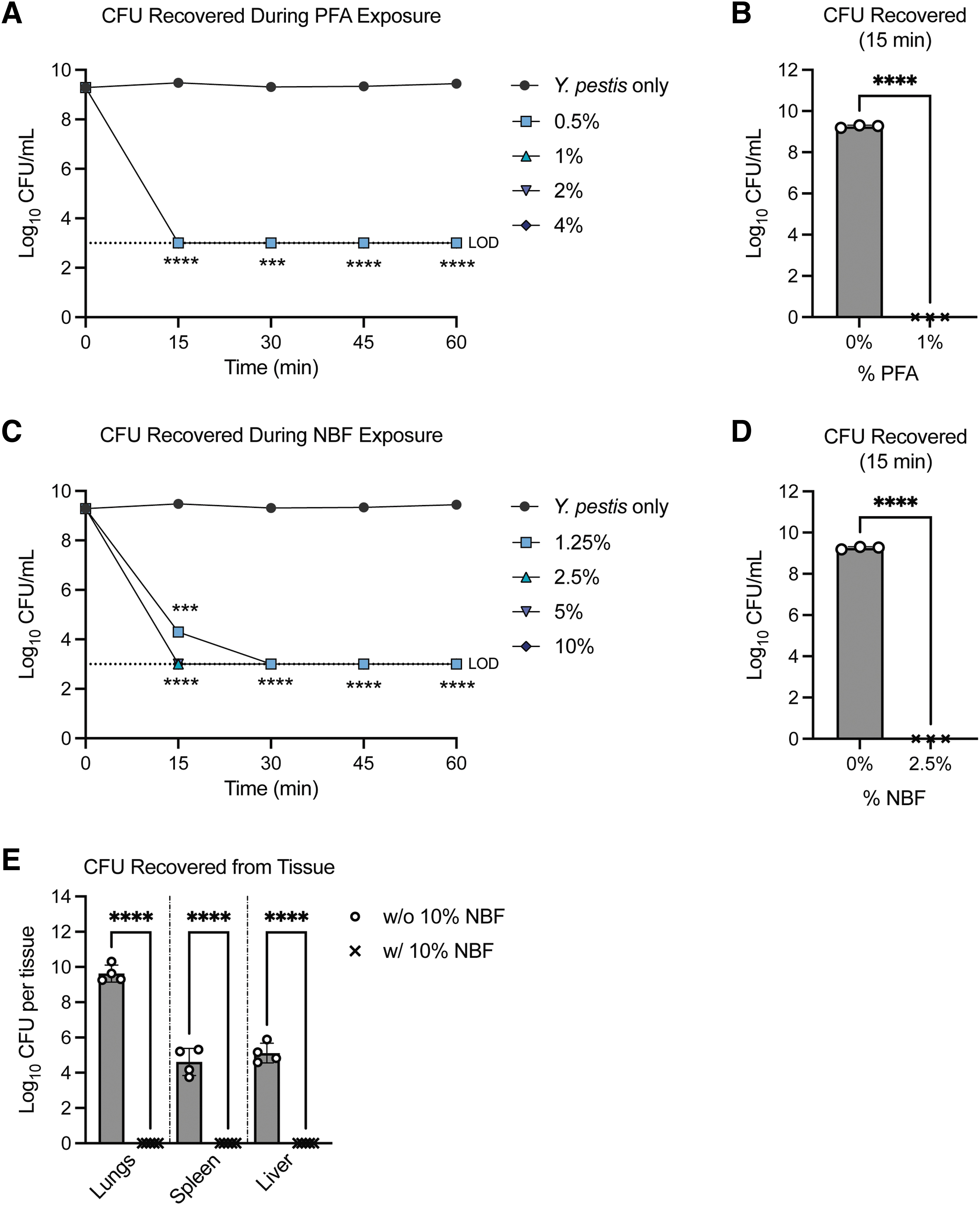
PFA and NBF inactivate biological samples by covalent crosslinking amines between proteins and nucleic acids.16 Typical protocols for microscopy and flow cytometry applications include incubation with 4–10% of fixative for 15–30 min.17 To define the kinetics of PFA inactivation, Y. pestis was incubated with 0.5%, 1%, 2%, or 4% PFA (final concentration), and CFU were enumerated every 15 min for 60 min. Regardless of concentration, all samples were below the limit of detection within 15 min of treatment (Figure 3A; p ≤ 0.0001). In a separate experiment, Y. pestis was incubated with 1% PFA for 15 min, and the entire sample was transferred to an agar plate to determine if any viable bacteria were present. No viable bacteria were recovered (Figure 3B; p ≤ 0.0001).
Low concentrations of PFA and NBF inactivate Yersinia pestis. Y. pestis was incubated with indicated concentrations of (A, B) PFA or (C, D) NBF, and bacterial survival was measured by CFU from (A, C) 10 μL aliquots every 15 min or (B, D) the whole sample at 15 min. Limit of detection was determined as (A, C) 103 or (B, D) 1 CFU. (E) C57BL6/J mice were infected intranasally with 10 × the LD50 of Y. pestis KIM5+ and lungs, spleen, and liver were harvested 48 h postinfection. Incubated with 10% NBF for 24 h. (A, C) Symbols represent the mean of three biological replicates and the error bars represent ± the standard deviation. Two-way ANOVA with Dunnett's post hoc test comparing to untreated. ***p ≤ 0.001, ****p ≤ 0.0001. (B, D) Each symbol represents an independent biological experiment, and the bars represent the mean of the group ± the standard deviation. Two-tailed unpaired T-test. ****p ≤ 0.0001. (E) Each symbol represents tissues from an individual mouse, and the bars represent the mean of the group ± the standard deviation. Two-tailed unpaired T-test within each tissue group. ****p ≤ 0.0001. NBF, neutral-buffered formalin; PFA, paraformaldehyde.
For NBF, bacteria were incubated with 1.25%, 2.5%, 5%, or 10% (final concentration). As observed for PFA, viable bacteria were below the limit of detection for all concentrations within 30 min of incubation (Figure 3C; p ≤ 0.0001), and all concentrations >1.25% were below the limit of detection within 15 min (Figure 3C; p ≤ 0.0001). To determine in a separate experiment if any viable bacteria were present at this concentration, bacteria were incubated for 15 min in 2.5% NBF, and the entire sample was transferred to an agar plate. No viable bacteria were recovered (Figure 3D; p ≤ 0.0001). Together these data indicate that incubation with ≥1% PFA or ≥2.5% NBF for 15 min is sufficient to inactive Y. pestis.
Formalin is also commonly used as a fixative for tissues for histological examination. To demonstrate that 10% NBF can inactivate Y. pestis in tissues, mice were intranasally infected with fully virulent Y. pestis. Forty-eight hours postinfection, bacterial numbers in the lungs were 7.12 × 10⁹ ± 2.76 × 10⁹ per tissue, in the spleens were 9.68 × 10⁴ ± 1.59 × 10⁴ per tissue, and the in the livers were 2.55 × 10⁵ ± 8.36 × 10⁴ per tissue (Figure 3E). After 24 h incubation with 10% NBF, tissues were cultured in BHI broth for 48 h. Cultures were not turbid after incubation, indicating the absence of viable bacteria. Sterility was confirmed by plating a portion of the broth on BHI agar plates. Again, no viable bacteria were recovered (Figure 3E). Together, these data demonstrate that 24 h incubation with 10% NBF can effectively inactivate Y. pestis in murine tissues.
MeOH Inactivation of Y. pestis
MeOH can be used to permeabilize and fix samples for microscopy and to extract lipids from biological samples for subsequent lipid identification by liquid chromatography tandem mass spectrometry (LC-MS/MS).18–20 To determine if MeOH inactivates Y. pestis, bacteria were treated with increasing concentrations of MeOH and bacterial viability was determined every 15 min for 60 min by enumeration. Y. pestis appeared relatively resistant to 25% MeOH but was sensitive to inactivation at concentrations ≥50% (Figure 4A). In a separate experiment, bacteria were incubated with 50% MeOH, and the entire sample was plated at 15, 30, and 60 min (Figure 4B). In this experiment, incubation for 1 h was required to completely inactivate >108 CFU of bacteria (p ≤ 0.0001).
MeOH inactivation of Yersinia pestis. (A, B)Y. pestis was incubated with indicated concentrations of MeOH, and bacterial survival was measured by CFU from (A) 10 μL aliquots every 15 min or (B) the whole sample at 15, 30, or 60 min. Limit of detection was determined as (A) 103 or (B) 1 CFU. (C) Enumeration of the bacteria recovered after lungs+Y. pestis was incubated with 75% MeOH +0.1% BHT. Limit of detection was calculated as 1 CFU. (D) Enumeration of bacteria recovered after lungs from Y. pestis-infected animals were incubated with 75% MeOH +0.1% BHT. Limit of detection was calculated as 50 CFU. (A) Symbols represent the mean of three biological replicates and the error bars represent ± the standard deviation. Two-way ANOVA with Dunnett's post hoc test. **p ≤ 0.01, ***p ≤ 0.001, ****p ≤ 0.0001. (B–D) Each symbol represents an independent biological experiment, and the bars represent the mean of the group ± the standard deviation. One-way ANOVA with Tukey's post hoc test. *p ≤ 0.05, **p ≤ 0.01, ****p ≤ 0.0001. BHT, butylated hydroxy toluene; MeOH, methanol.
To determine the ability of MeOH to inactivate Y. pestis in the presence of host tissue, mouse lungs were transferred to a 2 mL tube and a known concentration of bacteria (5.4 × 10⁹ CFU/mL) was added to the tissues. The tissues + bacteria were resuspended in 1 × PBS with ceramic beads for homogenization. Following tissue homogenization, bacterial viability decreased by ∼3-logs (Figure 4C; “After homogenization” vs. “Inoculum”; p ≤ 0.0001). Viability decreased slightly if the samples were further incubated at 4°C for 24 h (Figure 4C; “24 h PBS”). However, no viable bacteria were recovered from homogenized samples after 24 h of incubation in 75% MeOH + 0.1% BHT, final concentration (75% was chosen as this is a concentration applicable to lipid extraction) (Figure 4C; “24 h 75% MeOH”).
Based on these results, lungs were isolated from mice intranasally infected with fully virulent Y. pestis at 6, 12, 24, 36, and 48 h postinfection, and the tissues were homogenized. Prior to addition of MeOH, bacterial numbers were enumerated, and then samples were incubated with 75% MeOH +0.1% BHT for 24 h. Inactivation was verified by plating 5% of the sample, of which no viable bacteria were recovered (Figure 4D; p ≤ 0.0001). Together, these data demonstrate that 75% MeOH can effectively inactivate Y. pestis, even in the presence of host tissues.
Nucleic Acid Extraction Inactivates Y. pestis
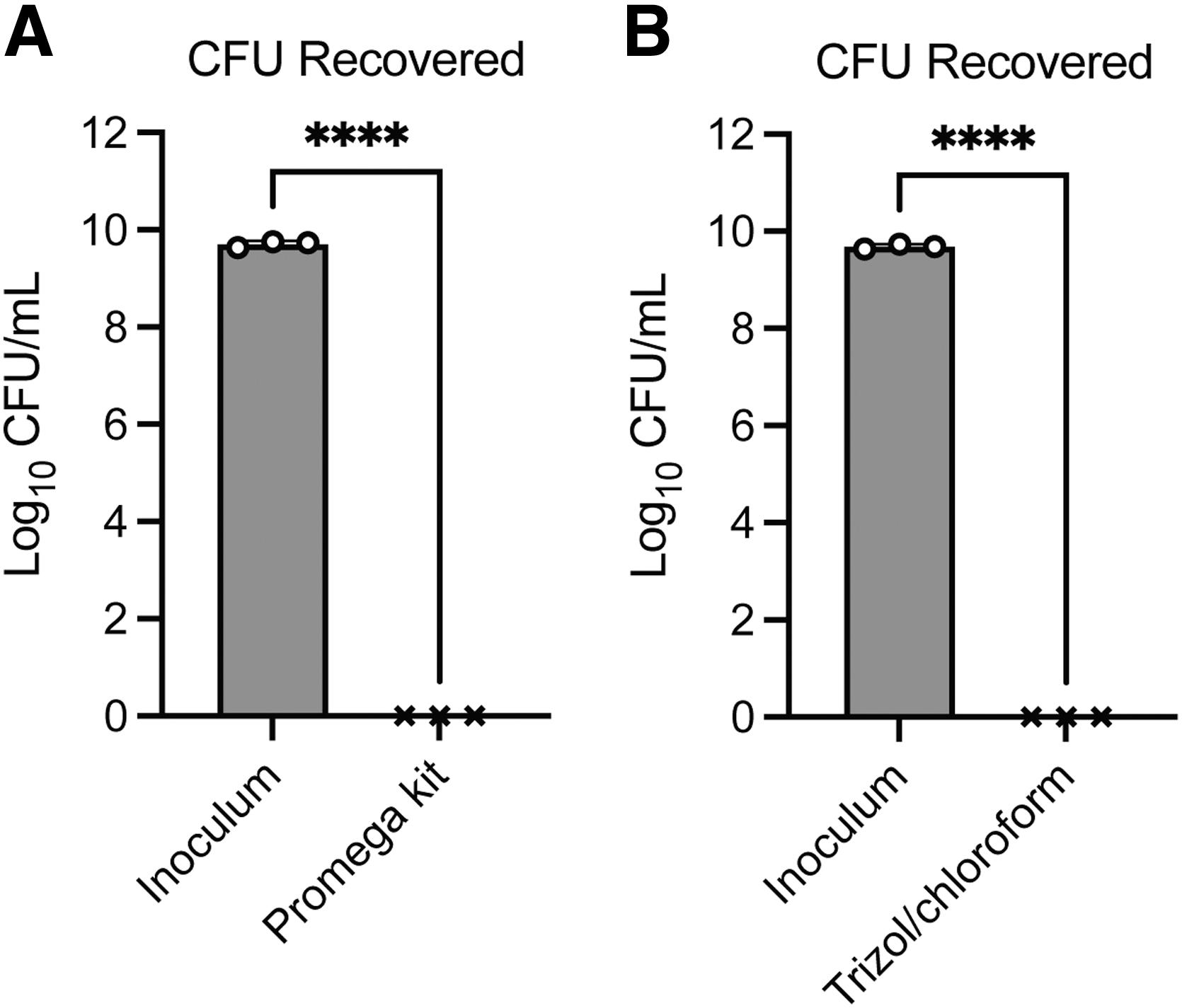
There are a variety of approaches to isolate nucleic acids from bacteria. Here, we chose two common approaches used to isolate genomic DNA or RNA from bacterial cultures. For DNA isolation, we used a commercial alkaline lysis approach following the manufacturer's protocol for gram-negative bacteria. No viable bacteria were recovered in the elution after extraction, indicating that this commercial kit completely inactivated the bacteria (Figure 5A). For RNA extraction, we used a TRIzol extraction approach and plated the entire aqueous phase after chloroform extraction. No viable bacteria were recovered from the aqueous phase, indicating that TRIzol/chloroform extraction completely inactivates Y. pestis (Figure 5B).
Alkaline lysis and TRIzol/chloroform extraction successfully inactivate Yersinia pestis. Y. pestis was treated for nucleic acid extraction using the (A) Promega Wizard Genomic DNA Purification Kit or (B) TRIzol/chloroform extraction. The limit of detection was determined as 1 CFU. Each symbol represents an independent biological experiment, and the bars represent the mean of the group ± the standard deviation. Two-tailed unpaired T-test. ****p ≤ 0.0001.
Discussion
BSL-3 containment facilities and procedures protect laboratorians from accidental exposure and infection by Y. pestis. However, many pieces of equipment needed for research may not be available or amenable to use within the BSL-3 laboratory. Thus, samples need to be inactivated and confirmed for inactivation for safe removal from BSL-3 for downstream experimentation. As such, the development of validated inactivation protocols is required to ensure that samples can be safely handled at lower containment.
For Y. pestis, there have been several studies published demonstrating the efficacy of common disinfectants,21,22 gas decontamination (both hydrogen peroxide and chlorine dioxide),23–27 and UV radiation.28–33 However, published data related to inactivation methods more amenable to research applications are limited.32,34–36 Here, we build upon these studies to provide a systematic analysis of several inactivation methods commonly used within the research field. Our goal was to provide others with approaches and data that can serve as the foundation for the development of in-house validated inactivation protocols.
Y. pestis is a mesophilic bacterium that thrives in a temperature range from 20°C to 40°C, temperatures of its native insect and mammalian hosts, but it can also grow efficiently at 4°C.37 Temperatures >45°C can negatively impact many cellular processes for mesophilic bacteria, including protein stability, membrane structure, metabolic activity, and DNA repair.36,38–40 Therefore, exposure to elevated temperatures can result in loss of viability over time.38
Wang et al. reported previously that incubation at 68°C for 10 h completely inactivated Y. pestis but also briefly mentioned that 1 CFU was recovered from treatments at lower temperatures for shorter periods of time.32 In our hands performing multiple biologically independent experiments, we were unable to recover viable bacteria from samples at a starting concentration of ∼2.65 × 10⁹ CFU/mL when the bacteria were incubated at 55°C for 2 h. Because of the specific details of the bacterial concentration of the samples, how many times the inactivation was performed, the amount of sample enumerated, and whether the single colony recovered was verified as Y. pestis were not provided by Wang et al., it is difficult determine why we observed differences in our evaluations. However, simple differences in the validation of the temperature of the heating elements or sample diluent (e.g., PBS vs. water) could explain the differences and highlight the need to have in-house validation of inactivation procedures.
Wang et al. also reported that incubation with 4% PFA at 4°C overnight inactivated Y. pestis.32 By applying both time- and dose-dependent kill curve analysis, we expanded on these data to show that concentrations of 1% PFA or 2.5% NBF can fully inactivate Y. pestis within 15 min when incubated at room temperature (Figure 3). These data support that standard treatments with formaldehyde that conserve cell morphology and retain fluorophore activity for confocal imaging and flow cytometry should be sufficient to inactivate Y. pestis.41–45
In the context of formaldehyde fixation of infected tissues, Chua et al.34 previously reported that incubation of tissues from Y. pestis-infected rabbits and guinea pigs with glutaraldehyde or formaldehyde fixatives for 6 or 13 days, respectively, resulted in bacterial inactivation. However, whether shorter incubation periods were sufficient was not reported. As tissues from mice are significantly smaller than those from rabbits or guinea pigs, we hypothesized that shorter incubation times would be sufficient to perfuse the tissues, and as predicted, incubation with 10% NBF for 24 h was sufficient to inactivate Y. pestis in murine tissues of different densities (lungs, spleens, and livers). However, as indicated by Chua et al.34 and Buesa and Peshkov,46 the time required for sufficient perfusion of tissues and bacterial inactivation may differ for other tissues like the skin. Therefore, fixation of tissues other than the ones tested here may require different incubation periods that will need to be empirically determined.
In addition to formaldehyde, alcohols are also commonly used to inactivate and fix biological samples for imaging.36,47–50 Moreover, these chemical fixatives are amenable to both lipid and proteomic analysis by LC-MS/MS.51 Lin et al. previously reported that incubation of Y. pestis with 40% ethanol for 30 min was sufficient to inactivate the bacteria without significantly impacting proteomic data quality.36 Wang et al. also reported that incubation with 100% MeOH at 4°C for 10 min inactivated Y. pestis while retaining cell morphology as assessed by atomic force microscopy.32 However, for both studies, bacterial concentrations were not reported, and only a portion of the inactivated cultures were plated for viability (10% and 1%, respectively).
We have expanded on these studies to show that while MeOH can rapidly inactive Y. pestis at a concentration of 50%, incubation for 1 h was required to completely inactivate ∼3.2 × 10⁹ CFU. We did not determine if shorter periods of time were required for 100% MeOH, but this time frame could easily be determined following a similar protocol. We also showed that incubation with 75% MeOH for 24 h in the presence of host tissue lysates was sufficient to inactivate Y. pestis. Moreover, we show inactivation in the presence of BHT, an antioxidant that prevents oxidation of lipids for downstream lipid analysis.52,53
Together these studies provide other researchers with a foundation as they develop their own in-house inactivation procedures. As protocols are being developed, careful considerations need to be made regarding the bacterial concentrations used in the validation process to ensure that protocols will not be applied later to experimental situations in which the bacterial concentrations are greater than the concentrations for which the protocols were validated. Moreover, as recommended by the CDC, protocols should also include a verification step to ensure inactivation was achieved each time the protocol is performed.
IAUCUC Approval
Use of animals in this work was approved under IACUC protocol number 22183.
Authors' Disclosure Statement
No competing financial interests exist.
Funding Information
This work was supported by the National Institutes of Health grants T32AI132146 (Amanda Brady), R01AI148241-S2 (Taylor M. Garrison), R01AI178106 (Matthew B. Lawrenz), R01AI148241 (Matthew B. Lawrenz), R21AI169423 (Matthew B. Lawrenz), P20GM125504 (Matthew B. Lawrenz), the Louisville Science Pathways Program (Maggie Tomaszewski), and in part by the Jewish Foundation for Excellence Research Funds (Matthew B. Lawrenz).
References
1.
FrithJ. The history of plague—Part 1. The three great pandemics. J Mil Vet Health, 2012; 20(2):11–16; doi: 11.2021-22485863/JMVH
2.
SahR, RedaA, MehtaR, et al.A situation analysis of the current plague outbreak in the Demographic Republic of Congo and counteracting strategies—Correspondence. Int J Surg, 2022; 105:106885; doi:10.1016/j.ijsu.2022.106885
3.
NguyenVK, Parra-RojasC, Hernandez-VargasEA. The 2017 plague outbreak in Madagascar: Data descriptions and epidemic modelling. Epidemics, 2018; 25:20-5; doi:10.1016/j.epidem.2018.05.001
4.
CarlsonCJ, BevinsSN, SchmidBV. Plague risk in the western United States over seven decades of environmental change. Glob Change Biol, 2022; 28(3):753–769; doi: 10.1111/gcb.15966
5.
Benavides-MontañoJA, VadyvalooV. Yersinia pestis resists predation by Acanthamoeba castellanii and exhibits prolonged intracellular survival. Appl Environ Microbiol, 2017; 83(13):e00593-17; doi: 10.1128/AEM.00593-17
6.
WheelisM. Biological warfare at the 1346 siege of Caffa. Emerg Infect Dis, 2002; 8(9):971–975; doi: 10.3201/eid0809.010536
7.
RiedelS. Plague: From natural disease to bioterrorism. Proc (Bayl Univ Med Cent), 2005; 18(2):116–124; doi: 10.1080/08998280.2005.11928049
OlsenSC, BoggiattoP, VrentasC. Inactivation of virulent Brucella species in culture and animal samples. Appl Biosaf, 2017; 22(4):145–151; doi: 10.1177/1535676017734202
10.
DarnellME, SubbaraoK, FeinstoneSM, et al.Inactivation of the coronavirus that induces severe acute respiratory syndrome, SARS-CoV. J Virol Methods, 2004; 121(1):85–91; doi: 10.1016/j.jviromet.2004.06.006
11.
Center for Disease Control. Select agents and toxins exclusions: Excluded attenuated strains of HHS select agents. Atlanta, GA; 2023. Available from: https://www.selectagents.gov/sat/exclusions/hhs.htm#print [Last accessed: December2, 2023].
12.
PalaceSG, ProulxMK, SzabadyRL, et al.Gain-of-function analysis reveals important virulence roles for the Yersinia pestis type III secretion system effectors YopJ, YopT, and YpkA. Infect Immun, 2018; 86(9):1–11; doi: 10.1128/IAI
13.
SunY, ConnorMG, PenningtonJM, et al.Development of bioluminescent bioreporters for in vitro and in vivo tracking of Yersinia pestis. PLoS One, 2012; 7(10):e47123; doi: 10.1371/journal.pone.0047123
14.
GongS, BeardenSW, GeoffroyVA, et al.Characterization of the Yersinia pestis Yfu ABC inorganic iron transport system. Infect Immun, 2001; 69(5):2829–2837; doi: 10.1128/IAI.67.5.2829-2837.2001
15.
PriceSL, VadyvalooV, DeMarcoJK, et al.Yersiniabactin contributes to overcoming zinc restriction during Yersinia pestis infection of mammalian and insect hosts. Proc Natl Acad Sci U S A, 2021; 118(44):e2104073118; doi: 10.1073/pnas.2104073118
16.
McDonnellGaR, DavidA. Antiseptics and disinfectants: activity, action, and resistance. Clin Microbiol Rev, 1999; 12(1):147–179; doi: 10.1128/cmr.12.1.147
17.
ConnorMG, PulsiferAR, ChungD, et al.Yersinia pestis targets the host endosome recycling pathway during the biogenesis of the Yersinia-containing vacuole to avoid killing by macrophages. mBio, 2018; 9(1):e01800-17; doi: 10.1128/mBio.01800-17
18.
HeipieperHJ, NeumannG, CornelissenS, et al.Solvent-tolerant bacteria for biotransformations in two-phase fermentation systems. Appl Microbiol Biotechnol, 2007; 74(5):961–973; doi: 10.1007/s00253-006-0833-4
19.
KabelitzN, SantosPM, HeipieperHJ. Effect of aliphatic alcohols on growth and degree of saturation of membrane lipids in Acinetobacter calcoaceticus. FEMS Microbiol Lett, 2003; 220(2):223–227; doi: 10.1016/s0378-1097(03)00103-4
20.
WeberFJ, de BontJAM. Adaptation mechanisms of microorganisms to the toxic effects of organic solvents on membranes. Biochim Biophys Acta, 1996; 1286(3):225–245; doi: 10.1016/S0304-4157(96)00010-X
21.
CalfeeMW, WendlingM. Inactivation of vegetative bacterial threat agents on environmental surfaces. Sci Total Environ, 2013; 443:387–396; doi: 10.1016/j.scitotenv.2012.11.002
22.
HilgrenJ, SwansonKMJ, Diez-GonzalezF, et al.Inactivation of Yersinia pseudotuberculosis, as a surrogate for Yersinia pestis, by liquid biocides in the presence of food residue. J Food Prot, 2009; 72(2):392–398; doi: 10.4315/0362-028x-72.2.392
23.
FalaiseC, BouvattierC, LarigauderieG, et al.Hydrogen peroxide vapor decontamination of hazard group 3 bacteria and viruses in a Biosafety Level 3 laboratory. Appl Biosaf, 2022; 27(1):15–22; doi: 10.1089/apb.2021.0022
24.
JohnJ. Lowe SGG, Peter C. et al. Decontamination of a hospital room using gaseous chlorine dioxide: Bacillus anthracis, Francisella tularensis, and Yersinia pestis. J Occup Environ Hyg, 2013; 10(10):533–539; doi: 10.1080/15459624.2013.818241
25.
PottageT, LewisS, LansleyA, et al.Hazard Group 3 agent decontamination using hydrogen peroxide vapour in a class III microbiological safety cabinet. J Appl Microbiol, 2020; 128(1):116–123; doi: 10.1111/jam.14461
26.
RogersJV, RichterWR, ShawMQ, et al.Vapour-phase hydrogen peroxide inactivates Yersinia pestis dried on polymers, steel, and glass surfaces. Lett Appl Microbiol, 2008; 47(4):279–285; doi: 10.1111/j.1472-765x.2008.02421.x
27.
ShamsAM, O'ConnellH, ArduinoMJ, et al.Chlorine dioxide inactivation of bacterial threat agents. Lett Appl Microbiol, 2011; 53(2):225–230; doi: 10.1111/j.1472-765X.2011.03095.x
28.
PaoliGC, SommersCH, ScullenOJ, et al.Inactivation of avirulent pgm(+) and Deltapgm Yersinia pestis by ultraviolet light (UV-C). Food Microbiol, 2014; 44:168–172; doi: 10.1016/j.fm.2014.06.002
29.
SommersCH, CookePH. Inactivation of avirulent Yersinia pestis in Butterfield's phosphate buffer and frankfurters by UVC (254 nm) and gamma radiation. J Food Prot, 2009; 72(4):755–759; doi: 10.4315/0362-028x-72.4.755
30.
SommersCH, NiemiraBA. Inactivation of avirulent Yersinia pestis in beef bologna by gamma irradiation. J Food Prot, 2011; 74(4):627–630; doi: 10.4315/0362-028X.JFP-10-421
31.
SommersCH, SheenS. Inactivation of avirulent Yersinia pestis on food and food contact surfaces by ultraviolet light and freezing. Food Microbiol, 2015; 50:1–4; doi: 10.1016/j.fm.2015.02.008
32.
WangC, StanciuCE, EhrhardtCJ, et al.Evaluation of whole cell fixation methods for the analysis of nanoscale surface features of Yersinia pestis KIM. J Microsc, 2016; 263(3):260–267; doi: 10.1111/jmi.12387
33.
YeZ, KoutchmaT, ParisiB, et al.Ultraviolet inactivation kinetics of Escherichia coli and Yersinia pseudotuberculosis in annular reactors. J Food Sci, 2007; 72(5):E271–E278; doi: 10.1111/j.1750-3841.2007.00397.x
34.
ChuaJ, BozueJA, KlimkoCP, et al.Formaldehyde and glutaraldehyde inactivation of bacterial tier 1 select agents in tissues. Emerg Infect Dis, 2019; 25(5):919–926; doi: 10.3201/eid2505.180928
35.
CoudercC, NappezC, DrancourtM. Comparing inactivation protocols of Yersinia organisms for identification with matrix-assisted laser desorption/ionization time-of-flight mass spectrometry. Rapid Commun Mass Spectrom, 2012; 26(6):710–714; doi: 10.1002/rcm.6152
36.
LinA, MerkleyED, ClowersBH, et al.Effects of bacterial inactivation methods on downstream proteomic analysis. J Microbiol Methods, 2015; 112:3–10; doi: 10.1016/j.mimet.2015.01.015
37.
TorosianSD, ReganPM, DoranT, et al.A refrigeration temperature of 4 degrees C does not prevent static growth of Yersinia pestis in heart infusion broth. Can J Microbiol, 2009; 55(9):1119–1124; doi: 10.1139/w09-060
38.
CebrianG, CondonS, ManasP. Physiology of the inactivation of vegetative bacteria by thermal treatments: mode of action, influence of environmental factors and inactivation kinetics. Foods, 2017; 6(12):107; doi: 10.3390/foods6120107
39.
LaskowskaE, BohdanowiczJ, Kuczynska-WisnikD, et al.Aggregation of heat-shock-denatured, endogenous proteins and distribution of the IbpA/B and Fda marker-proteins in Escherichia coli WT and grpE280 cells. Microbiology, 2004; 150(Pt 1):247–259; doi: 10.1099/mic.0.26470-0
40.
Mohácsi-FarkasC, FarkasJ, MészárosL, et al.Thermal denaturation of bacterial cells examined by differential scanning calorimetry. J Therm Anal Calorim, 1999; 57(2):409–414; doi: 10.1023/a:1010139204401
41.
PolliceAA, McCoyJ, PhilipJ, et al.Sequential paraformaldehyde and methanol fixation for simultaneous flow cytometric analysis of DNA, cell surface proteins, and intracellular proteins. Cytometry, 1992; 13(4):432–444; doi: 10.1002/cyto.990130414
42.
BornF, BraunP, ScholzHC, et al.Specific detection of Yersinia pestis based on receptor binding proteins of phages. Pathogens, 2020; 9(8):611; doi: 10.3390/pathogens9080611
43.
FosterB, PrussinC, LiuF, et al.Detection of intracellular cytokines by flow cytometry. Curr Protoc Immunol, 2007;Chapter 6(Chapter: Unit 624):Unit 6.24; doi: 10.1002/0471142735.im0624s78
44.
KrutzikPO, NolanGP. Intracellular phospho-protein staining techniques for flow cytometry: monitoring single cell signaling events. Cytometry Part A, 2003; 55(2):61–70; doi: 10.1002/cyto.a.10072
45.
TaddeseR, BelzerC, AalvinkS, et al.Production of inactivated gram-positive and gram-negative species with preserved cellular morphology and integrity. J Microbiol Methods, 2021; 184:106208; doi: 10.1016/j.mimet.2021.106208
46.
BuesaRJ, PeshkovMV. How much formalin is enough to fix tissues?. Ann Diagn Pathol, 2012; 16(3):202–209; doi: 10.1016/j.anndiagpath.2011.12.003
47.
Burnum-JohnsonKE, KyleJE, EisfeldAJ, et al.MPLEx: a method for simultaneous pathogen inactivation and extraction of samples for multi-omics profiling. Analyst, 2017; 142(3):442–448; doi: 10.1039/c6an02486f
48.
SatomiY, HirayamaM, KobayashiH. One-step lipid extraction for plasma lipidomics analysis by liquid chromatography mass spectrometry. J Chromatogr B, 2017; 1063:93–100; doi: 10.1016/j.jchromb.2017.08.020
49.
ZhaoZ, XuY. An extremely simple method for extraction of lysophospholipids and phospholipids from blood samples. J Lipid Res, 2010; 51(3):652–659; doi: 10.1194/jlr.D001503
50.
PattersonEI, PrinceT, AndersonER, et al.Methods of inactivation of SARS-CoV-2 for downstream biological Assays. J Infect Dis, 2020; 222(9):1462–1467; doi: 10.1093/infdis/jiaa507
51.
BradyA, ShenemanKR, PulsiferAR, et al.Type 3 secretion system induced leukotriene B4 synthesis by leukocytes is actively inhibited by Yersinia pestis to evade early immune recognition. PLoS Pathog, 2024; 20(1):e1011280; doi: 10.1371/journal.ppat.1011280
52.
MetherelAH, HoggRC, BuzikievichLM, et al.Butylated hydroxytoluene can protect polyunsaturated fatty acids in dried blood spots from degradation for up to 8 weeks at room temperature. Lipids Health Dis, 2013; 12:22; doi: 10.1186/1476-511X-12-22
53.
TranchidaF, ShintuL, RakotoniainaZ, et al.Metabolomic and lipidomic analysis of serum samples following Curcuma longa extract supplementation in high-fructose and saturated fat fed rats. PLoS One, 2015; 10(8):e0135948; doi: 10.1371/journal.pone.0135948