
Abstract
We introduce a label-free technology based on digital holographic microscopy (DHM) with applicability for screening by imaging, and we demonstrate its capability for cytotoxicity assessment using mammalian living cells. For this first high content screening compatible application, we automatized a digital holographic microscope for image acquisition of cells using commercially available 96-well plates. Data generated through both label-free DHM imaging and fluorescence-based methods were in good agreement for cell viability identification and a Z′-factor close to 0.9 was determined, validating the robustness of DHM assay for phenotypic screening. Further, an excellent correlation was obtained between experimental cytotoxicity dose–response curves and known IC50 values for different toxic compounds. For comparable results, DHM has the major advantages of being label free and close to an order of magnitude faster than automated standard fluorescence microscopy.
Introduction
In this article, we propose a noninvasive and label-free technology suitable for image-based screening using multiwell plates: label-free digital holographic microscopy (DHM). DHM relies on a signal that is proportional to both the cell thickness and the intracellular refractive index—a parameter linked to the protein content of the cell. 9,10 This signal is related to biophysical parameters, including absolute cell volume, dry mass, protein concentration, transmembrane water influx/outflow, and permeability, therefore making this noninvasive imaging technology suitable for sensitive measurements of various cellular events, such as cell migration, proliferation, death, and differentiation. Further, DHM provides extended depth-of-focus images, 11,12 which may be of interest for high-throughput applications. Practically, DHM was recently applied to live cell imaging, 9 determination of transmembrane ion fluxes in neurosciences, 13 early cell death diagnosis, 14 time-lapse studies of cancerous cell mitosis, 15 duct cell water permeability analysis, 16 and characterization of toxin-mediated morphology changes of single cells 17 to name only a few applications.
To validate the use of DHM for monitoring morphological cell changes in a HCS context, we focus on cell viability assays by comparing the experimental outputs of this technique with standard fluorescence microscopy methods. This work, using an automated holographic microscope, is to our knowledge the first demonstration and quantitative assessment of the applicability of DHM for image-based cellular screening in 96-well-plate format.
Materials And Methods
Cell Culture
HeLa cells (ATCC; cat. No. CCL-2) were maintained in Dulbecco's modified Eagle's GlutaMAX medium (Life Technologies Ltd.; cat. No. 32430) and CHO-K1 cells (ATCC; cat. No. CCL-61) were maintained in Dulbecco's modified Eagle's medium/F12 (Life Technologies Ltd.; cat. No. 31330). The cell culture media were supplemented with 10% fetal bovine serum gamma irradiated and heat inactivated (Life Technologies Ltd.; cat. No. 10101-145) and cells were grown at 37°C in 5% CO2.
All following measurements were then conducted at room temperature.
Cell Treatments with Toxic Compounds
The toxic compounds assayed, selected as they produce distinct cell death pathways, were HgCl2, Chloroquine (Sigma-Aldrich; cat. Nos. 429724 and C6628, respectively), and Gambogic acid (Tocris; cat. No. 3590). Cells were seeded at a density of 5,000 cells per well in 96-well imaging plates (BD Falcon; cat. No. 353219) or 100,000 cells per well in 6-well plates (BD Falcon; cat. No. 353046), and were grown in cell culture media supplemented with 10% (v/v) fetal bovine serum. After overnight incubation at 37°C in humidity-controlled environment containing 5% CO2, the above-mentioned toxic compounds were added to the medium. Finally, after 24 h of treatment, plated cells were then subjected to cell viability determination using the methods described in the following paragraphs.
Determination of Cell Viability with PrestoBlue
PrestoBlue is a modified molecule of the common Alamar Blue probe used to determine cell proliferation and cytotoxicity based on the ability of cell metabolism to reduce a nonfluorescent compound (resazurin) to a fluorescent molecule (resorufin). 18 After overnight incubation at 37°C with toxic compounds, pure PrestoBlue (Life Technologies Ltd.; cat. No. A13262) was added in each well to reach a final concentration of 10%. Following a 1-h incubation period, total well fluorescence was measured using the microplate reader Safire 2 (Tecan) with excitation 560±5 nm and emission 590±5 nm. The percentage of living cells was then computed by comparison with control wells.
Determination of Cell Viability by Fluorescence Microscopy
Dual-fluorescence microscopic image measurements of living/dead cells was performed using the following dyes: Hoechst 33342, which binds to DNA of all living/dead cells, and propidium iodide (PI), which only binds to DNA if able to penetrate through the altered membranes of dead cells. The cell viability was measured following 24 h of toxic treatment, with addition of both 1 μg/mL Hoechst 33342 (Life Technologies Ltd.; cat. No. H1399) and 5 μg/mL PI (Sigma-Aldrich; cat. No. 70335) dyes. Preliminary fluorescence images were recorded in a six-well plate format after 1 h of loading time using the fluorescence imaging channel of a DHM T-1001 system (Lyncée Tec SA) equipped with a 10×/0.30 NA microscope objective. For 96-well-plate fluorescence microscopy assays, we used a BD Pathway 435 automated microscope (BD Falcon) equipped with a 10×/0.30 NA microscope objective and dye-specific filter sets (Excitation: 377±25 nm/Emission: 435 nm long-pass for Hoechst 33342; Excitation: 542±11 nm/Emission: 593±20 nm for PI). The percentage of living cells was computed by comparison with control wells after image segmentation analysis.
Determination of Cell Viability with DHM
The acquisition of the preliminary six-well-plate DHM images was conducted on the same DHM T-1001 system that was used for the six-well-plate Hoechst/PI fluorescence recording process described previously, and with the same 10×/0.3 NA microscope objective. For automated 96-well-plate experiments, which represent most of the data exposed thereafter, the DHM T-1001 microscope was equipped with a motorized xy stage (Märzhäuser Wetzlar GmbH & Co.; cat. No. S429) (Fig. 1A).
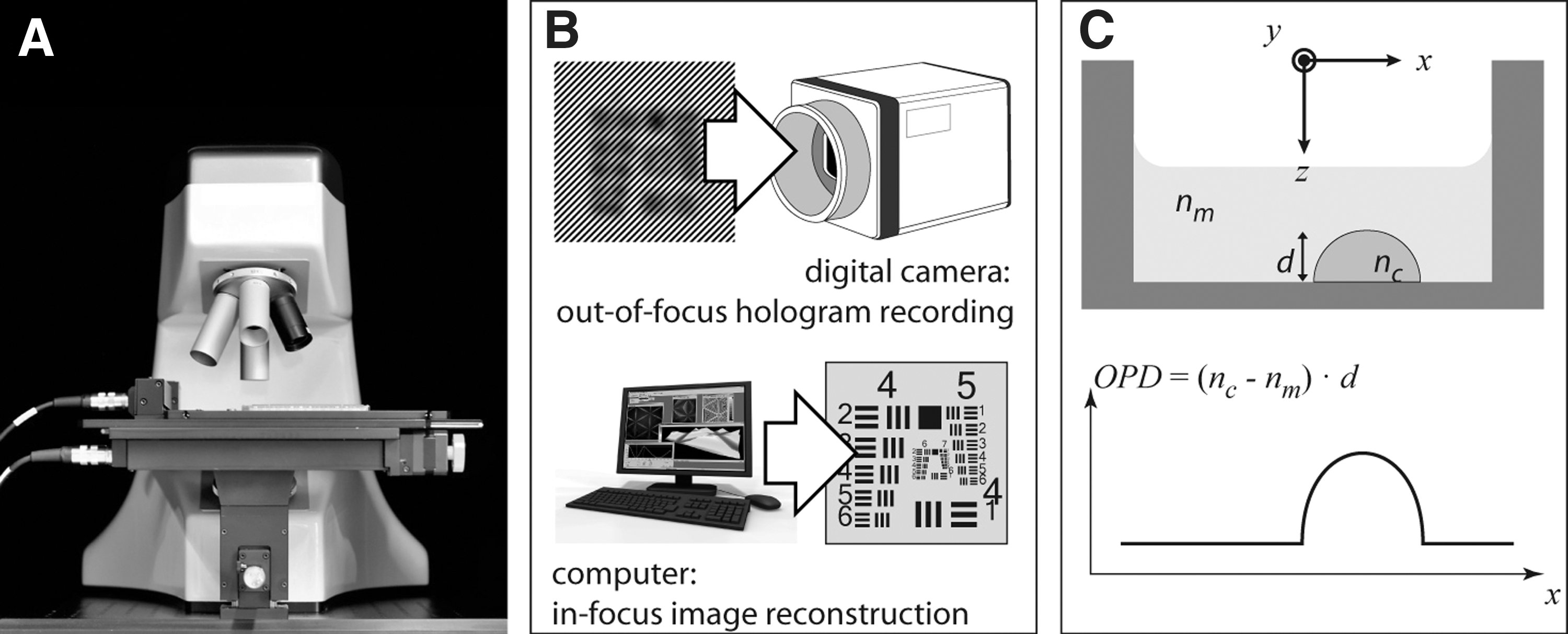
The digital holographic microscopy (DHM) technology.
Label-Free DHM Image Contrast
In essence, DHM is an interferometric microscopy approach to complex light field measurement and in particular provides a quantitative measurement of the optical path length. It uses a reference wave to encode specimen-induced amplitude and phase modulations of a coherent light source into an intensity-only hologram (a hologram is simply the interference pattern generated by the two mutually coherent waves). In the context of cell imaging in a transmitted-light configuration (Fig. 1C), the retrieved quantitative contrast is produced by the optical path difference (OPD) that may be expressed in terms of physical properties as follows:
where d=d(x, y) is the cell thickness, and nc=nc (x, y) is the mean z-integrated intracellular refractive of cell index immersed in a culture medium of uniform and known refractive index nm . The exposure time is 470 μs and the sample illumination is <200 μW/cm2 (>6 orders of magnitude less than with confocal fluorescence microscopy). The wavelength of the laser source is 682 nm.
The OPD signal is, by nature, absolute in scale for a given culture medium osmolarity, without any need for calibration procedures, which might be a potential advantage for automated phenotype recognition with machine-learning software.
Simply put, Equation (1) means that the label-free OPD signal is proportional to both cell thickness and intracellular refractive index—with respective accuracies of around 10 nm and 5×10⁻⁴ as far as cultured cells are concerned. 9
Image Segmentation and Data Analysis
Image segmentation, quantification, and analysis were performed using open-source software CellProfiler (Broad Institute;
We used supervised machine learning approach to determine the percentage of viable cells in each well with the classifier tool of CellProfiler Analyst software (Broad Institute;
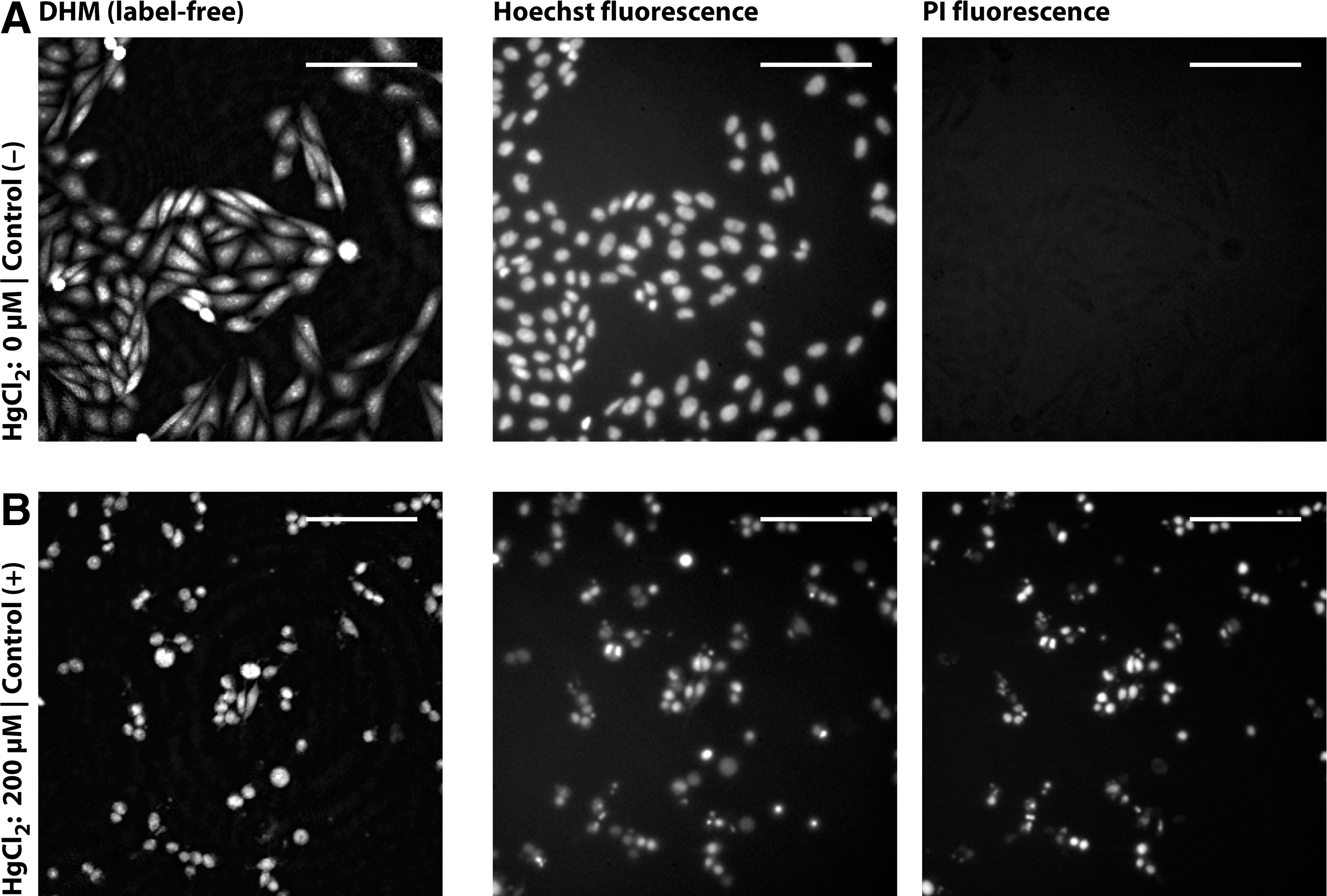
Image contrast comparison between DHM and fluorescence. CHO-K1 cells were plated in a six-well plate and treated
For statistical analyses, the mean viability was obtained for the different concentrations of the toxic compounds and the curves were fitted with Prism5 software (GraphPad). Finally, half maximal inhibitory concentration (IC50) values were calculated from a least squares minimization of variable slope dose–response curves (with four parameters).
Results and Discussion
While use of DHM for monitoring cellular responses had already been extensively reported by our research groups 9,13,14 and others, 15,16 its potential for screening applications had yet to be demonstrated by a passage to multiwell plates and automatic image recording.
In a first step toward this direction, preliminary cell viability experiments were carried out with CHO and HeLa cells in six-well-plate format for a variety of toxic compounds using a dual-mode DHM/fluorescence microscope setup for cell imaging. 21 As an example, Figure 2 depicts images of CHO-K1 cells treated with toxic compound HgCl2, showing a very high qualitative similarity between DHM and fluorescence images. The DHM label-free contrast highlights the morphological and local biomolecule (proteins and nucleic acids) concentration changes induced during cell death. Qualitatively, living cells appear as somewhat triangular or rhombus-like shapes with elongated ends, whereas dead cells form smaller, roughly circular structures. Overall, this demonstrates the suitability of DHM for discriminating living cells from dead ones, just as efficiently as a double fluorescence labeling using Hoechst and PI.
Subsequently, a DHM microscope was equipped with a motorized xy plate holder for automatic image acquisition of cell viability experiments using commercially available 96-well plates. At that point, we made sure that an image processing program (here, CellProfiler) was capable of segmenting and unambiguously detecting cells independently of their number. To this purpose, we plated HeLa cells at different densities and qualitatively evaluated the segmentation performances by overlaying segmented cell contours on DHM images (Fig. 3). As expected, CellProfiler had no trouble segmenting the cells, regardless of their degree of confluence.
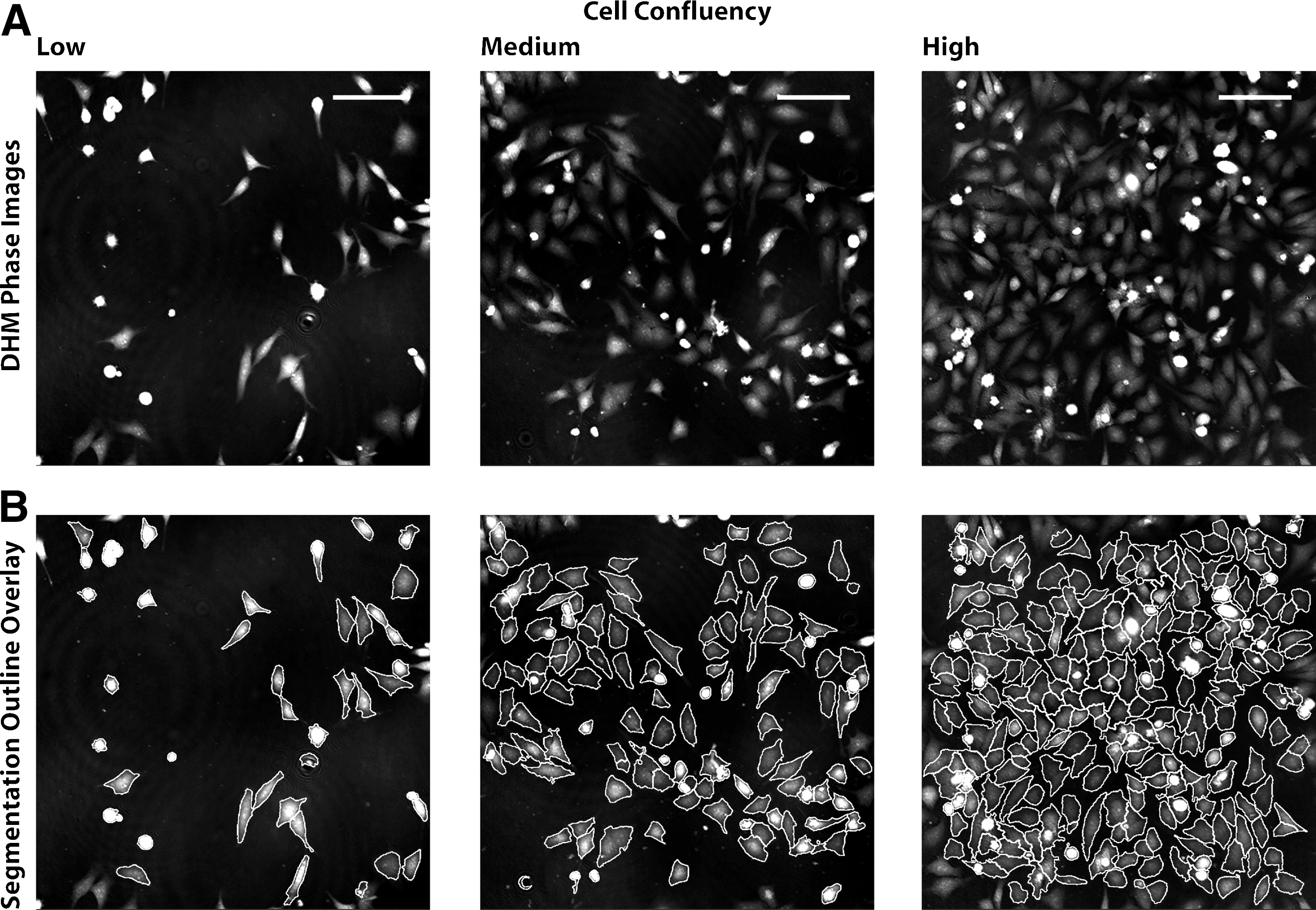
DHM phase images and cell segmentation for different cell confluency. HeLa cells were plated in a 96-well plate at low, medium, and high confluency. After 24 h, plate was imaged by DHM
Assay Performance Assessments
To measure the quality of the cell viability assay by DHM imaging, two separate experiments in duplicated plates have been carried out in 96-well-plate format. For each experiment, cells in half of the plate were treated with 200 μL of HgCl2 (cell death positive control), while those in the other half remained untreated (viable cells). For these experiments addressing the well-to-well and interplate variability, the window coefficient of the screen was estimated using the Z′-factor statistical parameter (a value close to 1 indicates an excellent screening window whereas a value below 0.5 reflects a marginal essay 22 ). We obtained a Z′-factor close to 0.9, reflecting the good quality of the assay for screening purposes (Fig. 4A).
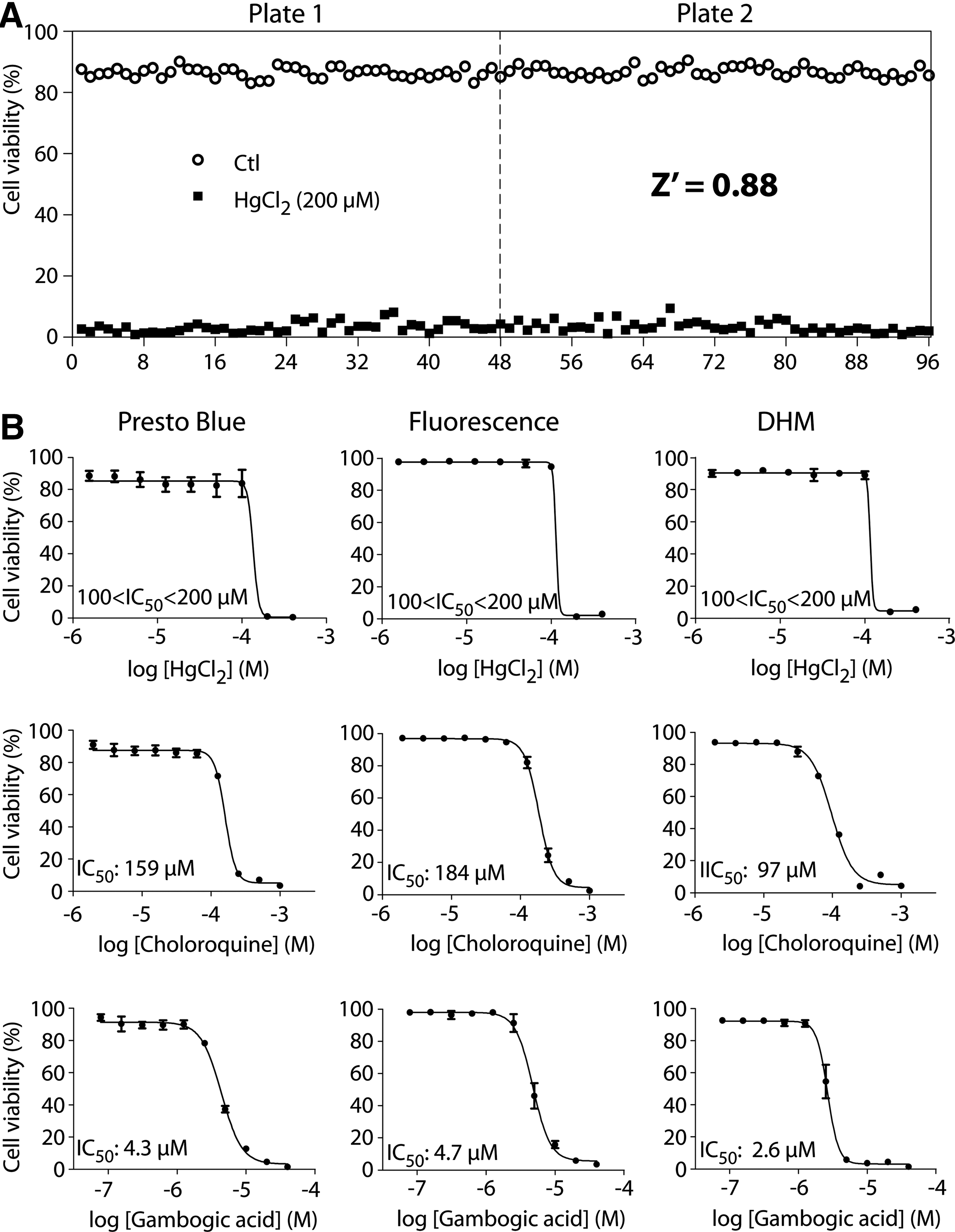
Cell viability assay: Z′-factor determination for DHM readout and dose–response curve comparison with established methods.
Additionally, three well-characterized toxic compounds, selected as they produce distinct cell death pathways, have been used for generating a series of cell viability dose–response curves for PrestoBlue assay, DHM, and PI/Hoechst fluorescence imaging, using HeLa cells. The curves obtained for the three methodologies used are reported in Figure 4B for three compounds: HgCl2, Chloroquine, and Gambogic acid. HgCl2 usually produces a rapid concentration-dependent induction of DNA single-strand breaks, as well as a rapid leakage of superoxide radicals, provoking cell death. 23 Chloroquine is a 4-aminoquinoline drug traditionally used as an antimalarial therapeutic agent but it was also shown to strongly potentiate the inhibitory effect of radiation on cancer cell proliferation by inducing autophagic vacuole accumulation and cell death. 24 Gambogic acid is a natural product shown to be a potent apoptosis inducer that inhibits the growth of a variety of tumors. 25 We have calculated the IC50 values for each of these drugs, inducing cell death by different mechanisms. In at least two independent experiments (n=4 for each drug concentration), IC50 values (Fig. 4B) obtained using the DHM imaging system were in a range of 100–200 μM,* 97.4±5.5 μM, and 2.65±0.11 μM for HgCl2, Chloroquine, and Gambogic acid, respectively, in agreement with the values obtained by the two fluorescence methods tested and with previously reported data for these compounds. 24 –26 Given the high assay quality, DHM appears not only appropriate for high-throughput screening campaigns but also suitable for quantitative characterization of active compounds, such as the accurate determination of IC50 or half maximal effective concentration (EC50) values in hits validation and optimization steps.
Assay Advantages and Limitations
DHM is an optical imaging technique capable of revealing spatially resolved morphological information, just like fluorescence microscopy and some other label-free techniques. To provide a deeper insight on what DHM can or cannot do in a screening environment, we propose to go over the main characteristics of the technology, and compare it to some more widely known techniques.
Multiwell plate compatibility
As this work may attest, DHM is compatible with commercially available multiwell plates designed for imaging applications. Actually, we have even been carrying out experiments with other commercially available multiwell plates that were not specifically designed for cellular imaging purposes (unpublished results). Furthermore, while most label-free sensors may be affected by low cell confluence levels, 7 DHM performs equally well at different degrees of cell confluence.
Digital focusing
The DHM approach has the advantage of operating faster, mostly thanks to the way image focus is performed. Indeed, DHM may be seen as a two-step process (Fig. 1B): during the acquisition process, the hologram is recorded by a digital camera; then, it is transferred to a computer for image (of amplitude or phase contrasts [PCs]) reconstruction. One key element in this workflow is that the hologram does not need to be recorded in focus; in fact, the microscope does not need a motorized z-stage. Image focusing in DHM is performed numerically during the hologram reconstruction step, by using diffraction-based algorithms, which use is made possible only because the technique retrieves both amplitude and phase. For the data disclosed in this article, image reconstruction was performed a posteriori, but existing commercial software like Koala (Lyncée Tec SA) allows up to 100 of such images reconstructions per second and makes live digital focusing much faster than its mechanical counterpart.
Acquisition speed
For the experiments reported here, the acquisition of 4 images per well for a whole 96-well plate takes <4 min in DHM—with yet plenty of room for speed improvement—compared with 30 min for the BD Pathway 435 automated fluorescence microscope. While other label-free microscopy techniques like bright field (BF), PC and differential interference contrast (DIC) microscopy also work with very short exposure times; they are necessarily slower than DHM because of their hardware image focusing. In fact, with typical exposure times in the microsecond range and digital focusing, DHM is ultimately limited in speed only by the plate-scanning mechanism.
Quantitative contrast
Unlike the vast majority of microscopy techniques (BF, PC, DIC, and fluorescence microscopy), DHM benefits from an OPD contrast that is quantitative in an absolute manner, meaning that it is totally independent of the microscope hardware and acquisition settings and it does not require any calibration procedures. In addition, the cellular OPD contrast is relatively monotonous and related to the cell thickness, which eases the isolation of individual cells from a larger population by simply using a threshold-based algorithm (Fig. 3) something not possible with BF, PC, and DIC.
Compared with fluorescence microscopy, DHM is significantly faster in terms of acquisition speed and boasts an intrinsically quantitative contrast. However, like most label-free techniques, it lacks the specificity provided by the wide range of fluorescent probes available. Therefore, both techniques can complement each other and could be combined in a DHM–fluorescence multimodal microscope. 21
In summary, we demonstrated the suitability of DHM, a label-free technology, for automated image-based screening. Results show that its intrinsic optical path length (optical thickness) contrast clearly highlights cellular morphological and local biomolecule concentration changes induced during cell death. Moreover, we have validated that DHM images had a satisfactory Z′-factor to support quantitative cell-based assay analysis. We believe that DHM may be of great interest for screening applications in which its high acquisition speed, ultimately limited only by the plate-scanning mechanism, and its label-free nature may reduce cost per data point. In addition, DHM is inherently free of photo-bleaching issues and, since it operates with a low-intensity light source and over very short exposure times, it does not induce phototoxicity. Obviously, this makes DHM an efficient future tool for time-lapse image-based experiments.
Footnotes
Acknowledgments
This work was supported by the Swiss Initiative in Systems Biology (SystemsX), through the Bridge-to-Industry project (BIP) grant entitled “High-Content Screening by Digital Holographic imaging.” Instrumental developments were supported by the CTI program (grant No. 12669.1 PFLS-LS). The authors thank the staff from Lyncée Tec SA for the technical support and for providing the DHM imaging system, as well as Sylviane Reymond and Nathalie Ballanfat from the BSF-EPFL for the cell preparation and culture. We also thank Dr. Nicolas Pavillon and Dr. Pascal Jourdain for many interesting discussions related to cell death and data interpretation topics.
Disclosure Statement
Christian Depeursinge, Pierre Magestretti, and Pierre Marquet are cofounders of Lyncée Tec S.A.
*
Due to the specific on/off toxic effect of HgCl2 and the sampling (∼100% cell viability at 100 μM and no viability at 200 μM), it is not possible to determine more precisely where the IC50 for HgCl2 stands within this range.