Abstract
Introduction
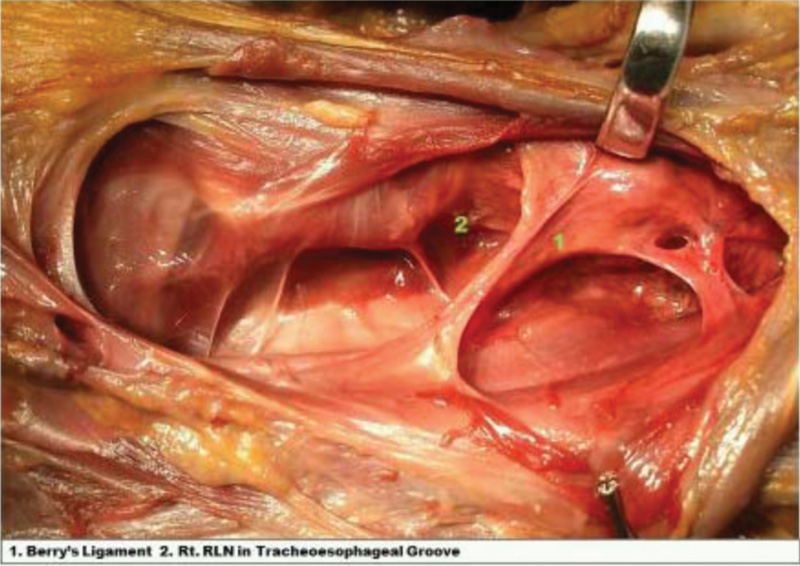
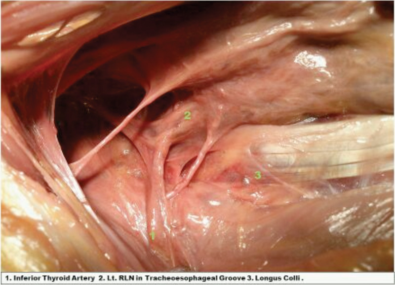
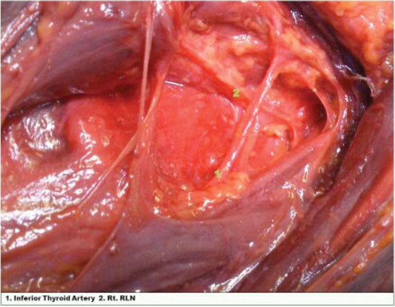
While most cadaveric studies of the Recurrent Laryngeal Nerve (RLN) have focused on course variations as a suitable guide for Right versus Left RLN, they have mostly been done on preserved (fixed) cadavers which renders the RLN immobile. Our aim was to perform anterior cervical exposures from C2 to T2/3 with particular attention to the course of the RLN on right and left sided exposures in fresh cadaveric specimens. In addition, we aimed to expose the entire course of the RLN. Finally, we wanted to show the position of the RLN in relation to the trachea-oesophageal groove, inferior thyroid artery and Berry's ligament.
Methods
Eight fresh cadavers had extensive layer by layer dissections performed by 2 surgeons (one of whom has extensive experience as an anatomy demonstrator and dissector). The RLNs were exposed in their entire length and relationship to different landmarks recorded. Photographs were taken at each stage of the exposure.
Results
In all specimens, we were able to demonstrate the entire course of both RLNs from origin to insertion. The RLNs were consistently associated with the inferior thyroid artery and Berry's ligament bilaterally with the RLNs passing at almost perpendicular to these structures.
Conclusion
The near horizontal direction of the Berry's Ligament in the cervical tissue planes exposed during anterior cervical exposures enables the surgeon to reliably identify the expected position of RLN at its medial end and hence avoid it prior to visual observation of the nerve on either side. We found that the most reliable anatomical landmark bilaterally for the RLN was the inferior thyroid artery and Berry's ligament both of which would be encountered in anterior surgical exposure prior to the nerve itself. We believe that this will help spinal surgeons refine their surgical technique to identify this nerve where necessary and thus reduce the incidence of iatrogenic injury.