
Abstract
Introduction
Back pain is strongly associated with degeneration of the intervertebral disk (IVD), which is associated with ongoing mineral deposition.1 The presence of calcium deposits and type X collagen (COL X) and the level of the indicators of calcification potential (alkaline phosphatase (ALP), Ca2 + ions and Pi) were consistently higher in degenerative and scoliotic discs. We also showed that in mesenchymal stem cells (MSCs), parathyroid hormone (PTH 1–34) inhibits the expression of COL X while promoting type II collagen (COL II) expression, thereby preventing endochondral ossification.2 In this study, we investigated the effect of PTH on expression of COLII, COLX, and ALP in human IVD cells and analyzed the potential mechanisms related to its effect.
Materials and Methods
Human lumbar IVDs from a donor without spinal pathology were obtained within 24 hours after death. Nucleus pulposus (NP) and annulus fibrosus (AF) tissues from the IVDs were digested and the corresponding NP and AF primary cells were isolated as previously described.3These cells were cultured in complete DMEM to 90% confluence. Then the cells were incubated overnight in serum-free medium followed by treatment with 100 nM PTH 30 minutes to 48 hours. Protein expression was analyzed by immunoblotting using specific antibodies to COL I and COL II (Abcam, Cambridge, MA), COL X, and PTH receptor 1 (Sigma Aldrich). Expression and phosphorylation of AKT and MAPKs was assessed by using specific corresponding antibodies (Cell Signaling, Danvers, MA). Alkaline phosphatase activity was measured colorimetrically using the StemTAG kit (Cell Biolabs, San Diego, CA, USA) and Ca2 + release from cells was measured using calcium assay kit (Cayman Chemical, Ann Arbor, MI, USA). Statistical analyses were done using one-way ANOVA, posthoc tests p < 0.05 was considered significant.
Conclusion
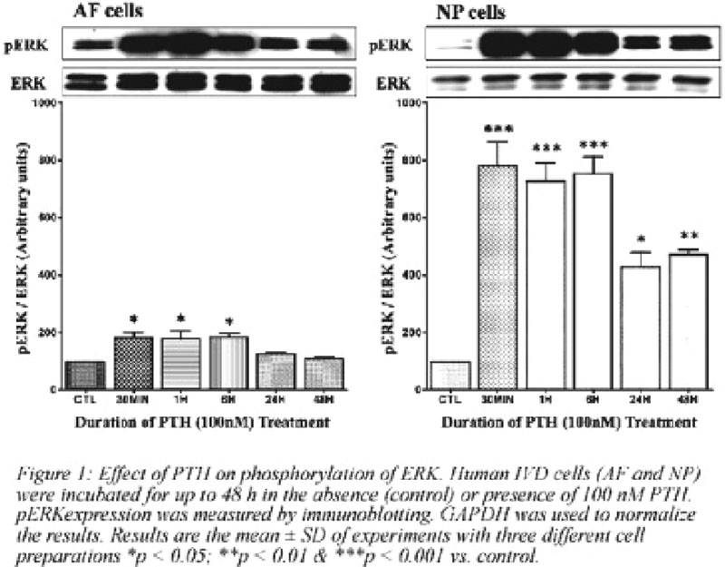
PTH has previously been shown to promote chondrogenesis and to inhibit the expression of COL-X in chondrocytes probably via the activation of MAPK signaling pathways.4 The present results demonstrate that PTH upregulates COL-II and downregulates COL-X in IVD cells, indicating that PTH has the potential of being able to stimulate disk repair and to improve nutrient supply in the degenerative disc. Our data also suggests that activation of MAPK pathway takes place much earlier than the alterations in COL-II or COL-X expression. Interestingly, COL-II expression inversely correlates with alkaline phosphatase activity in NP cells treated with PTH. Although understanding of IVD calcification would be of great value, not only for elucidation of its mechanism, but with an eye toward eventual therapeutic intervention. PTH can thus be used towards disk regeneration therapy.
Yes
None declared
Hristova, GI et al. Journal of Orthopaedic Research 2011; epub
Mwale F et al. Tissue Engeneering Part A (2010)16:3449–3455
Chelberg MK et al. Journal of Anatomy 1995;186 (Pt 1): 43–53
Datta, NS et al. Cell Signalling 2010;22: 457–46