
Abstract
Introduction
Cells of the immature nucleus pulposus (NP) are believed to originate from the notochord with many unique morphologic features,1–3 including larger cell size and vacuolated appearance. NP cells of immature IVD tissues are heterogeneous with a population of cells that are notochordal in appearance. While these morphological characteristics of notochordal cells are absent in NP cells of adult human, it is unknown if a distinct molecular phenotype for notochordal-like cells exists in the cells of human IVD. Previously, we discovered a set of novel cell surface proteins (CD24, CD54, CD155, CD166, CD221), one transcriptional factor (brachyury, T), and three neuronal-related proteins (brain abundant membrane attached signal protein 1, Basp1; Neurochondrin, Ncdn, and Neuropilin, Nrp1) in cells from immature rat NP that is enriched in notochordal-like cells.4,5 The goal of this study is to evaluate whether these proteins can serve as markers of notochordal-like NP cells in the human IVD and the change of these markers during aging by both flow cytometry and immunohistochemistry analysis.
Materials and Methods
Human IVD cells were isolated by a nonenzymatic protocol from NP tissues of patients undergoing surgery for scoliosis (juvenile group, n = 4, 6 to 17yo) and degenerative disk disease (adult group, n = 3, 43 to 71yo). Cells were allowed to become confluent before removal via trypsin and incubated with antibodies against human CD24, CD54, CD155, CD166, and CD221 (with isotype controls and secondary fluorescent antibodies). To evaluate brachyury expression, cells were first fixed and permeabilized before incubation with antibodies. The percentage of cells with positive protein and mean fluorescence intensity (MFI) was analyzed by flow cytometry (Accuri C6). Analyses were performed to detect differences in percentage of ( + ) cells between the two age groups (one-factor ANOVA). Frozen tissue sections were fixed and permeabilized, then incubated with specific antihuman antibodies for selected NP markers (CDs, brachyury T, Basp1, Ncdn, Nrp) and secondary antibodies, counterstained with propidium iodide and imaged using confocal microscopy.
Results
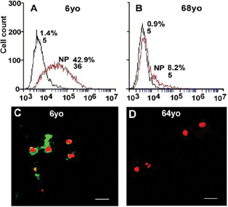
NP cells from the juvenile group stained positively for all studied cell surface antigens and brachyury (Table 1). A significant lower percent of adult NP cells stained positively for CD24, CD54, and brachyury (p < 0.05), as compared to the juvenile NP cells; in contrast, a higher percentage of adult NP cells stained positively for CD155 and CD166 (p < 0.05). No differences in CD221 expression was detected between the two age groups (TABLE). All CD surface proteins, brachyury T, Basp1, Ncdn, and Nrp1 were detected in juvenile NP tissues via immunostaining, however only Basp1 and CD24 stained intensively positive (Fig. 1C) and others were only moderately positive. In contrast, immunostaining only detected moderate expression for CD54 and CD166, but not brachyury T, CD24, CD155, CD221, Basp1, Ncdn, and Nrp1 in adult NP tissues (Fig. 1D).
Average % of Positive Cells and MFI Analyzed by Flow Cytometry for the Selected NP Markers (*Significant Difference Detected between Groups, One-Factor ANOVA, p < 0.05)
| NP Markers | Juvenile (n = 4) %(+) MFI | Adult (n = 3) %(+) MFI |
|---|---|---|
| CD24 | 3414 | 8*5 |
| CD54 | 82321 | 12*50 |
| CD155 | 26336 | 39*39 |
| CD166 | 25293 | 70*118 |
| CD221 | 3346 | 29298 |
| Brachyury T | 25293 | 5*45 |
Figure (A, B) Representative histograms of flow cytometry illustrated the relative fluorescence intensity of CD24 on X-axis for isolated NP cells (6 and 68 yo). The numbers appeared in each histogram indicate MFI and % positive fluorescence labeled cells. (black line: isotype control, red line: NP cells). (C, D)Immunostaining illustrated age-related changes in CD24 expression in NP cells (6 and 64 yo). Bar: 20 µm.
Conclusion
The results of this study suggest the utility of a new set of NP cell markers (CD24, CD54, CD155, CD166, CD221, brachyury T, Basp1, Ncdn, and Nrp1) for identifying notochordal-like NP cells in the human IVD. These findings are consistent with our previous findings for notochordal-like cells in the rat NP5. Our data confirm consistent expression of notochordal-like NP molecular phenotypes across species and in cases where the notochordal-like cell morphology is not preserved.
Yes
None declared
Rufai et al. Anatomy and Embryology (Berl) 1996;192:53
Trout et al. Tiss Cell 1982;14:359
Urban and Roberts. Molecular Medicine Today 1995;1:329
Chen et al. Trans ORS 2007;32:1104
Tang X et al. Trans ORS 2011;36:470