
Abstract
Introduction
In utero exposure to Diethylstilbestrol (DES) has been shown to adversely affect a number of estrogen sensitive tissues, including bone. Effects of in utero DES exposure have also been shown to affect the third generation. Environmental exposure to estrogens was shown to be higher than anticipated. The aim of this study is to determine the effect of in utero exposure to DES on the intervertebral disk (IVD) of adult mice.
Materials and Methods
Animal Manipulation - Pregnant C57/bl mice were injected with either vehicle (peanut oil) or one of the three doses of DES (0.1, 1.0, and 10.0 µg/kg/day) at 11 to 14 days of gestation. A minimum of three dames were set for each group. Pups were then allowed to grow to adulthood without further intervention until 3 months of age. At this point, mice were randomized into two groups; one group with a once daily swimming regimen which started as 5 minutes and was escalated to a maximum of 1 hour. The second group was left to their normal activity level and considered sedentary. All animals were sacrificed at exactly 4 months of age. Histology, the lumbar segment of the spines was dissected from three animals per group for histologic evaluation, sections were stained with 0.1% Safranin O and counter stained with 0.02% Fast Green. All slides were scanned and measurements of intervertebral disk height were taken using the NDPview software. Three measurements of disk height were taken per disk and the means compared for control versus DES exposed mice. Proteoglycan content-proteoglycans in the supernatant were measured using the DMMB assay. Statistical analysis-all statistical analyses were done using ANOVA and Fischer Least Significant Difference Post Hoc test, except for histological quantitative analysis where the Mann-Whitney test was used.
Results
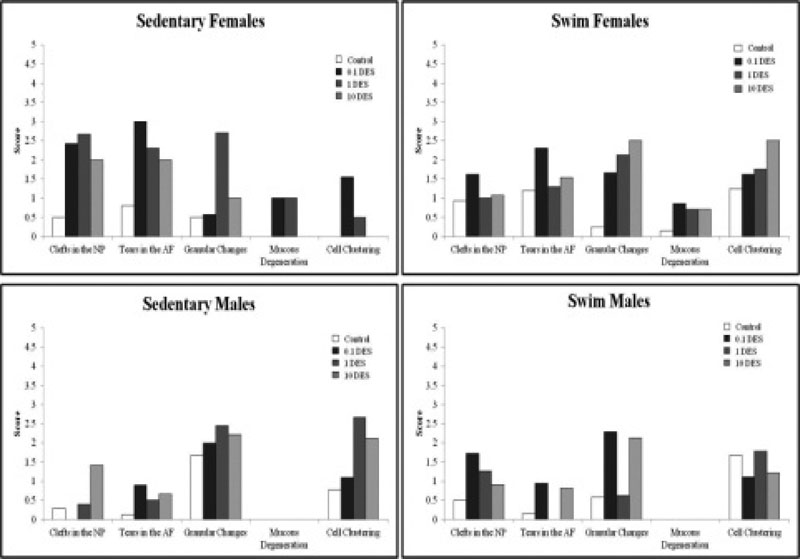
Parameters associated with IVD degeneration were found to have higher scores in DES exposed mice versus controls (Fig. 1). Clefts in the NP were only significantly increased in the female swim group at 0.1 µg/kg/day (p = 0.007) whereas the female sedentary group showed significant increases at all three doses of DES (p < 0.0001 for all three). NP clefting was less evident in males, where the male swim group showed a significant increase at 0.1 and 1.0 µg/kg/day (p = 0.002 and p = 0.02) and the male sedentary group showed a significant increase at 10.0 µg/kg/day (p = 0.002). Tears in the AF showed a similar pattern as NP clefting, where the female swim group was only affected at 0.1 µg/kg/day (p = 0.001) and the female sedentary group showed significant increases at all three doses (p < 0.001 for all three). The male swims showed increased AF tearing at 0.1 and 10.0 µg/kg/day (p = 0.02 and p = 0.0004) while the male sedentary group showed a significant increase at 10.0 µg/kg/day (p = 0.007). Granular changes were significantly increased at all three doses in the female swim group (p = 0.0003, p = 0.002 and p = 0.0003). The female sedentary group showed significant increases at 1.0 and 10.0 µg/kg/day (p = 0.0001 and p = 0.03). Male swims showed significant increases at 0.1 and 10.0 µg/kg/day (p < 0.0001 for both). Sedentary males showed no significant granular changes compared to control. Mucous degeneration was only increased in the female swim group at all three doses of DES (p = 0.004, p = 0.03 and p = 0.03). The female sedentary group showed nonsignificant increases at 0.1 and 1.0 µg/kg/day. No significant changes in mucous degeneration were seen in the males. Cell clustering was significantly increased in the female swim group at 10.0 µg/kg/day (p = 0.002). The female sedentary group showed significant increases at 0.1 and 1.0 µg/kg/day (p = 0.002 and p = 0.03). The male sedentary group showed significant increases at 1.0 and 10.0 µg/kg/day (p < 0.0001 and p = 0.0007). No significant changes were seen in the male swim group compared to control. DES had little effect on male IVDs than those of females. Swimming caused an overall worsening of scores in females. Males were less affected by swimming than females. Proteoglycan content in the intervertebral disk was significantly decreased at 0.1 µg/kg/day and 1.0 µg/kg/day. A significant increase was noted at 10.0 µg/kg/day as compared to control.
Conclusion
The intervertebral disk was recently shown to be an estrogen-sensitive tissue. This had a clinical effect in obese postmenopausal women undergoing hormone replacement therapy. Our study shows that DES in utero exposure can cause nuclear extrusion and affect the intervertebral disk. The mechanisms of the effects of DES are however not well understood and further studies are necessary. This study might shed light on the possible increased risk of disk degeneration in the sons and daughters of mothers exposed during gestation, as well as their children. Furthermore, this new found environmental exposure to estrogen agonists makes it important to understand the full effects that such estrogen agonists may have.
Yes
None declared