
Abstract

A four and a half-year-old castrated Persian was presented for trauma subsequent to having been missing for five days. There was avulsion of the skin on the ventral surface of the tail adjacent to the anus. This was associated with bruising, infection and necrosis distally (Fig 1). Other injuries noted were fairly superficial and included skin abrasions of the lateral right hock. Haematology, biochemistry and radiography of the thorax and pelvis were unremarkable.
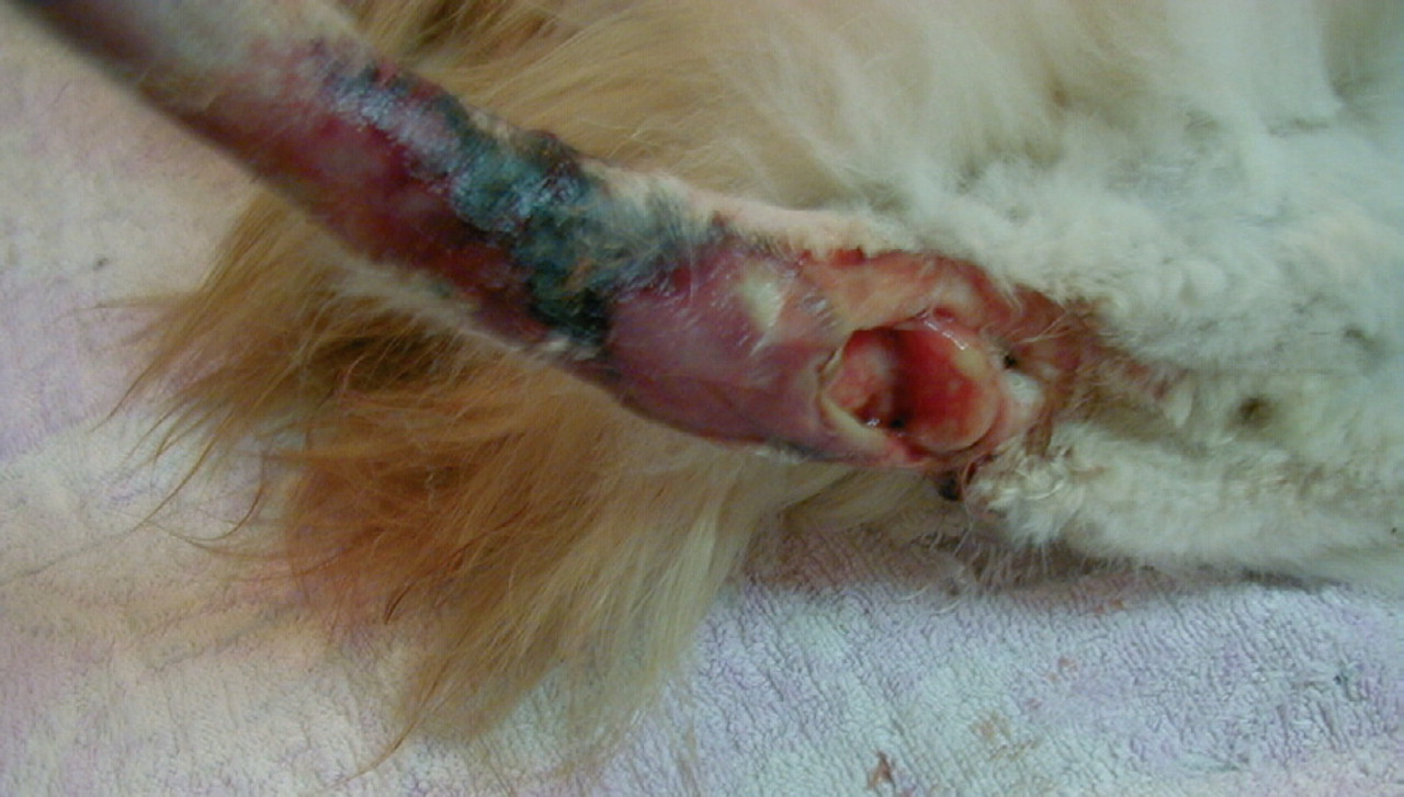
Appearance of the cat's tail at presentation
The tail was debrided, flushed with saline and bandaged. The cat was given amoxycillin/clavulanate (70 mg subcutaneously then 125 mg orally every 12 h) and metronidazole (100 mg orally once daily).
Two days later, the bandage was changed and the wound cleaned and debrided. Five days after presentation, infection and necrosis had extended to include the whole tail; distally the tail was oedematous and cold. The tail was amputated at S3/Co1. The same antibiotics regimen was continued.
Recovery was uneventful until two days post-surgery when an altered gait was first recognised. All muscle groups of the left hind leg were hypertonic (Fig 2) and the patellar reflex was exaggerated. The left hind leg was hyper-aesthetic and developed an intermittent tremor. The right hind leg subsequently became similarly but more mildly affected. Small amounts of faeces dribbled from the anus, although anal tone was present and the cat was also able to defaecate consciously. Bladder tone and micturition were normal. The surgical wound and hock abrasion were healing satisfactorily.

Note the extensor rigidity of the left pelvic limb
What is your diagnosis?
What further diagnostic tests could be considered?
What is this cat's prognosis?
(Answers on page 222)
Answers to questions on page 221.
What is your diagnosis?
The clinical signs that this cat showed were virtually pathognomonic for local tetanus, especially considering the history of a necrotic infection in a compatible anatomic position. Much less likely differential diagnoses might include localised meningitis, neuritis or myelitis of peripheral nerves or nerve roots due to toxoplasmosis, feline infectious peritonitis, neoplasia or unknown causes. Tetanus occurs subsequent to germination of Clostridium tetani spores following their introduction into wounds. C tetani produces an exotoxin, tetanospasmin, which inhibits release of the inhibitory neurotransmitters glycine and gamma-aminobutyric acid (GABA). This disinhibition of spinal motor neurons results in increased motor unit activity and muscle spasm (Bizzini 1979, Greene 1998). The incubation period can vary from three to eighteen days after an injury (Lee & Jones 1996, Greene 1998), depending on the proximity of the injury to the CNS, the degree to which local oxidation-reduction potential favours toxin production and the numbers of organisms present (Baker et al 1988). Cats are naturally resistant to tetanus especially compared to horses and man. This inherent resistance is related to the inability of the toxin to penetrate and bind to nervous tissue (Greene 1998), and likely accounts for the ability of the disease to remain local. Local tetanus has been noted after routine surgery (such as ovariohysterectomy) as well as trauma (Lee & Jones 1996).
What further diagnostic tests could be considered?
Definitive diagnosis of tetanus can be confirmed by isolation of C tetani from an infected focus (Fildes et al 1931), although neurological signs may not occur until initiating wounds have healed (Malik et al 1989, Lee & Jones 1996). Culturing anaerobic bacteria is difficult and is often unrewarding, thus failure to culture C tetani does not rule out the disease (Baker et al 1988, Greene 1998). Serum tetanus antibody concentrations may be useful (Baker et al 1988) but are of very limited availability. Electromyography (EMG) typically demonstrates characteristic persistent motor unit activity (Lee & Jones 1996, Greene 1998) but is not readily available. Diagnostic criteria for local tetanus have been developed for human and veterinary medicine (Millard 1954, Malik et al 1989, Lee & Jones 1996); Table 1. Extreme spasticity localised to one limb subsequent to penetrating trauma is considered virtually pathognomonic for this condition (Malik et al 1989, McKee 1994).
Diagnostic criteria for local tetanus

What is this cat's prognosis?
The prognosis is excellent. Clinical signs typically resolve entirely within two to three months (Malik et al 1989, McKee 1994, Lee & Jones 1996, Greene 1998). In this case, all neurological signs had resolved 4 weeks after their onset. Local tetanus can progress to generalised if there is inadequate wound management, inappropriate antibiosis or concurrent corticosteroid use. A case report from the pre-antibiotic era noted a cat that did progress from local to generalised tetanus (Bateman 1931), as did a cat in a more recent paper for which an inappropriate antibiotic (a fluoroquinolone) was used (Malik et al 1998). Antibiotic agents to be used against C tetani must have an anaerobic spectrum of activity. Metronidazole has been shown to be superior to penicillin G and tetracycline in clinical and experimental tetanus. It is bactericidal against most anaerobes and achieves effective therapeutic concentrations even in anaerobic tissues (Greene 1998).
Further to wound management and antimicrobial therapy, other potential treatments include tetanus antitoxin and muscle relaxants. Tetanus antitoxin cannot dislodge tetanospasmin after its entry into the peripheral nerve. Hence its main purpose would be to neutralise circulating toxin outside the nervous system (Lee & Jones 1996, Habibah et al 1998); this is unlikely to be necessary if signs are localised and nonprogressive. The main benefit of muscle relaxants would be to reduce discomfort during the prolonged recovery period (Lee & Jones 1996). Diazepam has been inconsistent in providing relief to cats with local tetanus, sometimes achieving noticeable relief (Malik et al 1989) and on another occasion, appearing to be of no benefit (Lee & Jones 1996). It is likely that in most cases, no treatment beyond wound management and antibiotics is required. This cat was given diazepam, as a centrally acting muscle relaxant, sporadically over the first seven days after the onset of neurological signs. Combination antimicrobials were given for a total of three weeks.