
Abstract
Therapy for many of the neocortical epilepsies remains unsatisfactory. Recent research has demonstrated that focal cooling, using thermoelectric (Peltier) devices, may be capable of terminating, or possibly even preventing, some types of seizures.
Local Cooling—A Potential Therapy for Seizure Control
Another promising experimental therapy is local cooling. Substantial physiological precedent exists for investigating local cooling, an alternative evidence-based technique, as a means to enhance the surgical management of neocortical epilepsy. The first studies to demonstrate that cooling results in reversible, focal inactivation of the brain were carried out independently by Stefani and Trendelenburg more than a century ago and were summarized by Brooks (8). Research reports and reviews, published as recently as 2002, confirm the ability of local cooling to reversibly deactivate cortex in vivo (9–11). Although not yet definitively shown, cooling likely exerts its effects through a variety of mechanisms, including the blocking of vesicular transmitter release and voltage-gated ion channels (12,13).
For centuries, clinicians have recognized a relation between seizures and body temperature, and modern accounts persist of terminating human seizures by brain cooling (14). Local application of cold saline can terminate both induced and spontaneous epileptiform activity that occurs during intraoperative mapping (15,16). During the 1970s, animal experiments showed diminished interictal bursting during slow cortical cooling with several epilepsy models (17). However, no attempt was made to stop seizures immediately in any of these experiments. Currently, several different laboratories are using brain-slice preparations to reexamine the therapeutic potential of cooling for epilepsy. For example, Smith et al. (18) used the potassium channel blocker, 4-aminopyridine, to induce spontaneous bursting in rat hippocampal slices. They then found that slowly cooling the perfusate to 24°C reduced both burst frequency (by more than 90%) and burst amplitude. Similarly, Javedan et al. (19) recently demonstrated that slowly cooling the perfusate bathing hippocampal slices from 34°C to 20°C reversibly abolished the rapid gamma oscillations (approximately 40 Hz) evoked by a burst of tetanic stimulation. The researchers reported preservation of normal evoked excitatory synaptic potentials when the slice was slowly cooled to 20°C. The dissociation between disappearance of gamma activity and maintenance of evoked synaptic responses suggests that cooling may limit paroxysmal activity without shutting down normal brain electrical activity.
The availability of small thermoelectric (Peltier) cooling devices, initially developed for the computer industry, has permitted the investigation of a different cooling technology as a therapy for intractable epilepsy. Thermoelectric heat pumps are composed of arrays of crystalline semiconductors, each of which generates a temperature differential when electrically polarized. The numerous semiconductors are arranged in parallel lines, connected electrically in a series, and sandwiched between metallized ceramic plates to create a wafer (see Fig. 1). When current flows, one of the plates rapidly cools, and the other heats. To maintain effective cooling of one side of the wafer, heat must be continually removed from the opposite or “hot” side by connecting it to a heat sink or heat pipe. Thermoelectric devices are commercially available in a variety of sizes, as small as 3 mm wide and 2.5 mm thick. Fortunately, substantial economic motivation exists to improve the design of thermoelectric heat pumps and heat pipes because they also are used to cool computer microprocessors (20).
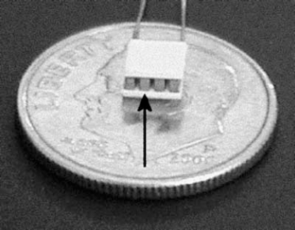
Example of a small thermoelectric cooling device used in research on seizure control. Arrow, one of the individual semiconductors, which is attached to the two ceramic wafers.
Hill et al. (21) were the first group to investigate the ability of thermoelectric devices to abort 4-aminopyridine–induced ictal activity in hippocampal slices. They implanted a small thermoelectric device into the base of their perfusion chamber and placed the slice on its surface. They found that seizures terminated within a few seconds when cooling was activated at burst onset. Seizures in slices cooled as low as 21°C stopped within about 8 seconds, whereas in the control slices, ictal events lasted 35 seconds. In some of the experiments, seizures automatically were detected by a thresholding algorithm, which was then used to trigger the cooling. Importantly, activation of the thermoelectric device terminated seizures only when it was in direct physical contact with the slice, making it unlikely that seizure termination was a result of the electrical field generated by the thermoelectric unit.
Yang et al. (22,23) moved on to investigate the effect of rapid cooling on in vivo seizures in anesthetized rats. A frontal bone window was removed, and focal seizures were produced by microinjecting 4-aminopyridine into the motor cortex. Within about 15 minutes of convulsant injection, discrete ictal events lasting 80 to 100 seconds were detected by standard bipolar EEG recording. Normal background EEG activity recovered between these events. A small thermoelectric device was then placed in direct contact with the dura overlying the site of drug injection. Once again, if the device was activated at seizure onset, seizures were abolished, typically within 10 seconds.
The cortical surface was never cooled lower than 20°C in these experiments, but had to be cooled below 26°C to have any effect on seizure duration. Although severe cooling definitely can damage the cortex, no injury was observed, and the cortical surface temperature returned to baseline within seconds of thermoelectric inactivation. In subsequent experiments, a thermocouple, mounted in a 33-gauge needle, mapped the temperature gradient immediately beneath the thermoelectric device. The temperature decreased rapidly but was no different from core brain temperature beyond 4 mm, suggesting that this method produces extremely restricted cooling. Moreover, recent experiments demonstrated that intermittent cooling, either before 4-aminopyridine injection or at regular intervals after injection, influences seizure development (24). In both instances, the subsequent seizure activity was significantly briefer and less intense.
Possible Clinical Applications for Thermoelectrical Cooling Devices
In at least three clinical situations, focal cooling with Peltier devices might improve therapy for patients with intractable focal seizures. First, a Peltier device potentially could be used as an ancillary tool during focal cortical resection. With patients for whom a restricted site of seizure origin has been identified by prior invasive monitoring or intraoperative electrocorticography, the specific area could be cooled to verify that local cortical inactivation has aborted the abnormal electrical activity. Furthermore, while patients are awake, temporary inactivation of a discrete region suspected of generating focal seizures offers the possibility of anticipating potential cognitive consequences before irreversible cortical resection. Although microelectrodes have been used for years to stimulate and inactivate specific brain regions focally, cooling potentially may provide a more accurate functional localization because it inactivates a more restricted region of cortex.
A second possible therapeutic improvement may be the addition of a thermoelectric device to the recording grids presently used for invasive monitoring. The grids are comprised of arrays of regularly spaced recording electrodes implanted in the subdural space and, are typically kept in place for several days to provide high-resolution localization of a patient's spontaneous seizures (3). The monitoring apparatus is especially useful for children too young to tolerate attempts at intraoperative localization. If an array of thermoelectric devices were added to the grid, these devices could be used to inactivate specific regions from which seizures appear to arise and would enable clinicians to identify definitively the cortical regions responsible for a patient's spontaneous seizures. Furthermore, reversibly cooling prospective areas of resection could reveal possible neuropsychological consequences of the procedure, before any permanent loss of cortex.
A third and most ambitious therapeutic improvement would be the development of an implantable cooling device as an alternative to neocortical resection. The device could be activated by a seizure-detection or -prediction algorithm (25–29). Resection would remain the most definitive and straightforward therapy for a seizure focus that is unambiguously localized to “nonessential” cortex. However, cooling would be extremely attractive if the epileptogenic focus were located in primary motor or language cortex because it would prevent the neurologic deficits associated with permanent cortical resection.
Technical Problems Associated with Thermoelectrical Cooling Devices
Despite the obvious benefits of focal brain cooling, technical problems must be overcome before cooling can be used as a diagnostic and therapeutic tool for focal epilepsies, especially those arising from the neocortex. First, it is necessary to address a possible physical limitation associated with thermoelectric devices—heat dissipation on the warm side of the wafer. In electrical appliances and computers, heat is removed with fans or large heat sinks. These options are not applicable to thermoelectric devices implanted in the cranium. However, significant advances have occurred in the fabrication of thin, bendable, and flexible heat pipes, some less than 0.5 mm thick, which could be attached to the warm side of thermoelectric devices. Theoretically, heat pipes could efficiently transfer heat to either the vascular dura or scalp, where blood flow would rapidly dissipate it throughout the body. These heat pipes would be thin enough to insert into a cortical sulcus and configured to allow cooling of epileptic cortex buried within a sulcus. In contrast, heat transfer is much less an issue for hand-held thermoelectric devices used for intraoperative mapping. For instance, the thermoelectric device previously described for use in rodents has a copper rod attached to serve as both a handle and a heat sink (22).
Second, the extent of cortical injury, if any, induced by cooling must be determined. It is conceivable that direct contact with a thermoelectric device could damage cortex, even if the temperature is kept above 20°C. However, research thus far has demonstrated that rats treated with thermoelectric devices do not have cortical injury in the first several days of cooling (22). Even more encouraging are the findings reported by Lomber et al. (9,10) that no evidence is found of anatomic or electrophysiologic neocortical damage after daily cortical cooling to below 20°C for 1.5 hours in the monkey and cat for 1 and 2.5 years, respectively (S.G. Lomber, personal communication, May 2002).
Finally, functional development of an implantable device will proceed more quickly when detection or anticipation algorithms, which are applicable to human seizures, have been designed. Much work already has been done to develop these programs, and although no unequivocally validated programs presently are available, some very encouraging preliminary results exist [30].
Chilling by the End of the Decade?
Although advances in technology clearly are necessary before focal cooling for epilepsy becomes a clinical reality, the goal is not overly ambitious. Several groups have made seizure detection and anticipation a major research focus and, concurrently, the necessity for controlling processor temperature in personal computers is rapidly driving research on thermoelectric devices and heat pipes. It is easy to imagine a hand-held device, applicable for intraoperative cortical cooling, available by the middle of the decade. If a hand-held device proved successful, an implantable device could be implemented by the year 2010—the centennial of Trendelenburg's first description of the functional effects of cooling the mammalian brain.
Footnotes
Acknowledgment
This work was supported by grants from Citizens United for Research in Epilepsy, Inc. (CURE), the Stein Fund for Pediatric Neurology Research, the McDonnell Center for Higher Brain Function, and the NIH (R01 NS 37773 and R01 NS 42936).