
Abstract
In Spain, snakebites are uncommon medical emergencies that cause barely 100 hospitalizations annually. Most of the venomous bites are by snakes of the Viperidae family. Venom from Vipera snakes is reported to have cytotoxic and hematotoxic effects, and neurological effects have also been described. Ptosis (cranial nerve III palsy) is the most common sign, although any cranial nerve can be affected. We describe isolated ptosis, which was very likely after a Vipera aspis bite in the East Catalonian Pyrenees. No antivenom was administered. The ptosis resolved spontaneously within 10 h. Although neurologic findings are usually mild, they indicate a moderate or severe envenomation. Treating snakebites can be challenging for clinicians, especially when there are uncommon clinical manifestations. A toxicologist at a poison center should be consulted to help guide management. Development of local protocols may provide clinical support.
Introduction
In Spain, bites of venomous snakes are uncommon and cause death only in 1% of all hospitalizations.1,2 Three Vipera species can be found in Spain: Vipera aspis, Vipera latastei, and Vipera seoanei. Two other species of venomous snakes, the non-front-fanged colubroids Malpolon monspessulanus and Macroprotodon brevis, occur in Spain. However, most reported cases of envenomation are caused by snakes of the family Viperidae. The most common location of bites is the upper extremities. In Spain, snakebites more commonly affect males than females. 3 Signs of neurologic envenomation after V aspis bite have been previously reported in the southeast of France. 4 Although neurologic findings are usually mild and transient, they may indicate moderate or severe envenomation. We describe a case in which the only neurologic manifestation was bilateral ptosis after a V aspis bite.
Case Report
An 85-y-old male patient was admitted to the emergency department in summer 2017 after a snakebite. He was bitten on the fourth finger of the right hand after he picked up a log while working in his orchard. The bite occurred in the East Catalonian Pyrenees at 1100 m in the small village of Fontanals de Cerdanya, near the city of Puigcerdà. V aspis is the only snake found at that altitude and location whose venom can cause neurologic manifestations.
The patient received no prehospital care. He was immediately driven to the hospital, a distance of less than 10 km, by his son. The family also brought in the dead snake. The emergency physicians identified it as V aspis based on the location at which the bite occurred and by identifying the most common features, including a length of <70 cm, a triangular head, solenoglyphous dentition (long, hollow fangs that fold into the roof of the mouth when the jaws are closed), carinate scales, and vertical pupils.
On presentation to the emergency department, the wound had already developed local edema and ecchymosis (Figure 1). The extent of the edema was outlined with a permanent marker. On the primary survey, the patient was alert and able to converse normally. The Glasgow Coma Score was 15. Vital signs were as follows: respiratory rate 12 breaths·min-1, heart rate 69 beats·min-1, blood pressure 128/62 mm Hg, and SpO2 92% on room air. The patient had full range of motion of all extremities. A peripheral line was placed, and a blood sample was obtained, which showed the following: hemoglobin 15.1 g·dL-1, leucocytes 5.4 × 10³·μL-1, platelets 42 × 109·L-1, prothrombin time ratio 1.05, international normalized ratio 1.08, activated partial thromboplastin time ratio 0.99, glucose 122 mg·dL-1, creatinine 0.84 mg·dL-1, glomerular filtration rate >60 mL·min-1, and potassium 4.3 mmol·L-1. The blood tests were repeated in 12 h and still showed normal coagulation parameters but a rising leukocyte count of 13.9 × 10³·μL-1. Antibiotic treatment with 1 g of amoxicillin/clavulanate was initiated because of suspected local infection, based on an expanding area of erythema and edema with warmth. A tetanus booster was given.
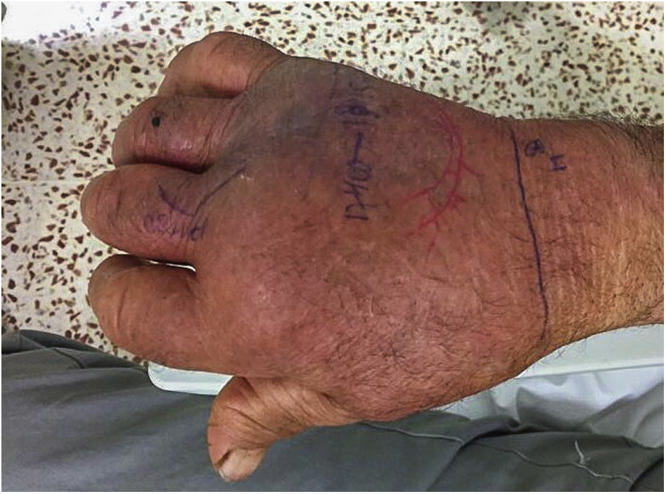
Bite on the fourth finger of the right hand.
The patient had sudden onset of bilateral ptosis during a repeat examination 6 h after the bite (Figure 2). The palpebral fissures were equally closed bilaterally. No other neurologic effects were observed. Cranial nerves II-XII were intact, except for the third cranial nerve palsies. Pupils were equal, round, and reactive to light. Extraocular muscles were intact. Because the patient was not able to blink, glabellar reflexes were not tested. Visual acuity was normal. Visual fields, tested by confrontation, were normal. He was unable to open his eyes for almost 10 h. The emergency physicians considered the risk-benefit ratio of administering antivenom. They did not administer antivenom because the only sign of toxicity was the isolated third cranial nerve palsies. They decided to wait, reassess the patient frequently, and administer antivenom if his condition worsened. The patient was admitted for observation. After 24 h of observation, he was discharged home without neurologic or other sequelae.
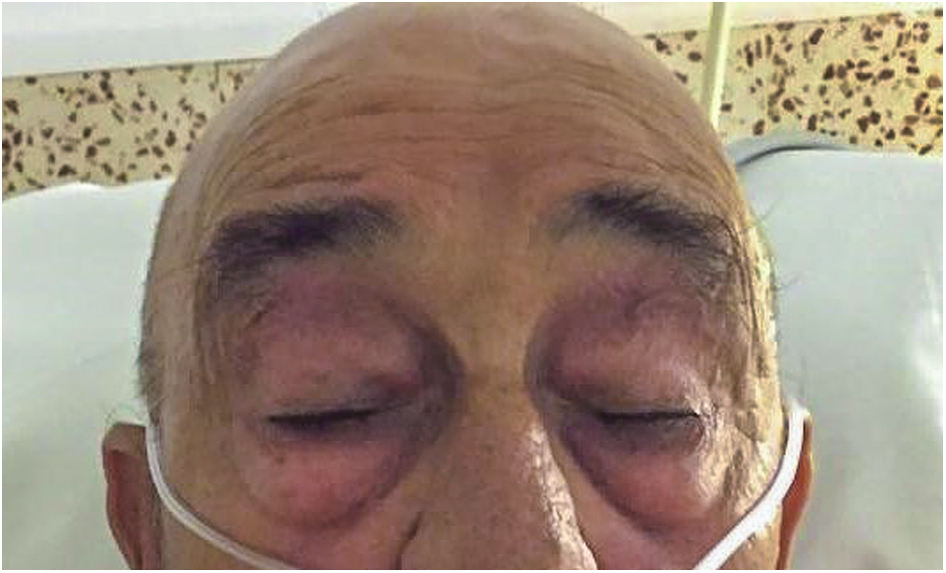
Bilateral ptosis.
Discussion
Most envenomations by snakes in the Pyrenees mountains are caused by V aspis. Neurologic manifestations of bites from both V aspis subspecies living in the Pyrenees, V aspis aspis and V aspis zinnikeri, have been described, 4 although no case report of a neurologic manifestation in Spain has been previously published as far as we are aware. Venom of V aspis contains phospholipase A2 neurotoxins that are the likely cause of neurologic manifestations affecting pre- or post-synaptic neuromuscular transmission. 4 -7 Neurologic manifestations can include generalized weakness, paralysis of extraocular muscles, diplopia, ophthalmoplegia, dysarthria, dysphagia, and flaccid paralysis. Up to 15% of European Vipera spp. bites can cause cranial nerve palsies. 5 Cranial nerve palsies are usually seen in the first 4 to 12 h after the bite and can be the only neurologic manifestation. The most common sign is ptosis, followed by ophthalmoplegia.
The presence of neurologic manifestations indicates moderate or severe envenomation. 8 Standard treatment for moderate or severe envenomation is immunotherapy with Fab or F(ab')2 fragments. 8 -10 Viperfav, an antivenom that contains F(ab')2 fragments of purified equine antibodies, is effective against the neurologic injury caused by V aspis. Viperfav is most effective when administered in the first 10 h. 8
General management includes local wound care, monitoring the spread of local edema, monitoring vital signs, and treating pain. Routine use of corticosteroids is contraindicated. Antibiotics should only be given if an infection is suspected. Secondary infection after European Vipera bite occurs in fewer than 4% of cases. 5 A tetanus booster should be given if indicated.
Conclusions
Envenomation by V aspis can cause a broad range of manifestations, including neurologic effects. Although abnormal neurologic findings are usually mild, their presence indicates moderate or severe envenomation, requiring treatment with antivenom. The decision whether to administer antivenom can be challenging, especially when there are uncommon clinical manifestations. A toxicologist at a poison center should be consulted to help guide management. Development of local protocols may provide clinical support.
Footnotes
Acknowledgements
Acknowledgments: We thank Dr. Santiago Nogué and Dr. Emilio Salgado from Hospital Clínic, Barcelona, for providing supplementary information in this case.
Author Contributions: All authors wrote and revised the manuscript.
Financial/Material Support: None.
Disclosures: None.