
Case Report
A 47-y-old man presented to the emergency department 3 h after being bitten by a snake. The patient had been searching in bushes for a golf ball when he was bitten on the left hand by a pit viper (likely Gloydius spp). He received the appropriate antivenom (6000 units intravenously) at a local hospital 90 min before being transferred to our referral hospital for further management. On arrival in the emergency department, he reported worsening pain in the left hand. Physical examination revealed 2 fang marks, with swelling and tenderness, over the left second metacarpophalangeal joint (Figure 1).
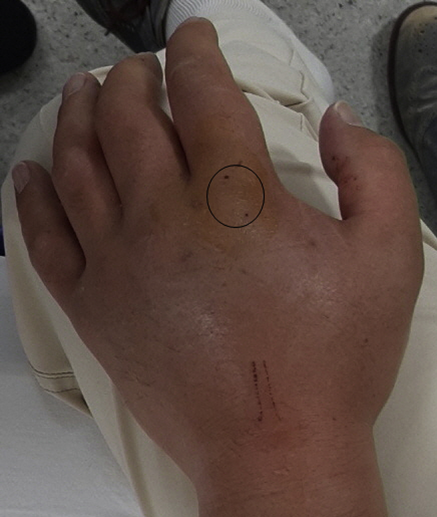
The patient’s left hand. Two fang marks are visible in the circled area.
What is the diagnosis? How would you manage this patient?
Diagnosis
Retained tooth or tooth fragment.
A plain x-ray of the left hand revealed a tiny foreign body in the soft tissue near the left second metacarpophalangeal joint (Figure 2). A retained tooth or tooth fragment was removed from the wound using fluoroscopy-assisted surgery (Figure 3).
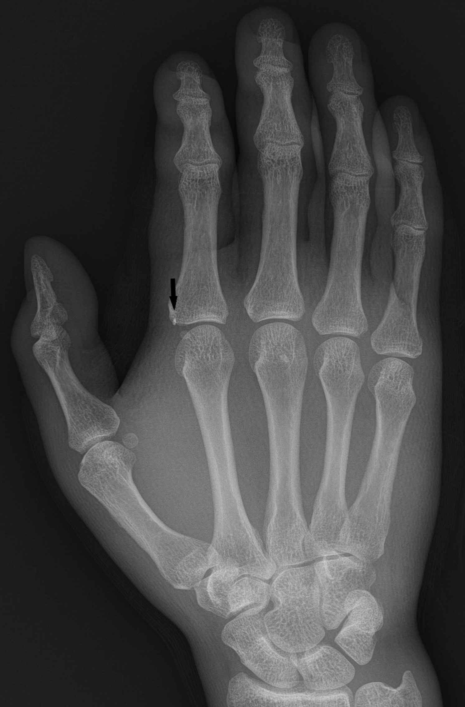
A tiny foreign body (solid arrow) was embedded in the soft tissue near the second metacarpophalangeal joint.
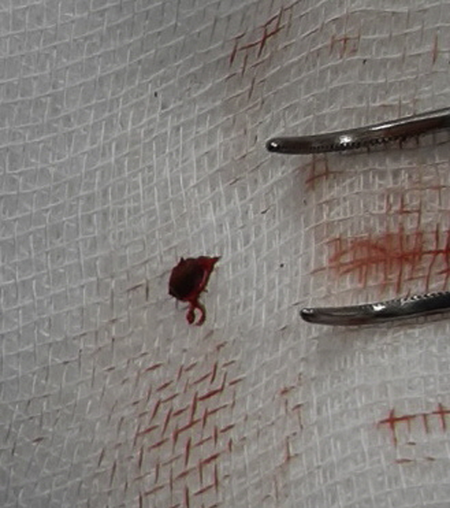
A retained tooth or tooth fragment was surgically removed from the wound.
Discussion
X-rays are not necessarily indicated for all snakebites. One indication for obtaining x-rays might be worsening pain and tenderness on delayed presentation. 1 In our patient, worsening of localized pain prompted x-ray evaluation to look for a retained foreign body. Healthcare providers should examine snakebite wounds carefully and obtain x-rays if there is worsening pain or tenderness.
Footnotes
Acknowledgements
Author Contributions: Conceptualization (SC, HK); investigation and visualization (YSJ, KCS); writing original draft (YSJ, HK) writing revision and editing (YSJ, SC); approval of final manuscript (all authors).
Financial/Material Support: None.
Disclosures: None.