
Abstract

To the Editor:
Adequate laceration management continues to challenge providers in resource-poor settings. Even minor lacerations in wilderness scenarios can rapidly lead to significant morbidity. Most noncontaminated lacerations, once appropriately irrigated, can be treated safely with acute primary closure.1,2 Multiple commercial options for wound closure are available, including tissue adhesives, surgical tapes, staples, and sutures. However, if wilderness providers do not have ready access to such supplies, they may need to depend on improvised techniques using resources found in the environment.
There may be value in a “natural” method of laceration closure that has not been previously mentioned in recent wilderness medicine literature. Leaf-cutting ants, members of 2 genera—Atta and Acromyrmex (subfamily: Myrmicinae; tribe: Attini)—have been used to close wounds in Central and South America for centuries. 3 These ants are found in the tropics and subtropics approximately between latitudes 33°N and 44°S, including regions of Texas and Louisiana. 4 Leaf-cutting ants are usually red-brown in color with 11-segmented antennae, 3 pairs of thorax spines, and a 2-segmented petiole (narrow waist). Nests are made from soils and can extend up to 1200 m2 in surface area and more than 7 m deep. These colonies are typically easily identified by their numerous surface-level, crescent-shaped mounds. 5 Leaf-cutter ants, like many insects, are polymorphic, meaning that within each species there are a variety of body types, with each suited to a specific task necessary for colony survival. The soldier caste of a leaf-cutting colony is the largest of all castes and defends the colony against threats. Members of this caste are most suitable for the application of wound closure given their larger mandible size.
When primary closure is necessary, both sides of the leaf-cutting ant's head are held firmly between the first and second digits (Figure 1). Gentle pressure is applied, forcing the ant’s oral cavity to open. The ant’s head is placed perpendicularly to and transversely across the wound. On contact with a surface, such as the skin, the oral cavity quickly closes, bringing the 2 sides of the wound together in apposition. The thorax and abdomen of the ant are subsequently detached, leaving the head and mandible behind. The decapitated head reportedly stays in place for approximately 3 d 3 ; however, there is no literature detailing such information or the rates of dehiscence.
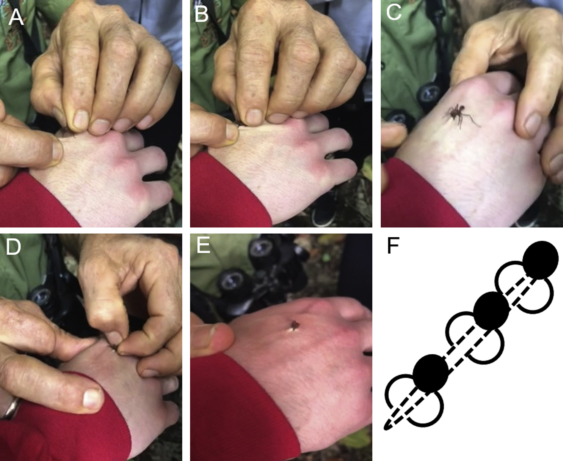
Attachment of a leaf-cutter ant: (A) positioning of ant; (B) closure of ant’s mandible; (C) ant secured on skin; (D) decapitation of ant; (E) completed ant suture; and (F) pictorial of multiple ant sutures to close a wound.
Some anecdotal reports indicate that wounds closed with leaf-cutter mandibles are less prone to infection than untreated lacerations. This may be secondary to the leaf-cutter ant’s natural chemical defenses against parasites. The metapleural glands of this type of ant secrete bioactive components that inhibit a range of microorganisms.6,7 When the metapleural glands of Acromyrmex octospinosus were artificially occluded, the ants experienced a subsequent increase in susceptibility to invading bacteria. 8 Unfortunately, there are no studies available to determine whether the metapleural secretions of leaf-cutter ants are active against human skin pathogens.
Leaf-cutter colonies can contain millions of individuals, allowing ants to be a readily available and renewable resource. Although there is not enough evidence to recommend this means of wound closure over commercially available devices, in appropriate situations with limited supplies, this may be a worthwhile option. Additional investigation into this means of laceration repair is certainly warranted.