
Abstract

A 75-year-old man was gored by a bull as he was attempting to load the animal on a truck. He presented to the emergency department with focal swelling, ecchymosis, and pain in the right lower quadrant (Figure 1). He appeared uncomfortable but was hemodynamically stable. Bowel sounds were present over the area of bruising, and his abdomen was diffusely tender to palpation. He did not have peritoneal signs or gross abdominal distention. Laboratory results showed an elevated white blood cell count of 13.2; serum chemistries were normal, and his urinalysis was negative for blood.
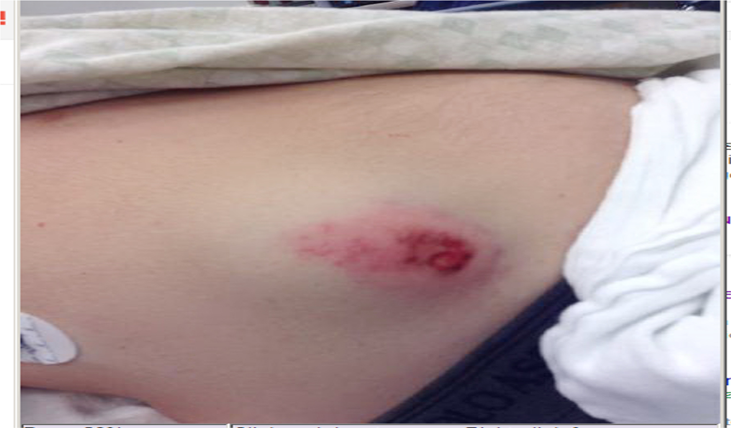
Focal swelling and ecchymosis of the right lower quadrant of the patient’s abdomen. Photograph by Michael N. Ofori, MD.
A computed tomography scan of the abdomen and pelvis was performed (Figure 2).
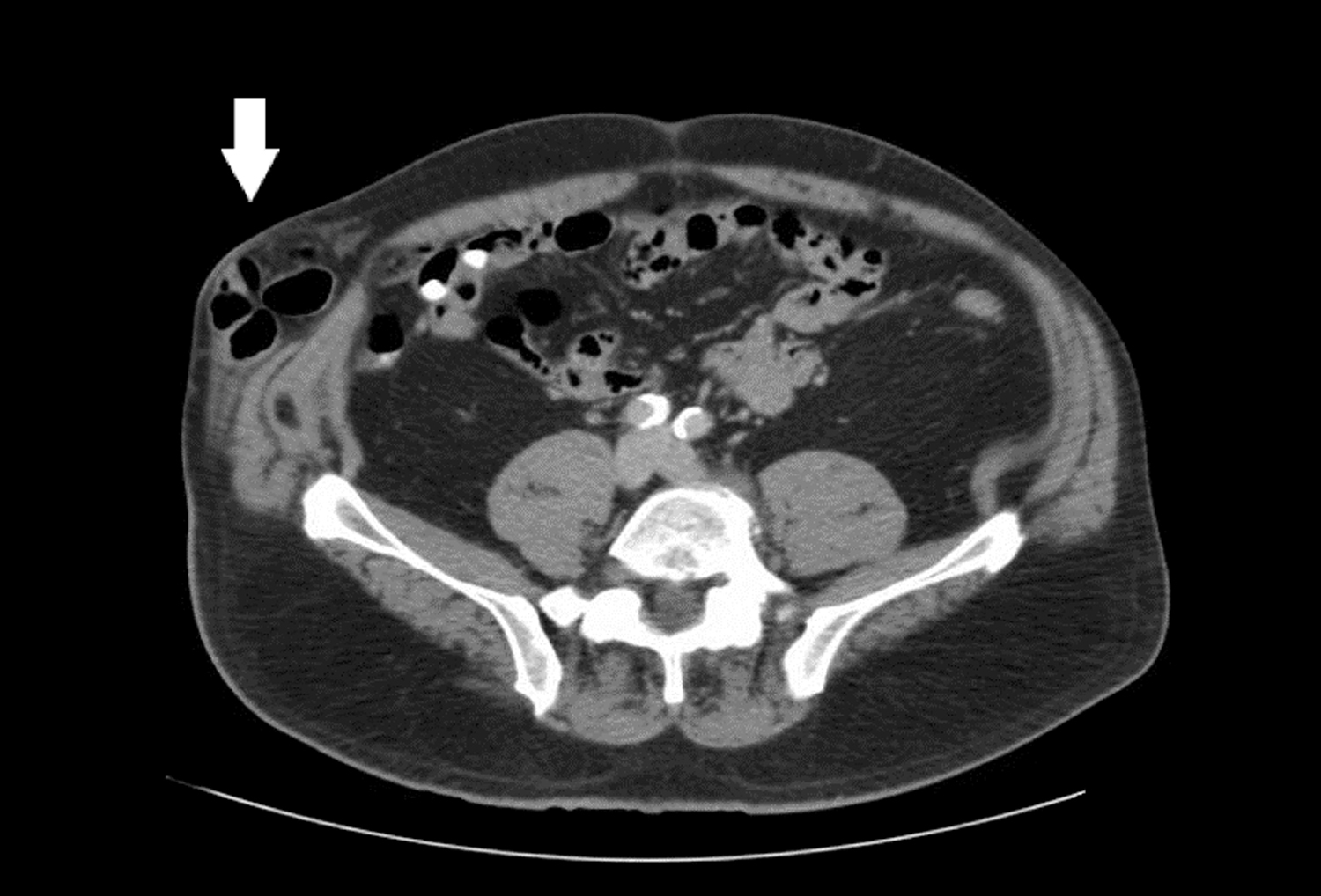
Computed tomography image showing bowel in the extraperitoneal soft tissue (arrow).
Diagnosis
Traumatic Abdominal Wall Hernia
This patient developed a traumatic abdominal wall hernia due to blunt force trauma from bull horns. After imaging, he was taken to the operating room for exploration. No other injuries were identified, the defect was repaired with mesh, and he was discharged on postoperative day 2. He was doing well at 2-week follow-up.
The majority of bull goring victims will have penetrating injuries, but isolated blunt trauma is present in up to 25% of patients. 1 The lower extremities are most commonly involved (58%), followed by the groin/perineum (15%), upper extremities (9%), abdomen (5%), head (5%), neck (5%), and thorax (3%). 2 Computed tomography scanning is useful for evaluating associated injuries, but point-of-care ultrasound has also been used to confirm the diagnosis of traumatic abdominal wall hernia. 3 The majority of patients will require operative intervention, including debridement of soft tissue injuries, repair of vascular injuries, urogenital reconstruction, and open reduction-internal fixation of bony injuries.1,2 Wound infections occur in up to 20% of patients. 2
Most animal-related fatalities in the United States occur from farm animals, 4 but similar patterns of injury have been described in wild animal attacks. A case series of 56 injuries sustained in bison attacks in Yellowstone National Park described 1 traumatic abdominal wall hernia, as well as instances of cardiac contusion, diaphragmatic rupture, and splenic injury. 5 As with bull goring injuries, the majority of bison goring injuries occurred in the buttock/thigh/pelvis region; this pattern was also noted in a series of attacks by wild boars. 6 A man was fatally gored by a mountain goat in Olympic National Park in 2010. Based on lay media reports, it appears injury to the great vessels of the thigh caused exsanguination. 7 A case report of a fatal moose attack described bilateral pneumothoraces, multiple rib fractures, lower extremity fractures, and extensive soft tissue injuries. 8 Skull penetration by deer antlers has been reported. 9 Injuries caused by animals with horns or antlers can be categorized as penetrating trauma, direct blunt trauma from horns or trampling, and deceleration injuries from being thrown or knocked to the ground. These injuries differ in mechanism from injuries caused by predators, which most commonly result in bite wounds. 10 Knowledge of injury patterns may assist practitioners in anticipating needs of patients who have suffered potentially devastating injuries.
(Images presented at the American Academy of Emergency Medicine meeting, Orlando FL, March 16–20, 2017.)