
Abstract

Mystery case
One dry summer day after several rainy days disrupting their camping trip, four 10-year-old campers were throwing a Frisbee in a field when one observed a tennis ball–sized brown ball loosely attached to the moist grass (Figure 1). He decided to stomp on the ball, which made a popping noise and released a powdery cloud of brown dust when crushed. He summoned his 3 friends over to gather several of the brown balls as pretend hand grenades to pelt each other with. One of the boys was hit in the upper chest with an exploding “grenade,” which covered his neck and chin with brown dust. Shortly after the war games were over and the group returned to Frisbee games, the “wounded” boy became dyspneic and walked back to his tent to retrieve his inhaler. He had a history of nonsteroid-dependent asthma. He went home, and over the next 2 days, he became febrile and developed a nonproductive cough with end-expiratory wheezing. Because the inhaler did not improve his wheezing and his fever continued despite oral acetaminophen, his mother called his pediatrician, who recommended a medical examination in the closest emergency department.
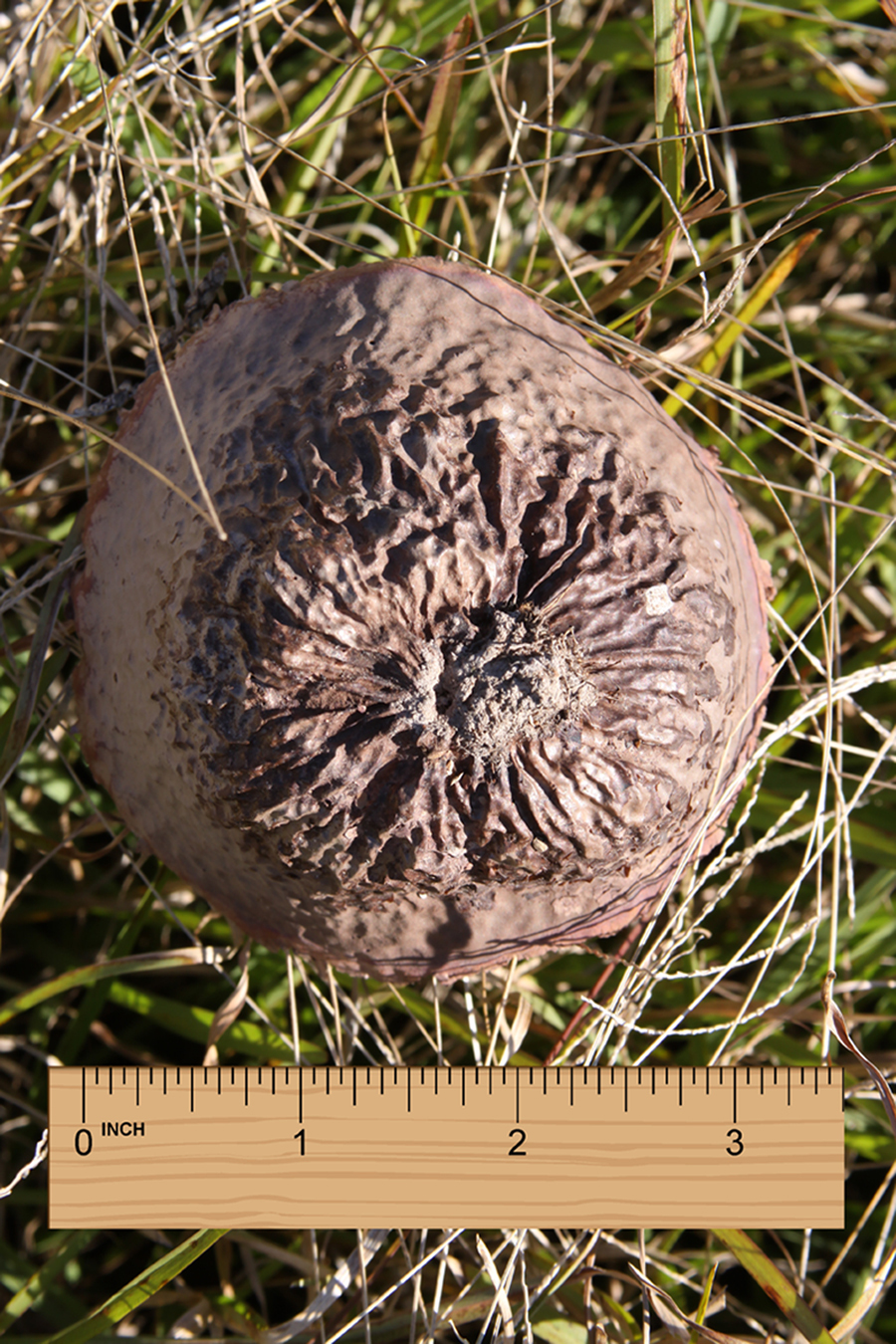
A tennis ball–sized brown ball loosely attached to the moist grass in an upland meadow. Source: Photograph courtesy of David K. Lirette, PhD.
What is your diagnosis? How would you manage this case?
Diagnosis
Lycoperdonosis, extrinsic hypersensitivity alveolitis after inhalation of spores (conidia) from the common puffball, Lycoperdon utriforme.
Management
On arrival in the emergency department, the child was febrile (38.5°C), dyspneic, and coughing. His respiratory rate was 36 breaths·min−1, and oxygen saturation by pulse oximetry was 94% on room air. Chest auscultation demonstrated crepitant rales at end-exhalation. The chest radiograph demonstrated bilateral reticulonodular infiltrates. An intravenous catheter was inserted, and nasal cannula oxygen was instituted at 2 L·min−1. The child was admitted to the hospital with a presumptive diagnosis of pneumonia. Blood cultures were drawn, but sputum cultures could not be obtained. Empiric antibiotic therapy with intravenous amoxicillin was instituted. Labored breathing continued over the next 12 h. The blood cultures revealed no growth.
A bronchoalveolar lavage (BAL) sample was obtained for additional cultures, including acid fast and fungal cultures. On microscopic examination of Giemsa-stained BAL slides, a consulting pathologist described nonbudding yeast-like cells. A consulting mycologist also reviewed the BAL slides and confirmed the yeast-like cells as Lycoperdon species spores. Intravenous antibiotic therapy was discontinued, and intravenous corticosteroid and antifungal therapy were instituted with dexamethasone, 4 mg twice a day, and voriconazole, 6 mg·kg−1 twice a day. Rapid improvement in breathing was observed over the next 12 h. The patient’s fever resolved, and the chest became clear to auscultation. The infiltrates cleared on the chest radiograph. All medications were discontinued after 2 days. The patient was discharged home on hospital day 5 for follow-up with his pediatrician.
Discussion
Lycoperdonosis is an extrinsic hypersensitivity alveolitis caused by the inhalation, insufflation, or ingestion of spores released by puffballs, primarily from the genus Lycoperdon. Puffballs are mushrooms without gills and stems; they release spores when they dry, decay, and rupture. If the puffballs are forcibly crushed, the spores can be aerosolized. Puffball spores have been administered medicinally as folk remedies for epistaxis. 1 Puffball spores have also been intentionally ingested and insufflated by adolescents seeking hallucinogenic effects. 2
Taxonomy, distribution, and ecology
Puffballs belong to the mushroom family Lycoperdaceae and are distributed worldwide. They may be found during warm seasons in clusters in moist shaded areas and grassy fields. Puffballs are similar to truffles and are edible seasonally when young and white. Unlike truffles, puffballs grow above ground and not below (Figure 1). Puffballs turn brown with age, desiccate, and fill with brown spores (Figure 2). They later decay and spontaneously burst, releasing trillions of spores (Figure 2).

The carefully opened Lycoperdon utriforme puffball initially seen intact in Figure 1. Note the fine brown powder filling the puffball and composed of trillions of microscopic spores. Source: Photograph courtesy of David K. Lirette, PhD.
Most species of puffballs that have been implicated in case reports of lycoperdonosis have belonged to the genus Lycoperdon and have included L perlatum, L pyriforme, L gemmatum, and L utriforme. 3 Inhalation of spores from the family relative giant puffball (Calvatia gigantea) has also been implicated less commonly in cases of lycoperdonosis. 3
Toxicology and toxicity in humans and animals
The first human cases of lycoperdonosis were reported by Strand et al in 1967 in an adolescent and a 4-year-old who were prescribed Lycoperdon spore insufflation as home remedies for epistaxis. 3 Their condition was characterized by the rapid onset of nasopharyngitis, nausea, vomiting, coughing, and pneumonitis. This prodrome progressed over a few days to wheezing, dyspnea, myalgias, fever, and chest pain mimicking pneumonia. The chest radiograph demonstrated a reticulonodular pattern of apical infiltrates that resembled Pneumocystis carnii pneumonia or histoplasmosis.
In 1994, Taft et al reported a case series of 8 teenagers aged 16–19 years in Wisconsin who inhaled and chewed puffball mushrooms at a party. 2 The puffballs were later expert-identified as L utriforme. No illicit drugs were used at the party. Three patients reported nausea and vomiting within 6–12 h of exposure. All patients developed cough, fever (maximum 38.9°C), dyspnea, myalgia, and fatigue within 3–7 days of exposure. Two patients had a history of asthma and were using steroid inhalers. Five patients were hospitalized, and 2 of them required endotracheal intubation for mechanical ventilation. The chest radiographs of all 5 hospitalized patients demonstrated bilateral reticulonodular infiltrates. Two patients had transbronchial lung biopsies, and one underwent open lung biopsy. Although the fungal cultures of the lung biopsies were all negative, the histopathological examination of the lung biopsies demonstrated inflammatory processes with the presence of yeast-like spores, later identified as Lycoperdon spores. All hospitalized patients received intravenous corticosteroids, and 4 received antifungal therapy with either amphotericin B or an azole. The authors concluded from the series that the pulmonary pathology was the result of a hypersensitivity reaction, but they could not rule out a concomitant fungal infection caused by infective spores. All patients recovered normal lung function whether treated with antifungals or not.
In 1997, Munson et al described 2 more cases of lycoperdonosis in 18-year-old men in Wisconsin. 4 In 1 case, the patient presented with fever, chills, nausea, vomiting, and dyspnea after riding a bicycle through a cloud of dust. The chest radiograph demonstrated bilateral diffuse pulmonary infiltrates. Blood, sputum, and urine cultures were all negative for bacteria, mycobacteria, and fungi. A methenamine silver stain of a sputum specimen did demonstrate intracellular round bodies 2.5 micrograms in diameter that resembled Histoplasma capsulatum spores but were not budding. The patient was treated empirically for pneumonia of undetermined etiology with intravenous corticosteroids and erythromycin. The patient improved quickly and was discharged on hospital day 6. Another 18-year-old man was also hospitalized at the same time with severe pneumonia requiring endotracheal intubation and mechanical ventilation for respiratory failure. Blood and sputum cultures were negative. The chest radiograph demonstrated bilateral diffuse pulmonary infiltrates that resembled Pneumocystis carinii pneumonia. A methenamine silver stain of a sputum specimen demonstrated intracellular round bodies 2.5 micrograms in diameter that resembled nonbudding fungal spores, later expert-identified as Lycoperdon spores. The patient was treated with intravenous corticosteroids and antibiotics and recovered normal pulmonary function over a month-long hospital course.
In 2010, Alenghat et al reported 2 fatal cases of lycoperdonosis in domestic dogs observed playing or digging in areas with puffballs before developing clinical signs of pneumonia. 5 Transtracheal aspirates demonstrated intrahistiocytic yeast-like spores 3–5 micrograms in diameter. In 1 case, polymerase chain reaction confirmed DNA from Lycoperdon pyriforme spores in a lung specimen. The authors concluded that it remained unclear whether lycoperdonosis represented an inflammatory reaction to a foreign material, an allergic reaction to a foreign antigen, an active fungal infection or mycotoxic poisoning, or a combination of all of these processes.
Conclusions
Lycoperdonosis is an inflammatory hypersensitivity reaction to the inhalation, insufflation, or ingestion of puffball spores, primarily from the genus Lycoperdon. The clinical findings of lycoperdonosis are consistent in animals and humans and include nausea, vomiting, myalgia, fatigue, and cough followed by febrile pneumonia. Bacterial and fungal sputum and blood cultures are negative, but sputum specimens often demonstrate intrahistiocytic and free nonbudding yeast-like spores 2.5–5 micrograms in diameter, consistent with puffball spores. The chest radiographic findings are also consistent with bilateral reticulonodular infiltrates. It remains unknown whether patients with pre-existing atopy or asthma are predisposed to lycoperdonosis after puffball spore inhalation. In most cases, normal lung function is restored after short-term corticosteroid therapy with or without antifungal therapy. To prevent the aerosolization and inhalation of puffball spores, puffballs should not be forcibly crushed near the face.
Acknowledgments: The author formally acknowledges the photographs in Figures 1 and 2 were taken by David K. Lirette, PhD, a former assistant professor of Environmental and Occupational Health Sciences at the LSU School of Public Health, New Orleans, LA, and provided as a courtesy to the author.
Author Contributions: Sole author contributed 100% to this article.
Financial/Material Support: None.
Disclosures: None.
Footnotes
Disclaimer: The case reported in this article is fictitious.