
Abstract
Stingray envenomation is a common occurrence. X-ray evaluation of stingray wounds is an unnecessarily misunderstood diagnostic concept. We present the case of a patient stung by a stingray with a prolonged and complicated course and permanent disability due to a retained barb. The patient had undergone multiple medical evaluations before an X-ray was obtained.
Introduction
Retained teeth, claws, and spines are an important source of long-term morbidity in animal-related wounds. These items serve as a nidus for polymicrobial infections and prolong and complicate wound healing. Stingray injuries have traumatic, venomous, and infectious potential. 1 We present the case of a patient with retained stingray barb and debilitating sequelae from extensive tenosynovitis.
Case report
While fishing in the Atlantic Ocean off the South Carolina coast, a 44-year-old man sustained a whip injury to the ulnar aspect of his right (dominant) wrist while removing a stingray from his fishing line. Initially the patient noted bleeding and pain at the site of injury. He presented to a local emergency department where it was noted he had a puncture wound to his wrist with a small amount of bleeding. Neurologic function was intact with normal strength, normal sensation, and full range of motion. The wound was washed with soap and water, a dressing was applied, and he was prescribed ciprofloxacin for wound infection prophylaxis. The wound was not explored, hot water immersion was not applied, tetanus immunization was not provided, and no imaging was performed. The patient adhered to the prescribed local wound care, which included washing with soap and water, and took his antibiotics as directed. The pain slowly resolved; however, he gradually developed an inability to extend his right ring finger, although flexion was intact. The patient returned to his home in Pennsylvania and on the eighth day after the injury, he saw his primary care physician who treated the patient with a second course of ciprofloxacin; again no imaging was performed. The patient subsequently and gradually developed an inability to extend his right middle finger. There was also moderate swelling of the hand, wrist, and distal forearm without pain, redness, fever, or discharge from the puncture site (Figure 1).

Stingray barb entrance wound with extensor dysfunction of the middle and ring fingers.
One month after the initial injury, the patient presented to our emergency department as the swelling had progressed to his right elbow. Physical examination showed a puncture wound over the ulnar aspect of the wrist with moderate edema of the hand, wrist, and distal half of the right arm. The wound had spontaneously closed, and a large eschar covered the puncture area, which was without drainage or redness. Radial and ulnar pulses were intact. Sensation was intact. He was unable to extend the third and fourth digits (Figure 1). A radiograph of the wrist revealed 2 foreign bodies that had the appearance of stingray spines on the extensor aspect of the wrist (Figures 2 and 3). The patient was taken to the operating room by hand surgery and found to have a complete laceration and rupture of the extensor digitorum communis (EDC) and extensive tenosynovitis with copious amounts of purulent gray material that required prolonged intraoperative irrigation and an interposition tendon graft repair. The patient remained hospitalized for 1 day, receiving intravenous ceftazidime (1 g every 8 hours) and tobramycin (1 mg/kg every 8 hours) and was discharged to home with doxycycline (100 mg twice daily for 14 days). He underwent prolonged physical and occupational therapy and regained adequate but not full function of his hand. The patient demonstrates good grip strength but persistent weakness with extension of his middle and ring fingers.
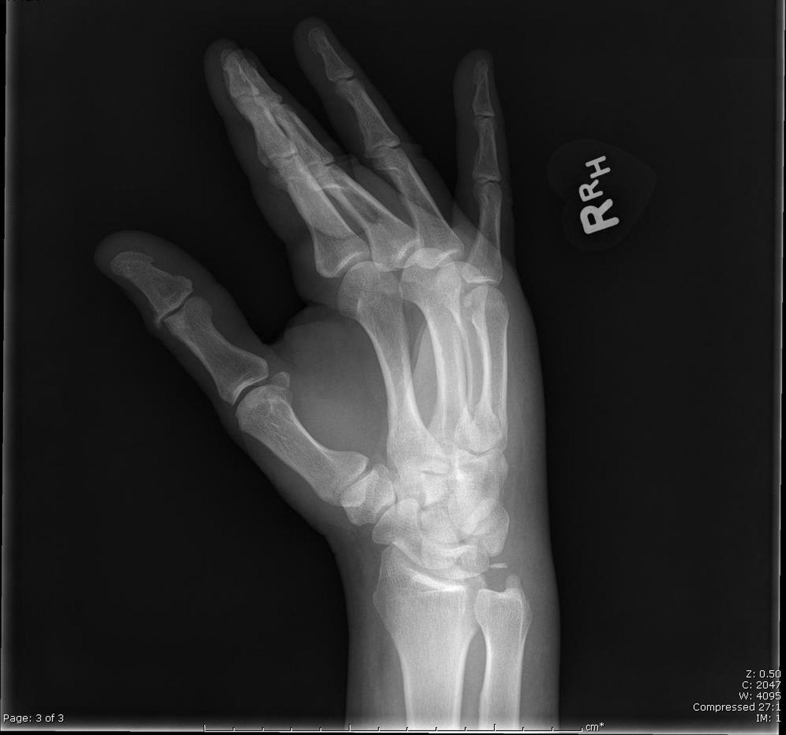
Oblique angle x-ray demonstrating radiopaque stingray barb.

Lateral angle x-ray demonstrating radiopaque stingray barb.
Discussion
Epidemiology
Stingrays are the most common group of fish involved in human envenomation, but the wounds are rarely fatal. 2 In the United States, there are approximately 2000 stingray injuries per year. 3 Although accurate statistics are very hard to find, fatalities from stingray injuries range from 1 to 2 or fewer per year in Indo-Pacific countries and the United States to as many as 8 per year in South American countries. 4 Fatal injuries are usually because of thoracoabdominal puncture wounds causing lethal cardiopulmonary compromise. 4
Stingrays are cartilaginous fish of the Chondrichthyes class. There are approximately 150 freshwater and saltwater species of stingrays divided into 2 families: Dasyatidea (true stingrays) and Myliobatidea (true rays). Myliobatidea rays are large, rhomboid-shaped animals, and are often found in open ocean water and not commonly associated with envenomation. 5 The majority of stingray injuries are caused by dasyatid and urolophid stingrays of the Dasyatidea family. 6 Stingrays of the Dasyatidea family are usually found in tropical, subtropical, and warm temperate oceans, generally in shallow water, hidden under sand. They have flattened, pancake-shaped bodies with long tails. At the base of their tails are spines. The spine of the stingray is a bilaterally serrated structure enveloped in an integumentary sheath with 1 to 4 spines located on the dorsal surface. 7 Toxin or venom is concentrated inside the integumentary sheath. The venom contains a variety of noxious substances and more than 10 proteins such as 5′-nucleotidase and phosphodiesterase. These toxins have been shown in animal studies to have cardiotoxic effects including bradycardia, tachycardia, atrioventricular block, ischemic Q and ST-T wave abnormalities, and asystole. 8 Additionally they can cause ataxia, vasoconstriction, central respiratory depression, seizure activity, coma, and death. 3
Injuries
Stingray injuries are unique and challenging because they are at the same time traumatic injuries and envenomations and can present unusual infectious complications. Injuries caused by stingrays produce intense local pain in the presence of edema, erythema, and necrosis, which suggests an extracellular destructive property of the toxin. An analysis of the proteins found in the venom of Potamotrygon falkneri (Largespot river stingray) identified enzymes that act directly on the extracellular matrix components contributing to protein breakdown and tissue necrosis. 9 These toxins are heat labile.3,6
Generally, stingrays are docile and nonaggressive and attack as a purely defensive gesture. Common activities that expose individuals to stingray injuries include wading, snorkeling, scuba diving, and bottom fishing; divers, fishermen, fish handlers, and aquarium workers are also at risk. 4
Injuries occur as the stingray reflexively whips its tail upward and forward, thrusting the spine into its victim. This response is usually elicited by physical touch such as occurs when the stingray is stepped on. The injury produces a puncture wound or laceration. Additionally venom may be released into the wound or the spine may break off leaving either fragments or an entire spine in the wound. Injuries to the lower extremities (foot and ankle) are more common than to the upper extremities, abdomen, and thorax. Clark et al 10 reported that 94% of stingray injuries involve the lower extremities, most probably because of the fact that most stingray injuries occur when an unsuspecting person wades into an area where a stingray is lying hidden under sand or in shallow water and steps on the animal. Fishermen who are removing stingrays from nets or fishing lines are more likely to sustain injuries to the upper extremities.
In rare cases, death occurs when a vital organ is injured. The incidence of death caused by stingray envenomation is not known but has been reported to be fewer than 20 cases. 11 A review of the American Association of Poison Control Center Annual Reports from 2002 to 2013 under the search heading “Bites and Envenomations: Aquatic” does not reveal a single death report; however, these data only represent the geographic area of North America and Hawaii. 12
Envenoming stingray wounds are characterized by lacerations causing severe lancinating pain for 15 to 90 minutes. 6 As with other envenomations, pain from stingray trauma is typically out of proportion to what would be expected from the wound alone and may last up to 48 hours without treatment.6,13 Initially, the wound is erythematous but may progress to hemorrhagic discoloration with surrounding edema and petechiae. Venom toxicity and secondary infections from bacteria contribute to delayed wound healing. Secondary infections from bacteria present in seawater, including the genera Vibrio, Aeromonas, and Mycobacterium, are more common than the usual pathogens associated with wound infections. 14 Systemic manifestations of the toxin, although not common, have been reported and include anxiety, diaphoresis, nausea, vomiting, diarrhea, hypotension, syncope, tachycardia, headache, arrhythmia, and potentially cardiogenic shock and death. 6
Treatment
Treatment of stingray injuries is directed at identifying traumatic damage to the involved anatomy, combating the effects of the venom, alleviating pain, and preventing infection. Initial management should begin at the scene. Victims should be removed from the water and assessed for cardiopulmonary stability. The spine should only be removed under medical supervision if superficially embedded, and it should be left in place if the spine penetrates vital organs, such as the neck, thorax, and abdomen.
The wound should be cleaned with fresh water or sterile irrigating solutions if available. As soon as possible, the wound should be soaked in hot water for 30 to 90 minutes to reduce pain and wound necrosis. The water should be as hot as the patient can comfortably tolerate without causing thermal injury (42°–45°C). 10 The hot water is thought to accelerate the breakdown of the heat-labile toxins released into the wound, although this has never been proven in vivo. Tying half a raw onion onto the site of the envenomation has been reported to help alleviate pain in one case report. 15 Tetanus prophylaxis should be administered, and the wound should be explored thoroughly after appropriate analgesia. Use of local infiltration to facilitate removal of the spine and irrigation have been described in numerous case reports 11 ; epinephrine-containing anesthetics should be avoided to prevent possible ischemic insult to already vulnerable tissue, and regional nerve blocks may be considered for analgesia. 16 Hyperbaric oxygen treatment was reported to improve the survival of marginally viable tissue and may have been helpful in one case report, 17 but given the multifactorial nature of injuries from stingray wounds, no recommendations can be generalized from a single case report. The victim should be observed for at least 3 hours for systemic side effects before discharge in uncomplicated injuries.
Prophylactic antibiotics are recommended in stingray injuries because of the high incidence of ulceration, necrosis, and secondary infection. In a retrospective review of 119 cases of stingray envenomation, there was a relatively high rate of return visits for infection in patients not initially treated with antibiotics. 13 Antibiotic selection is important as most penicillin derivatives and first-generation cephalosporins that are commonly used for routine skin infections are inadequate for stingray puncture wounds. The choice of prophylactic antibiotic should include coverage for skin bacteria and gram-negative Vibrio species for saltwater stingray species and Aeromonas for freshwater species. A 5-day course of imipenem, a fluoroquinolone, or trimethoprim-sulfamethoxazole is an appropriate antibiotic choice.6,13
Imaging
The decision on whether to perform diagnostic imaging for stingray envenomation is unnecessarily controversial. Clark et al 10 in their large case series noted that “radiographs were largely unhelpful”; however, routine radiographs of stingray injuries should not be interpreted as unimportant. This case and multiple other case reports have demonstrated stingray barb visualization in wounds. 6 ,10,18 Radiographs may reveal hyperdense, radiopaque fragments of spines or barbs but unfortunately do not reveal hypodense fragments of integumentary and glandular tissues. 6 There are numerous investigations demonstrating the utility of ultrasonography in the identification of radiolucent foreign material in wounds,19,20 and magnetic resonance imaging may help locate hypodense foreign bodies but may not be cost effective for routine evaluation of all wounds. 10
Wounds that are properly treated may require a few months to fully heal with complete resolution of local tissue swelling; however, wounds that are not properly debrided or explored are likely to develop wound complications, as in this case. Our patient’s initial visit to the emergency department was managed with local wound care and prophylactic antibiotics. At that time, the patient demonstrated no evidence of traumatic tendon injury. His course was complicated by an indolent infection with associated tissue necrosis and tendon rupture with resultant partial loss of function of his dominant hand, which suggests that the venom and infectious complications were the primary causes of the patient’s subsequent disability. The lack of severe pain, redness, and drainage may be explained by the multiple courses of antibiotics and the resultant partially treated local infection.
Although local wound exploration may have identified a foreign body, and early treatment with hot water immersion may have mitigated the necrotic effects of the stingray toxin, a radiograph is noninvasive and has little to no risk of the potential for further injury associated with wound exploration. A radiograph taken at the time of injury would have demonstrated the retained animal parts, and the patient might have been speared the prolonged and complicated course. As a result, we recommend that all stingray wounds undergo radiograph imaging.
Conclusions
Most stingray injuries are nonfatal and heal without complication with proper wound care, wound exploration, debridement if necessary, prophylactic antibiotics, and tetanus immunization updating if necessary. Stingray venom is heat-labile, and hot water immersion immediately after injury may reduce pain and skin necrosis. Stingray spines and barbs can be radiopaque, and radiographs or ultrasound can be helpful in detection.
Footnotes
Acknowledgment
The authors would like to thank Dr. Charles Fasano for his assistance in the preparation of this manuscript.