
Abstract
Overuse and acute injuries to the upper body are common in rock climbing. Such injuries primarily affect the fingers; but shoulder problems are increasingly common, especially among more experienced and older climbers who climb at a high ability level. Such shoulder problems are often due to subacromial impingement, shoulder dislocations with bankart lesions, hyperlaxity, SLAP lesions or irritations of the long biceps tendon. In contrast to these known conditions, we describe a case of an ambitious female rock climber who trained intensively and developed a coracoid impingement caused by hypertrophied subscapularis tendon and muscle following sport-specific training. Diagnosis was made through clinical evaluation and confirmed by magnetic resonance tomography. Coracoid impingement syndrome is a less common cause of shoulder pain and occurs when the subscapularis tendon impinges between the coracoid and the lesser tuberosity of the humerus. The patient was treated successfully with a conservative therapy and returned to full activity within 6 weeks.
Introduction
Injuries and overuse syndromes of the shoulders are common pathologies in rock climbers. 1 The rise in reporting of such problems may also reflect the increased global participation in sport climbing and bouldering since the 1980s that gave rise to the establishment and prevalence of indoor climbing walls. Since this time, rock climbing has become more technically and physically demanding, with the maximal climbing grade increasing over time. The most frequently reported shoulder conditions are subacromial impingement syndromes, bankart lesions after dislocations and hyperlaxity. 1 Nevertheless, over the last decade there has been a rise in the number of lesions to the long biceps tendon, the biceps tendon pulley, and the tendon-labrum complex (superior labrum anterior to posterior [SLAP]) reported and these conditions also need to be considered.1,2 Less well known, but also described shoulder conditions in rock climbers include a nerve entrapment syndrome of the suprascapular nerve and thoracic outlet syndrome. 1 Here we present a previously unreported shoulder pathology in rock climbers.
Case Presentation
We examined an ambitious female 28-year-old rock climber (climbing level 8 Union Internationale des Associations d'Alpinisme [UIAA], 5.11+ Yosemite Decimal System [YDS]) for anterior shoulder pain in our sports orthopedic clinic. She reported that she started rock climbing 2 years ago and focused all her training on the development of upper body strength. She presented during winter, so most of her activity in the previous 2 months was indoor lead climbing with some indoor and outdoor bouldering. She began working at an indoor climbing gym in the previous 3 months where she undertook very intensive climbing training 4 times a week. Additionally, she performed intensive sport-specific training on the “campus board,” and extra weight training for upper body strength. Her shoulder pain was positively correlated to her training schedule. 1 The onset of pain began in the evening after climbing, and persisted for 24 hours or more when she would begin training yet again. She was previously treated elsewhere with subacromial corticoidsteroid injections without improvement.
Physical examination revealed shortened pectoralis muscles bilaterally, hyperkyphosis of the thoracic spine, and inwardly rotated shoulders bilaterally. Impingement signs for subacromial impingement were weakly positive, meaning slight complaints with the Kennedy-Hawkins, crossover- and Neer-tests. Tests for instability, SLAP provocation, and the O′Brian test for SLAP lesion were all negative. Active and passive range of motion was within normal limits for all of flexion/extension, abduction/adduction, medial/lateral rotation, horizontal flexion/extension, and circumduction. Manual muscle tests did not show weakened muscle function for supra- and infraspinatus or subscapularis. Subscapularis muscle test (medial rotation against resistance) was slightly painful. No instability or hyperlaxity was found. Anterior apprehension sign was positive. Palpable point tenderness was noted at the lesser tuberosity and the coracoid process. Ultrasound did not show any definitive abnormality; therefore, an magnetic resonance image (MRI) was performed.
MRI
The MRI showed normal supra- and infraspinatus tendons and a slightly narrowed subacromial space (acromion type I) with minimal osteoarthritis (Kellgren Lawrence I) of the acromioclavicular joint (Figure). The biceps tendon, pulley, labrum, and biceps tendon insertion were all normal. The subscapularis muscle appeared hypertrophied with impingement of the muscle and tendon between the coracoid and the lesser tuberosity of the humerus. Minor edema was present next to the subscapularis tendon. The coracoid index was moderately increased (8.7 mm, normal: 8.2 mm), the coracohumeral interval was normal (8.9 mm, normal: > 6.8 mm).3,4
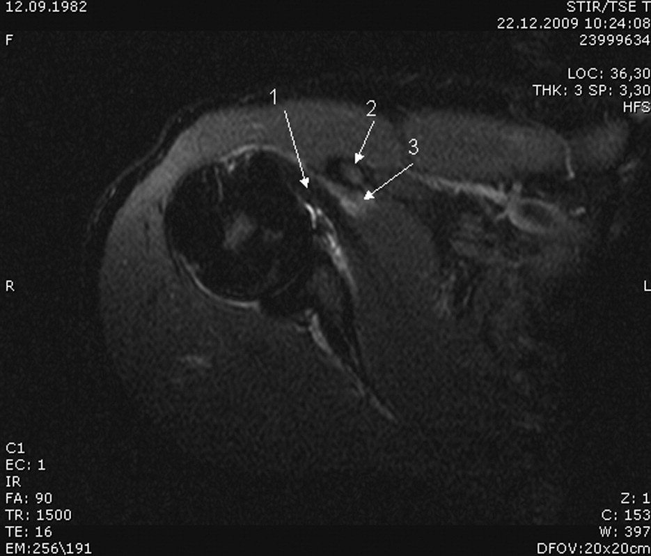
Axial MRI scan of the right shoulder. Note the thick subscapular tendon (1) and muscle with edema (3) within the muscle posterior to the coracoid process (2). The subscapular tendon and muscle almost completely fill the subcoracoidal space.
Therapeutic Course
After diagnosing a coracoid impingement syndrome, the patient underwent conservative therapy in accordance with the treatment algorithm described in the literature. 4 She received physiotherapy to detone the subscapular muscle, center the humerus head (training of the external rotators, abductors, and balancing scapulothoracic stabilization), stretch the muscles, perform proprioreceptive neuromuscular facilitation (PNF), exercises, and train the antagonists. Physiotherapy was provided 3 times per week initially, then reduced to twice weekly. Stretching exercises were prescribed twice per day. Postural training to decrease the thoracic kyphosis and to lengthen the shortened pectoralis muscles was also adopted. Non-steroidal anti-inflammatory drugs (NSAID) were administered for 2 weeks. The symptoms quickly resolved; after 3 weeks she was able to resume climbing training, and after 6 weeks she was climbing to her previous high ability level. Six and 12 months later she remained asymptomatic.
Discussion
Frequently reported shoulder problems in rock climbers include subacromial impingement, shoulder dislocations with bankart lesions, hyperlaxity, SLAP lesions, or irritations of the long biceps tendon.1,2 While a subacromial impingement syndrome is a common condition, a subcoracoidal impingement is not. In most cases, coracoid impingement occurs due to variation in the height and length of the coracoid process that alters the space between the coracoacromial arch and the rotator cuff, specifically the subscapularis muscle and tendon. 5 In our case, the subscapularis muscle and the tendon were hypertrophied, resulting in functional impingement. The training history of our patient revealed an intense focus on muscular development over the last 2 years, with little prior sport activity. Thus, over a comparable short interval the muscle diameter increased. The time factor may be of importance, as not all hypertrophic subcapularis muscles lead to such a condition. Because the muscle increased over a relatively short time, soft tissue and neural adaptation processes might have been delayed or restricted.
A recent cadaver study found that the problem of coracoid impingement may be functional in combination with anterior instability leading to a narrowing of the coracohumeral distance. 6 Our patient demonstrated a positive apprehension sign suggesting an anterior instability, which is a common condition in rock climbers.1,2 The cause of the condition in our athlete is likely threefold: (1) a relatively small subcoracoidal space; (2) an increase of muscle tendon diameter over a relatively short time; (3) a minor anterior instability together with shortened pectoralis muscles and an inwardly rotated shoulder girdle. Thus, we postulate that our patient's condition was functional and not anatomic.
Anatomic changes due to rock climbing have been described as the cause of several pathologic conditions. Förster et al characterized and objectively measured a postural adaptation sometimes found in elite male climbers—a condition referred to as “climber’s back.” 7 Förster et al. reported an increased kyphosis of the thoracic and an increased lordosis of the lumbar spine, accompanied by shortened pectoralis muscles. 7 The subject's climbing ability level was strongly correlated to these postural adaptations when these men climbed routes graded UIAA 10- or above. 7 When analyzing the functional pathology of the “climber’s back,” there was an observed imbalance between the strong inwardly and weak outwardly rotating muscle groups of the shoulder girdle. In addition, there was a habitual postural adaptation with anteriorly positioned shoulders that was possibly due to shortened pectoralis muscles and an increased thoracic hyperkyphosis. 7 Our female patient, who climbed grade UIAA 8, also presented with a slight increased thoracic hyperkyphosis in addition to her inwardly rotated shoulders and shortened pectoralis muscles. In a chapter in reference 1, Schlageter describes a link between shoulder disorders and high ability climbers with shortened pectoralis muscles and thoracolumbar spine postural adaptations. Shortened pectoralis muscles also have been linked to thoracic outlet syndrome in rock climbers. 1 Russo and Togo 8 observed that coracoid impingement is most common after a history of chronic overuse with multiple episodes of micro trauma, especially if the shoulder is in an internally rotated position.
The diagnosis of coracoid impingement is suggested by a positive coracoid impingement test, similar to Kennedy-Hawkins sign, except that the shoulder is in a cross arm adduction, forward elevation, and internal rotation position. Plain radiographs may reveal anatomic abnormities, and ultrasound may show subscapularis tendon irritation or muscular hypertrophy. Final diagnosis can be confirmed by magnetic resonance imaging (MRI) or computed tomography (CT) scans to assess the coracoid index. 4 The coracoid index is the measurement of the lateral projection of the coracoid process beyond a line tangential to the articular surface of the glenoid. 4 It is a linear measurement in millimeters and averages 8.2 mm. A minimum distance of 6.8 mm between the coracoid tip and the closest portion of the proximal part of the humerus (coracohumeral interval) is considered normal for axial MRI measurements in maximal internal rotation. 9 In our patient, the coracoid index was 8.7 mm and the coracohumeral interval was 8.9 mm; thus, the coracoid index was minimally abnormal, and the condition of the patient is functional rather than anatomical.
Conservative therapy is the appropriate initial therapy, but if this fails, a surgical subcoracoid decompression should be the next option. 4 Because the condition was functional in our patient, her physiotherapy placed a special emphasis on joint balancing and centering the humerus head, strength exercises for the anterior joint capsule, and detoning and stretching the subscapularis muscle. Our patient responded well to conservative therapy, so no surgical intervention was necessary.
Conclusion
Coracoid impingement syndrome should be considered in the differential diagnosis of anterior shoulder pain in rock climbers. Rock climbers may be especially prone to this condition due to the sport-specific muscular hypertrophy that frequently accompanies anterior shoulder instability 1 possibly combined with postural deficits such as an inwardly rotated shoulder girdle and shortened pectoralis muscles. 7 Since this may be primarily a functional rather than anatomic disorder in climbers, conservative therapy is initially recommended. “Easy” climbing can form part of the rehabilitation.