
Abstract
The aim of this study was to describe the use of cone beam computed tomography as an auxiliary method to diagnose changes to the temporomandibular joints in cats. We used five cats of various ages, breeds and genders that showed clinical signs consistent with changes in the temporomandibular joint. Cone beam computed tomography enables a complete and thorough examination of the temporomandibular joints by allowing the evaluation of selected images as a whole. It also enables the identification of all anatomical structures and any changes that may be present. The results showed that this method is effective in confirming or ruling out changes in the temporomandibular joint in cats, such as disjunctions of the palatine raphe; fractures of the mandibular symphisis, zygomatic bone and condylar; and dental resorption.
Injuries to the temporomandibular joint (TMJ) are common in cats, and trauma is the most common major cause. The diagnosis is complex and is not often accomplished using conventional radiographs. 1 The major acquired changes observed in this joint are luxation and fractures of the condyles or zygomatic processes; the major developmental abnormality is dysplasia. 2
With continuing advancements in veterinary dentistry, new diagnostic methods have emerged as options for assessing the bucco-dental changes that are essential for diagnostic imaging and improving examination quality. Among the diagnostic tests, computed tomography (CT) has been used successfully for imaging the oral cavity, especially in dogs and cats. 3
CT is a radiographic procedure in which millimetre-sized sections of the body are made in the transverse, sagittal and dorsal planes. According to the geometric shape of image acquisition, a CT scan can be divided into a fan beam, which results in conventional CT, or a cone beam, which results in cone beam CT. Both methods provide sliced images of the dentomaxillofacial region using X-rays. However, the equipment, the method for obtaining and processing the images, the radiation dose and the cost of the equipment are quite different. The amount of electricity needed to operate the cone beam CT scanner is significantly lower; the tomograph can be plugged into a surge protector, and a normal 110 V or 220 V single-phase outlet can be used. 4−6
Cone beam CT is a diagnostic method in which the X-ray detector performs a 360° scan around the patient's head and acquires images that are later analysed by a specific computer. 5−7 The volumetric data obtained by the tomograph comprises a three-dimensional block of small cuboidal structures called voxels, which are isometric (equal height, width and depth dimensions) with cone beam CT. 7
The advent of cone beam CT represents the development of a tomograph that is relatively small and inexpensive and is especially suited to the dentomaxillofacial area (Fig 1). The development of this new technology provides dentistry with a three-dimensional image of mineralised maxillofacial tissues. 5
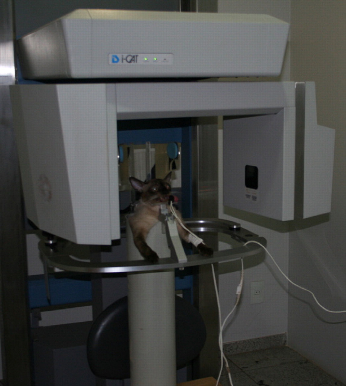
A cat placed in the PVC device for the cone beam tomography examination.
The aim of the present study is to describe the use of cone beam CT as an auxiliary method in the diagnosis of temporomandibular changes in cats.
Material and methods
The study was conducted at the Centro Veterinário do Gama, in Brasilia, DF, Brazil, between April 2008 and March 2009. The Ethics Committee on Animal Use of the Institute of Biological Sciences, University of Brasilia (UNB) approved this project under the protocol, UNBDOC 12339/2008. All ethics and animal welfare guidelines recommended by the Brazilian College of Animal Experimentation were followed. In this study, five cats of different ages, breeds and genders that showed signs consistent with temporomandibular changes were used.
The TMJs of all animals were evaluated by cone beam CT using a CT scanner i-CAT (Xoran Technologies, Ann Arbor, Michigan and Imaging Sciences International, Hatfield, PA, EUA). The CT scanner with the following parameters was used for the acquisition of images: a height of 6 inches, a period of 40 s, a voxel size of 0.2 mm, grey scale, 14 bits, a focal spot of 0.5 mm and a single 360° rotation in an amorphous silicon flat panel image detector. We used 120 kilovolts (kV) and 46.72 milliamps per second (mAs). A total of 430 slices were generated.
To perform the examination, the cats were anaesthetised and placed in a tube made of polyvinyl chloride (PVC) with a diameter that was compatible with the girth of the patients. Due to the short duration of the procedure, patients were anaesthetised by midazolam at a dose of 0.2 mg/kg and ketamine at a dose of 10 mg/kg; both were administered intramuscularly.
The cats were then placed vertically so that the height of the tube matched their cervical–lumbar length. In cases where there was a need to increase the height of the patient, the base of the tube was padded with a high-density foam. 3
Immobilisation of the cat within the tube was accomplished by inserting high-density foam between the animals’ body and the wall of the tube. After preparing the cat for the CT scan (Fig 1), the device was turned on, and images were captured and transferred to a computer that was located in a nearby room, where it was possible to view the patient through leaded glass. The images were analysed by a computer programme installed on the tomograph, which selected the appropriate slices and reconstructions for the visualisation of the TMJ.
Results
Cone beam CT enabled a complete and thorough examination of the TMJs by allowing the evaluation of selected images that allow for a view of the entire mouth.
The images enabled the identification of all anatomical structures of the TMJ and any changes that were present. The results showed the effectiveness of cone beam CT for confirming and ruling out changes in the TMJ in cats.
The examination was quick, painless and did not require repetition. The evaluation of the TMJ using the cone beam CT did not require extensive anaesthetic protocols, and the use of drugs was limited. This method provides a calm awakening and discharge shortly after the procedure in cases that did not require immediate treatment.
Some of the patients with no changes in the TMJ (Fig 2 a and b) showed pain upon manipulation of the oral cavity during the clinical examination. In the three patients (60%) who showed this clinical sign, lesions that did not involve the TMJ were observed.
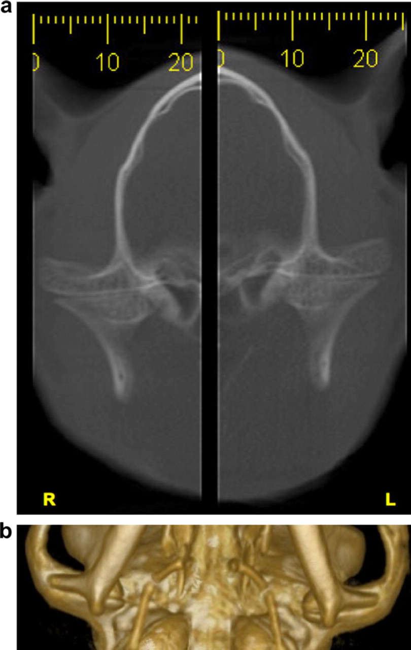
Normal aspects of the right (R) and left (L) TMJs in a cat. (a) Transverse section. (b) Ventral view of a 3D reconstruction.
The first patient presented with a history of anorexia. The owner reported that the cat tried to eat but could not grasp the food. The patient did not permit the manipulation of the oral cavity during the clinical examination. The diagnosis of dental resorption (Fig 3) was possible because the cone beam CT acquired images of the entire mouth of the patient and not just the TMJs.

Oblique view of transverse section of the cat dental element 309, showing dental resorption in the distal root (FDRL).
Patient 2 was a cat whose owner reported that it left home 2 days prior to its presentation and returned with its jaws misaligned; this patient also demonstrated pain upon manipulation of the oral cavity. The diagnostic hypothesis was dislocation of the TMJ. The patient was referred for cone beam CT that showed disjunction of the palatine raphe, bilateral zygomatic arch fractures and no alterations in the TMJs (Fig 4a–c).
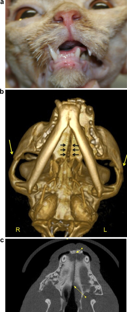
Mandibular misalignment and disjunction of the palatine raphe in a cat. (a) Cat presenting with misalignment of the mandible and a dental fracture of left inferior canine tooth (309). (b) Ventral view of a 3D reconstruction showing disjunction of the palatine raphe (black arrows) and bilateral fractures of the zygomatic arches (yellow arrows). (c) Dorsal plane image showing disjunction of the palatine raphe (yellow arrows).
Patient 5 had a misaligned mandible, which did not allow oral manipulation. Oral examination under general anaesthesia showed a left mandible fracture and disjunction of the mandibular symphysis (Fig 5), as confirmed by cone beam CT.

Dorsal plane image of a cat skull showing a fracture of the left mandibular ramus (red arrow).
The other two patients (40%) showed changes in the TMJ that resulted from bilateral fractures of the condyles (Fig 6a and b) and a fracture of the right condyle (Fig 7).
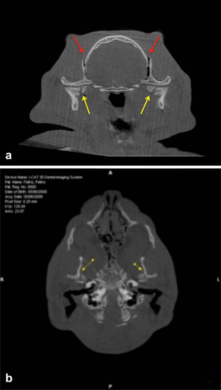
Cone beam CT cross-section at the level of the TMJ showing bilateral fractures of the mandibular condyles and temporal bone. (a) Transverse plane image at the level of the TMJ showing bilateral fractures of the mandibular condyles (yellow arrows) and temporal bone (red arrows). (b) Dorsal plane image showing bilateral fractures of the mandibular condyles (arrows).

Transverse image of a cat skull showing fracture of the right mandibular condyle (red arrow).
Discussion
Cone beam CT enabled a complete and thorough examination of the TMJs by allowing the evaluation of selected images that provide a view of the entire mouth, including three-dimensional reconstruction. Diagnosis of the changes in the TMJ is complex and is not always possible using conventional radiographic examinations. 3,8
Cone beam CT enabled the identification of all anatomical structures and any changes. The results showed that it is effective in confirming and ruling out the changes in the TMJ in cats (Table 1). This finding is consistent with the American Academy of Oral and Maxillofacial Radiology (AAOMR) parameters of care, which recommend streamlining the diagnosis, treatment, planning and monitoring of various aspects of dental status. 9 These conditions include TMJ disorders, maxillary diseases and dental implants planning.
Feline patients who were treated and underwent the cone beam CT between April 2008 and March 2009 at Centro Veterinário do Gama, Brasilia, DF, Brazil.

In humans, cone beam CT is recommended for the diagnosis of injuries in this region and for the evaluation of the TMJs, especially through the use of three-dimensional reconstructions. 9,10
Cone beam CT incorporated the images of the entire mouth into the same test session and provided the accuracy needed to rule out diagnoses of TMJs in cats. These results confirm the indications for the use of this kind of tomography in humans. 5
The criteria used in this study for cone beam CT evaluation of the TMJ included pain upon manipulation of the oral cavity, reluctance to closing the mouth and mandibular misalignment. These criteria are based on a study that recommended extraoral radiography of the TMJ in patients with reluctance to closing the mouth, dental malocclusion and reluctance or inability to access this joint if involved in mandibular trauma or trauma to another nearby region. 2 Our criteria were also based on a study that indicated that injuries to this joint may result in severe masticatory dysfunction and may potentially cause closed mandibular syndrome. 11
Although the literature is scarce regarding cone beam CT in the diagnosis of changes in the oral cavity of cats and dogs, this examination enables the detailed evaluation of the TMJ. 3 The images obtained provided the ability to check the morphology of the condyles and investigate for the presence of bone changes in the structures of the TMJ. These results are consistent with observations from a study conducted in humans. 12
Standardising the placement of animals in CT scans was fundamental to the use of this diagnostic tool in evaluating the dental changes in the cats used in this study. 3 Because the scanner has a chair where human patients are examined in the seated position, it was necessary to develop the PVC device that kept the animals in a standing position so that the images could be acquired. Similarly, the anaesthetic protocol enabled the safe and reliable acquisition of images because the animals remained immobile during the examinations, which obviated the need for repeat testing. Therefore, in addition to providing greater assurance for the procedures and making it safe for use with animals of any age and with any level of debilitation, this method minimises the exposure of animals and professionals who are involved in the procedure. 13,14
The use of cone beam CT in private practice has a number of advantages over conventional CT: reduced exposure to X-ray beams (minimises ionising radiation), greater accuracy of the images, shorter duration of image acquisition, decreased display of artifacts and better adaptation to the maxillofacial region. 5
The effective radiation dose of cone beam CT varies with the manufacturer of the apparatus and with the technical specifications selected during the image capture (ie, field of view, exposure time, kilovoltage and milliamperage), but generally, it was shown to be significantly reduced compared to traditional CT. 5,15
Although cone beam CT has been used in routine diagnosis of dental diseases in human patients, 6 only one citation was found where cone beam CT was used in veterinary medicine. 3 The authors of that study standardised the positioning of the animal during the CT scan and described techniques to assist in the diagnosis of bucco-dental changes in dogs and cats. Even though these results are encouraging, developing further scientific research could help to consolidate the method, which is especially useful in diagnosing diseases that are difficult to diagnose using conventional radiographs.
In the tomographic evaluations, there was no need to repeat the tests, and the images of the two TMJs were acquired in the same examination session. This characteristic of the examinations reduced the time required for the examination, reduced the occupational and patient exposure to ionising radiation and reduced the anaesthetic requirements, and it provided a fast and safe recovery of the patient. Because there have been measurements of the absorbed doses of radiation in previous studies, we can confirm that the dose of radiation emitted by cone beam CT is significantly reduced compared to conventional CT and periapical radiographs of the entire mouth. In addition, the results are rapid and accurate. 5,6,16–18
Finally, based on the results obtained and compared with the limited scientific literature, we believe that the use of cone beam CT in veterinary dental care is feasible and does not demand higher requirements for its installation and operation. Therefore, we believe that the investment is fully justified by the widest possible use of the technique in the diagnosis of a large number of bucco-dental diseases with speed, safety and accuracy.
Conclusion
Cone beam CT is an appropriate method to accurately identify or rule out fractures, luxations and structural changes in the TMJ in cats.