
Abstract
A domestic shorthair cat presented with a progressive history of polydipsia, lingual swelling and ulceration. The tongue was firm and grossly enlarged with associated regional lymphadenopathy. Surgical biopsies revealed lymphoma of the tongue. Following the procedure, the cat developed respiratory distress and was subsequently euthanased. Necropsy confirmed the diagnosis of lingual lymphoma and also identified lymphoma within the left kidney. This is the first report of lymphoma within the feline tongue in the literature.
An 11-year-old 4.5 kg female neutered domestic shorthair cat presented with a history of polydipsia and oral pain. Two years prior to this episode, renal insufficiency was diagnosed and was being satisfactorily managed medically. Previous examination by the referring veterinarian revealed progressively deteriorating lingual swelling and ulceration.
The lesions were initially presumed to be infectious in origin. Combinations of enrofloxacin, cefovecin, carprofen and meloxicam were prescribed over a 2-month period. Aspirates of the tongue were non-diagnostic and a wedge biopsy of the tongue revealed severe necrotising pyogranulomatous glossitis with abundant bacterial proliferation. Pasteurella multocida and Enterococcus species were cultured, which were susceptible to the chosen antibiotics. However, the lesions worsened and the cat developed anorexia and an inability to prehend food. The cat was referred to the Royal Veterinary College for further investigation and management.
On presentation, the cat was quiet but alert and responsive. Vital parameters and major body systems assessment were within normal limits. The body (cranial two-thirds) of the tongue was markedly thickened and was firm upon palpation. Superficial ulcerative lesions were present on both the dorsal and ventral surfaces of the tongue. Local lymphadenopathy was noticed, with a marked enlargement of the left medial retropharyngeal lymph node. Biochemical analysis revealed azotaemia, with creatinine of 350 μmol/l (reference interval (RI) 50–140 μmol/l) and urea 20 mmol/l (RI 3–10 mmol/l). Fluid therapy was initiated at 4 ml/kg/h with Hartmann's solution (Compound Sodium Lactate; Dechra Veterinary Products).
The following day, the azotaemia had improved; creatinine was 282 μmol/l and urea was 17.5 mmol/l. The cat underwent general anaesthesia. The cat was premedicated with methadone 0.2 mg/kg (Physeptone; Martingdale Pharmaceuticals), induced with midazolam 0.2 mg/kg (Hypnovel; Roche) and propofol 3.3 mg/kg (Propoflo; Abbott Animal Health) and maintained with isoflurane (Isoflo; Abbott Animal Health) in 100% oxygen. A computed tomography (CT) scan of the oropharynx, cervical area and thorax was performed. This revealed hyperattenuating, contrast-enhancing conspicuous lesions within the tongue and localised lymphadenopathy of both mandibular and retropharyngeal lymph nodes bilaterally. There was no evidence of thoracic metastatic disease. Multiple fine needle aspirates of the tongue were performed which revealed neutrophilic inflammation with bacterial sepsis and a mesenchymal cell proliferation. Excisional biopsy of the left medial retropharyngeal lymph node was performed (Fig 1) along with incisional biopsies of the mandibular lymph nodes and the ventral surface of the tongue (Fig 2). Cytological evaluation of impression smears of the tissues biopsied was consistent with neutrophilic inflammation with bacterial infection and mesenchymal cell proliferation. An oesophagostomy tube was also placed. Due to the glossal enlargement and associated oropharyngeal inflammation, the cat was unable to be woken from the anaesthetic due to development of cyanosis and progressive asphyxiation. A tracheostomy tube was placed and 0.1 mg/kg dexamethasone (Dexafort; Schering-Plough Animal Health) was administered intravenously. Recovery was tolerated with the tracheostomy tube and the following morning it was removed without subsequent deterioration in the cat's breathing. At this stage, although the main differential diagnosis was a primary lingual neoplasia, infection could not be ruled out. Analgesia was provided with buprenorphine at 0.02 mg/kg (Vetergesic; Alstoe) and antibiotic treatment consisted of trimethoprim sulfadiazine (Trimicare: Animal Health) at 15 mg/kg q 12 h.

Excisional biopsy of the enlarged left medial retropharyngeal lymph node.
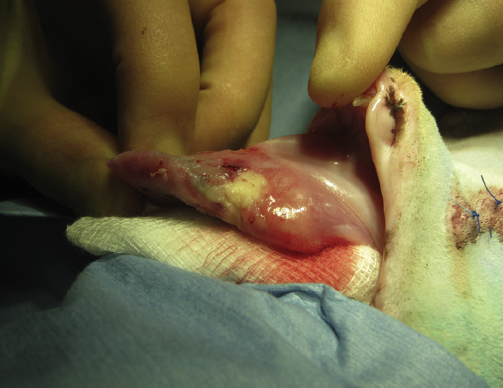
The glossal enlargement was extensive with areas of erosion and ulceration.
Histopathological evaluation of the lingual biopsies revealed a round cell neoplasm, with lymphoma the favoured diagnosis. There was a low mitotic index and superficial secondary ulceration. Lymph node biopsies revealed moderate to severe follicular hyperplasia which was likely due to the concurrent inflammation. Aerobic culture of the lymph node revealed profuse growth of an Enterococcus species. Mycobacterial culture was negative.
Twenty-four hours later, the cat became dyspnoeic. Thoracic radiographs revealed a diffuse alveolar pattern within the pulmonary parenchyma and mild pleural effusion. This was potentially attributable to cardiac failure, secondary to fluid therapy although fairly conservative rates had been maintained throughout the hospitalisation. It was possible that the cat had a subclinical cardiomyopathy; or possibly lymphoma affecting the myocardium. The radiographic findings could also have been consistent with aspiration pneumonia. There was a partial response to frusemide at 0.1 mg/kg q 8 h (Dimazon; Schering-Plough Animal Health). At this stage however, considering the guarded prognosis and need for further medical/surgical management, the owners elected for euthanasia and necropsy was performed.
On gross examination, there was mild enlargement of both the left and right superficial cervical lymph nodes. There was a full-thickness cream-coloured soft mass towards the root of the tongue; the lingual surface had multifocal erosions and ulcerations. The pleural cavity contained approximately 43 ml of serosanguinous fluid; the heart and lungs were grossly unremarkable. The capsular surface of the left kidney was irregular in outline and contained a 2 cm diameter cream nodular mass within the cortex and medulla at its cranial pole. The right kidney was small and irregular and there was mild hydronephrosis.
Histopathological examination confirmed the clinical suspicion of a neoplasm and the previous biopsy diagnosis of lymphoma within the tongue and an additional tumour within the left kidney. Within the tongue, there were large areas of myofibre disruption by a dual population comprising larger, neoplastic lymphocytes forming broad sheets, interspersed with smaller, more mature, lymphocytes (Fig 3). The mass within the left kidney was also composed of a similar dual population of lymphocytes. It was impossible to ascertain which of the lingual or renal location represented the primary location of the tumour.

Microscopic images of the tongue at 20× (a) and 100× magnification (b). On histopathological examination of the tongue, there were areas of myofibre disruption (arrow) by sheets of neoplastic lymphocytes. There was extensive ulceration of the tongue surface, including superficial infiltration with neutrophils and approximately one-third of the examined sections were composed of necrotic tissue. There were populations of large and small lymphocytes with moderate anisocytosis and anisokaryosis in the large neoplastic cells, and minimal anisocytosis in the smaller cells (reactive T-cells). Mitotic figures averaged 1.5 in 10 40× fields.
Immunohistochemical staining with CD3 and CD79a antibodies revealed diffuse intense cytoplasmic staining of the population of larger, more blastic appearing neoplastic lymphocytes with CD79a and strong cytoplasmic staining of the scattered smaller lymphocytes with CD3, thus the latter likely representing an inflammatory component.
The right kidney had an irregular contour throughout the capsular surface and diffusely the cortex and medulla were thinned. Numerous tubules were dilated by protein casts. These changes were consistent with mild chronic interstitial lymphoplasmacytic nephritis. There was multifocally extensive myocardial degeneration in the heart and areas of fibrosis. The pleural fluid was consistent with a transudate. The degeneration and fibrosis within the myocardium was likely to be incidental; however, a mild cardiomyopathy is a possibility. The changes within the contralateral kidney were considered incidental age-related findings.
Lingual disease in cats is rare with trauma representing the most common cause. Injury to the tongue can be secondary to burns, foreign bodies, laceration from linear foreign bodies, from self-trauma following seizures and anaesthesia 1 and the feline oropharyngeal pain syndrome. Linear foreign bodies can become entrapped at the lingual frenulum and cause a large granulomatous mass which can be difficult to distinguish from a neoplasm or an eosinophilic granuloma. In addition, unusual foreign bodies can cause granulomatous lesions, eg, grass awns and fish bones. Other causes of ulceration include calicivirus or herpesvirus infection, cryptococcosis, abscessation, severe periodontal disease or uraemia. Other reports of lingual lesions include ectopic thyroid tissue 2 and calcinosis circumscripta. 3
Oncological diseases of the tongue are rare in veterinary patients. The most common canine lingual tumour is squamous cell carcinoma (SCC) and accounts for almost half of all cases. 4 In feline patients, SCC is also the most common neoplasm and is most commonly located on the ventral surface near the frenulum. Ulceration is usually associated with these tumours. Clinical signs associated with tongue neoplasia include halitosis, ptyalism, dysphagia, anorexia, haemorrhage and respiratory stridor. Other feline lingual neoplasms reported include papillomas, 5 granular cell tumours 6 and haemangioma. 7 Oral lymphoma has been reported in the cat and mostly affects the tonsils. 4,8 A retrospective study conducted on 371 oral neoplasms identified 11 cases with oral lymphoma. 9 The lesions were single or multiple raised submucosal masses composed of unencapsulated sheets of neoplastic lymphoid cells. To the authors’ knowledge this is the first report of lymphoma affecting the tongue of a cat.
Lymphoma is the most common haematopoietic neoplasm affecting small animals 10 and is the most common neoplasm in cats. 11 Renal lymphoma is a form of extranodal lymphoma and can be primary or associated with involvement of other tissues often associated with extension to the central nervous system and is most commonly bilateral. 10,12,13 Approximately 50% of affected cats have signs related to renal insufficiency. 12,13 A recent study of 149 cases of feline lymphoma 8 identified 11 cases of concurrent laryngeal and one case of concurrent pharyngeal lymphoma.
Although in our case, the cat exhibited chronic signs of renal insufficiency, it was the lingual mass that was the most conspicuous clinical problem. However, given the level of azotaemia in our cat, an abdominal ultrasound scan or abdominal CT scan may have been performed to assess the renal parenchyma and would most likely have identified the renal mass; an ultrasonographically-guided aspirate of the mass may have allowed a more prompt diagnosis.
If the cat in this report had not developed pleural effusion and respiratory distress, treatment would have involved a systemic chemotherapy protocol involving agents such as prednisolone, vincristine, cyclophosphamide, l-asparginase, likely without doxorubicin because of the chronic renal insufficiency. 12 Renal lymphoma is associated with shorter survival times than for lymphoma in other locations; median survival ranges from 3 to 6 months. 10,13,14 Radiotherapy as a component of therapy may have been of great benefit to this cat to rapidly reduce the size of the primary lesion.
Surgical resection is indicated for certain lingual tumour types such as SCC, or for tumours without evidence of metastasis. Rostral neoplasms have a favourable prognosis as they are detected earlier and are easier to remove surgically. Excision of the majority or the entire tongue was well tolerated by five dogs in a case series. 15 To the authors’ knowledge, no such study has ever been performed in cats. One can presume that a subtotal glossectomy would be poorly tolerated in this species due to their grooming habits. Additionally, as lymphoma is a systemic disease, surgery might not have been indicated in this case, except for controlling the complicating tissue necrosis and secondary infection.
This case demonstrates the first case of lingual lymphoma in the cat and illustrates how their diagnosis can be challenging. Although rare, neoplastic disease should be considered for any tongue lesion and can be associated with tumours in other locations, prompting extensive staging.