
Abstract
Sporotrichosis is caused by
Sporotrichosis is an infectious disease caused by the dimorphic fungus
The definitive diagnosis of sporotrichosis requires the isolation of
For cytopathological examination, specimens can be obtained by aspiration of nodules, impression smears of ulcerated skin or exudate and by fine-needle aspiration of the lesion. 15 Smears are air-dried and stained with Romanowsky-type stains (eg, Wright or DiffQuik). 16 These stains are used in veterinary practice because of its low cost and simple preparation. 15 Analysis of exudate from skin lesions of cats with sporotrichosis often reveals numerous round, oval or cigar-shaped yeast-like structures inside macrophages and neutrophils or in the extracellular medium. These structures measure 3–5 μm in diameter and 5–9 μm in length, and are surrounded by a clear halo (Fig 1). 16

Photomicrograph of impression smear from ulcerated skin lesion in cat with sporotrichosis showing numerous cigar-shaped to oval or round budding yeast-like organisms fulfilled with blue cytoplasm with a single round pink nucleus surrounded by a non-staining cell wall within macrophages and extracellularly (Quick Panoptic stain, 100×).
The rapid, practical and reliable diagnosis of feline sporotrichosis is important for the early treatment of cats and prevention of zoonotic transmission. Although cytopathology is considered to be the fastest, most straightforward, and least expensive method for the diagnosis of feline sporotrichosis, 15 its sensitivity has not been investigated. Therefore, the objective of the present study was to establish the sensitivity of cytopathological examination in the diagnosis of feline sporotrichosis using mycological culture as the gold standard.
Materials and methods
Medical records from a cat cohort treated at the Laboratório de Pesquisa Clínica em Dermatozoonoses em Animais Domésticos (Lapclin-Dermzoo), IPEC/Fiocruz, Rio de Janeiro, Brazil, between 2004 and 2006 were reviewed. Criteria for inclusion were a diagnosis of sporotrichosis by isolation of
For culture, the material collected with a sterile swab was seeded onto Sabouraud dextrose agar and Mycobiotic agar (Difco) and incubated at 25°C. Dimorphism was confirmed by conversion to the yeast phase in brain-heart infusion broth at 37°C. For cytopathology, impression smears of the skin lesion were prepared on clean and dry glass slides and stained by the Quick Panoptic method (Instant Prov; Newprov), a Romanowsky-type stain similar to DiffQuik. The slides were analyzed by light microscopy using 40 and 100× objective lenses for the identification of yeast-like structures suggestive of
Results
A total of 806 cases of feline sporotrichosis from the metropolitan region of Rio de Janeiro were included in the study (Fig 2). Cytopathological examination of the skin lesions of these cats revealed the presence of yeast-like structures suggestive of
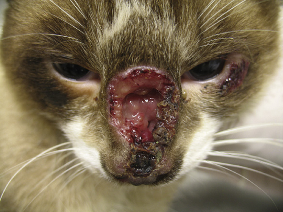
Feline sporotrichosis: ulcer on the bridge of the nose.
Discussion
This study represents the largest series of feline sporotrichosis cases diagnosed by fungal isolation and cytopathological examination. Several investigators reported the observation of abundant yeast-like structures by cytopathological examination of impression smears prepared from lesions of cats with feline sporotrichosis 11,17–23 , but the sensitivity of the method was not evaluated.
The sensitivity of cytopathology was considered to be satisfactory. This value was higher than that reported for histopathology (62.2%) using hematoxylin–eosin, Grocott's silver stain and periodic acid-Schiff for the diagnosis of feline sporotrichosis in 90 cats compared to isolation of
In order to obtain adequate samples for cytological examination for feline sporotrichosis thin smears of lesions with more vital-appearing tissue rather than those consisting of primarily dried, crusted exudate should be sampled. 15 Additionally, at least two slides with three impressions on each of them are recommended to better obtain positive specimens.
For biosafety purposes the collection of cytology samples should be undertaken with standard safety procedures including disposable gloves, long sleeved impermeable gowns and optimal physical restraint of the animal. 26
Despite its lower sensitivity compared to mycological culture and the fact that the fungal elements observed might be confused with poorly encapsulated
Considering the sensitivity of cytopathology found in the present study, a positive result in a cat with a clinical suspicion of sporotrichosis permits the immediate initiation of antifungal treatment and the establishment of measures to prevent the transmission of
Footnotes
Acknowledgements
The authors thank the staff of Mycology Laboratory of IPEC/Fiocruz, Raquel de Oliveira and Ingrid Paes for technical support. This study was partially supported by the Health Strategic Research Support Program (PAPES V) – Fiocruz and by Grant Programme for the Study of Neglected and Reemerging Diseases/The Carlos Chagas Filho Foundation for the Support of Research in the State of Rio de Janeiro (FAPERJ). TMPS has a fellowship from the Technological and Scientific Development National Council (CNPq).