
Abstract
The aim of this study is to characterise the feline mammary echotexture using B-mode ultrasonography, which is not routinely used to examine the feline mammary gland. Using a 5–9 MHz linear transducer the ultrasonographic appearance of non-stimulated and stimulated mammary glands was determined in 35 mature intact non-pregnant, pregnant and lactating queens aged from 16 months to 8 years. In intact non-pregnant queens, mammary glands are fairly underdeveloped and on the ultrasonograms they appear with a regular hypoechoic texture and generally show a thickness of less than 2.0 mm. The stimulated mammary tissue typically presents a more hyperechoic appearance compared to the non-stimulated gland and a fine granular echotexture. Maximum echogenicity of the mammary gland is reached during lactation. In late pregnancy, the mammary glands reach 6–9 mm in thickness. During lactation, the size of the glands depends on the existence of a suckling stimulus, with the suckled glands reaching about 11 mm in thickness. Ductal structures can only be imaged during late pregnancy and lactation. Ultrasonographic evaluation of the feline mammary gland can become a valuable diagnostic tool to characterise physiological changes and may further contribute to a better characterisation of diseased mammary tissue.
Sonographic breast imaging, along with mammography, is currently used as part of the standard procedure in the diagnosis of human breast masses (Svensson 1997, Schroeder et al 2003, Sehgal et al 2006). This rapid, simple and non-invasive technique provides useful information on the characteristics of a tissue, using a grey scale and two-dimensional image. The use of ultrasonography in the study of the mammary gland has been also developed for farm animals (Nudda et al 2000, Franz et al 2004). However, information on sonographic characteristics of small animal mammary glands is scarce. In the dog, some reports have been published on the sonographic features of the normal mammary gland (Trasch 2006) and of mammary tumours (Gonzalez de Bulnes et al 1998, Marquardt 2003, Nyman et al 2006). To the authors' knowledge, studies on the sonomorphological characteristics of the feline mammary gland have not been previously published.
Female cats usually have four pairs of mammary glands arranged in two rows, extending from the caudal part of the pectoral region to the inguinal region (Barone 1978). They are usually identified as thoracic, cranial and caudal abdominal and inguinal mammary glands, or simply as two thoracic pairs and two abdominal pairs of mammary glands (Johnston et al 2001).
Histologically, the non-stimulated mammary gland in mature intact or neutered queens possesses a very small amount of dense stroma and fat, and the glandular tissue is almost absent, with the thoracic glands demonstrating less development than the abdominal glands. The teat is rich in smooth muscle that histologically shows a three-layer arrangement. In the teat, one to eight ducts may open in an almost circular, irregular pattern (Barone 1978, Johnston et al 2001). Each teat duct connects to a small teat sinus, which continues into a lobule of the gland. Within the lobule, smaller branching ducts lead to the alveolar opening (Johnston et al 2001).
In the absence of previous studies on this subject the aim of this work is to establish the ultrasonographic appearance of the normal, healthy feline mammary gland and to assess the efficiency of ultrasound evaluation in clinical mammary examination. Hence, transcutaneous ultrasonographic imaging has been used to characterise the sonomorphological features of the feline mammary gland, using both pregnant and lactating animals (stimulated gland), and mature non-pregnant queens (non-stimulated gland).
Material and methods
In this study, 35 healthy intact domestic queens aged from 16 months to 8 years old were evaluated with owner consent. Experiments were conducted over a 6-month period at the Veterinary Teaching Hospital of University of Trás-os-Montes and Alto Douro (UTAD). Animals were selected from those presented for pregnancy diagnosis or routine procedures. The mammary glands of selected animals evidenced diverse functional states and the animals were grouped in mature, cyclic, non-pregnant animals (n=13), in pregnant animals (n=14) and in lactating females of less than 4 weeks post partum (n=8). Pregnant queens were further divided into early pregnant (until 40 days of pregnancy; n=6) and late pregnant (with more than 40 days of pregnancy; n=8) females. Pregnancy was staged using the ultrasonographic characteristics of the embryo and fetus as described by Zambelli and Pratti (2006). This study was developed with 16 Persian females and 19 mixed-breed females, evenly distributed between the diverse groups.
Ultrasonography was performed using a linear-array transducer of 5–9 MHz and a Philips HD3 scanner (Philips Medical System, USA). The animals were maintained in dorsal recumbency with the rear limbs in the frog-leg position for ultrasonographic examination. Generally, the ultrasound procedures were made without hair clipping. A sufficient amount of acoustic gel was used after alcohol impregnation of the cat's coat. No pressure was needed using the transducer to obtain good-quality images. In the non-pregnant and early pregnant animals only the abdominal mammary glands were evaluated, due to the insufficient development of the thoracic glands. In pregnant and lactating females all the mammary glands were evaluated. Each mammary gland was evaluated using longitudinal and transverse scans. The glands' echogenic pattern was described in terms of the homogeneity (homogeneous, heterogeneous) and echogenicity (hypoechoic, isoechoic or hyperechoic) and also for the presence of anechoic structures corresponding to the ductal structures of the mammary gland. The size of the mammary gland and the existence of artefacts and its appearance were noted.
All examinations were recorded for later analysis on a digital video camera (Sony Handicam, DCR-HC40E, Sony Corporation, Tokyo) as MPEG1 files (25 frames per second; 320×240 pixels). On the scanner two still-images (freeze-frames on the monitor) for each location were additionally captured and recorded as a TIFF file. In order to reduce any examination bias, a single operator performed all the ultrasound evaluations.
Basic statistical analysis of the mammary gland thickness (average, minimum, maximum and standard deviation) was performed using the Statview software for Macintosh (version 5.0.1, SAS Institute).
Results and discussion
In mature intact non-pregnant queens, the mammary gland is almost non-existent (Johnston et al 2001) and in ultrasound images the mammary tissue frequently shows a smaller thickness than the abdominal wall. In ultrasonograms the non-stimulated mammary gland is evidenced as a homogeneous, hypoechoic tissue of less than 2.0 mm thickness (Table 1; Fig 1A and B). Its echogenic pattern reflects the small amount of low-density stroma and glandular tissue is almost absent (Fig 1B). When non-stimulated, the mammary ducts remain narrow and are not usually visualised during ultrasonographic examination. Consequently, no anechoic structures are apparent on ultrasonograms (Fig 1).

Ultrasonograms of the non-stimulated feline mammary gland. (A) In the non-pregnant queen, ultrasonograms evidence a homogenic and hypoechoic parenchyma (arrowhead) above the abdominal wall (arrow). (B) The mammary parenchyma shows a discrete thickness of about 1.4 mm (delimited with the cursors). The arrow points to the abdominal wall (A and B).
Basic descriptive statistics of the queen mammary gland
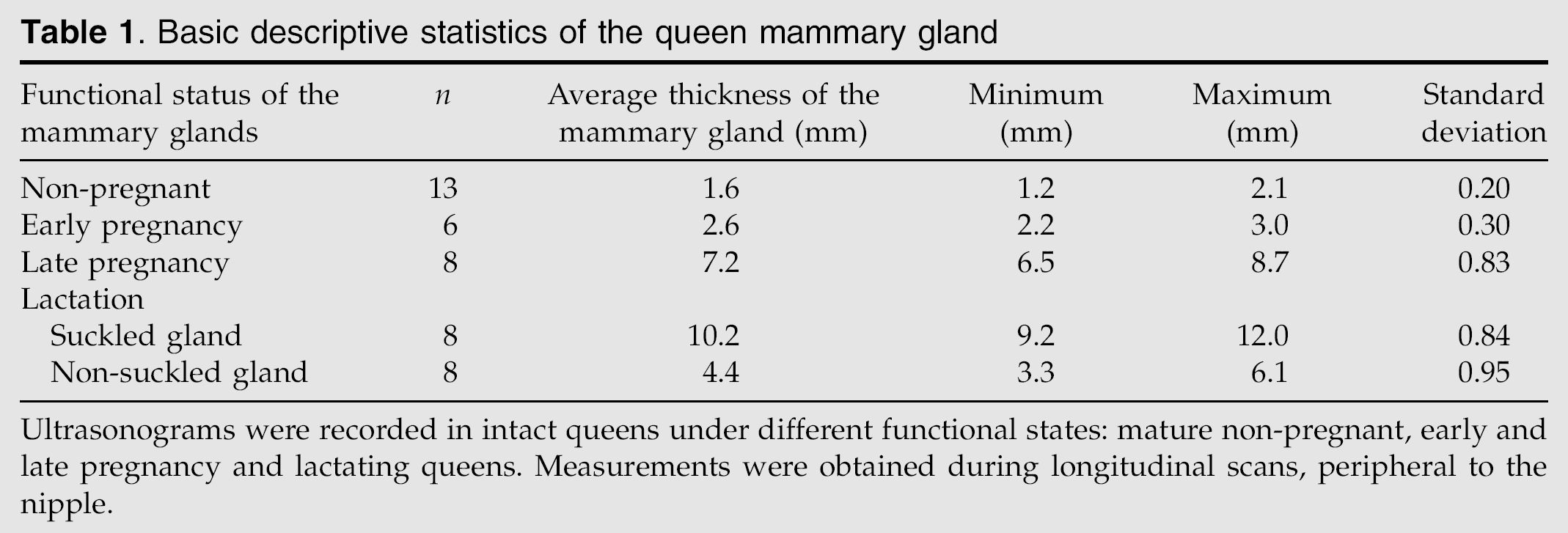
Ultrasonograms were recorded in intact queens under different functional states: mature non-pregnant, early and late pregnancy and lactating queens. Measurements were obtained during longitudinal scans, peripheral to the nipple.
The stimulated mammary gland gradually increases in size and in echogenicity relative to the non-stimulated gland as pregnancy lengthens, reaching maximum echotexture during lactation (Table 1). In the woman and the bitch, the non-stimulated mammary tissue evidences a lesser echogenicity than the stimulated gland (Svensson 1997, Trasch 2006). Compared to those species, the female cat is unique in having a very small thickness of the mammary gland.
In early pregnant queens with less than 40 days of pregnancy, a slight increase in the mammary gland dimensions is observed, but it rarely exceeds a 3-mm thickness (Table 1; Fig 2). The echoic pattern of the mammary gland at this stage is similar to that observed in non-pregnant females. Its echotexture remains uniformly hypoechoic, demonstrating its low density (Fig 2).

Mammary ultrasonograms of early pregnant queens. A moderate increase in the size of the mammary gland (arrowhead) is observed (A), whereas the echogenicity of the parenchyma remains hypoechoic (A and B). The large arrow points to the abdominal wall (A and B).
In the later pregnancy, after day 40, a more pronounced increase of the mammary size is demonstrated, and the glands reach 6–9 mm in thickness (Table 1; Fig 3A). Additionally, an increase in the echogenicity of the gland is observed. Its echogenic pattern is coarsely granular and small anechoic areas are evidenced, possibly corresponding to the ductal structures of the mammary gland (Fig 3B). It is also observed that the dorsal margins of the mammary glands are more hyperechoic and distinct than during early stages of pregnancy (Fig 3A and B).

Ultrasonograms of the late pregnant feline mammary gland. (A) The mammary parenchyma (arrowhead) evidences a marked increase in width (between cursors) and is easily discernible from the adjacent abdominal structures (large arrow). (B) Additionally the mammary tissue (arrowhead) acquires a more echogenic pattern and some small anechoic areas are evidenced in the parenchyma, possibly corresponding to ductal structures of the mammary gland (small arrowhead). An increase definition is also observed between the dorsal margins of the gland and the adjacent abdominal wall (large arrow).
Only at the time of parturition do all the mammary glands demonstrate similar echogenic features and achieve a higher thickness. From that moment on, in the lactating queen the size and the ultrasound features of the mammary glands reflect the existence or the absence of a suckling stimulus. The suckled mammary gland is moderately echogenic with a coarse granular pattern (Fig 4A and B). Furthermore, well-defined anechoic structures are easily evidenced and seem to correspond to the ductal milk collecting system (Fig 4A). It is also possible to observe very small anechoic areas over the nipple that may correspond to teat ducts. Consequently, the parenchyma is now rather heterogeneous (Fig 4B). Similar to late pregnancy, the dorsal margins of the mammary glands appear strongly hyperechoic (Fig 4A–D). Compared to the former the non-suckled gland parenchyma evidences a more hypoechoic and slight granular echotexture, achieving characteristics similar to those evidenced during late pregnancy (Fig 4C and D). They are also smaller than the suckled mammary glands (Table 1; Fig 4C and D).

Mammary gland ultrasonograms in the lactating queen. The echogenic appearance of the mammary gland can vary depending on whether a suckling stimulus exists (A and B) or not (C and D). (A) The suckled mammary gland (arrowhead) reaches its maximal physiological dimension as well as the nipple; the mammary gland and the nipple are delimited with + cursors and with × cursors, respectively. Some ductal structures are evidenced in the parenchyma (small arrowheads). (B) The mammary tissue (arrowhead) is rather echoic, with some anechoic areas, giving a heterogeneous appearance to the mammary gland. The dorsal margins of the mammary gland are clearly visible. The nipple usually induces a reverberation artefact (asterisk) on the adjacent parenchyma. (C and D) On the sonograms of the non-suckled mammary gland a small development of the glandular parenchyma (delimited with cursors) is easily seen. The echogenicity of the parenchyma (arrowhead) is hypoechoic, similar to that exhibited during late pregnancy. The nipple (asterisk) is evidenced at the top of the ultrasonogram (C) being clearly observed a hypoechoic area within this structure. Some small anechoic areas corresponding to ductal structures (small arrowheads) can be observed.
The queen's mammary gland ultrasonographic images are fairly distinct from those described for women and bitches. When compared to the appearance of canine mammary ultrasonographs, the non-stimulated queen mammary gland seems to remain less echogenic, possibly due to its small size. Also the stimulated queen mammary gland shows a relatively hypoechoic echotexture compared to the canine gland, although showing a similar granular pattern. In a pregnant and lactating human breast, a classic grey-scale ultrasound shows that the breast parenchyma has a homogeneous pattern with low-level echoes, while in a non-stimulated breast the mammary parenchyma is replaced by fat at varying rates (Svensson 1997). In contrast to women, the canine lactating mammary gland has been described as having a moderately echogenic and coarse granular pattern, while the non-stimulated gland shows a more hypoechoic tissue with a fine granular appearance (Trasch 2006, Trasch et al 2006). According to Kealy and McAllister (2000), on ultrasonographies the dog mammary tissue is echogenic and homogeneous, but no reference is made to the functional state of the mammary glands that corresponds to these features. Nevertheless, according to Marquardt (2003) due to the anatomical and histological differences of the canine and human mammary tissue, it is not advisable to directly compare the ultrasonographic findings between these two species. The cow appears to be similar to the dog; on B-mode ultrasonographic images, the glandular parenchyma of pregnant cows producing 20–25 kg of milk daily is described as homogeneous and hyperechoic. In the cow it is also possible to visualise anechoic alveoli, and the gland cistern is anechoic even when filled with milk (Franz et al 2004).
On ultrasonographic images taken over the teat of both suckled and non-suckled feline glands, either on longitudinal or transverse scanning, a reverberation artefact can be visualised, consisting of two to three hyperechoic layers (Fig 4B). This artefact could be produced by the laying of the nipple over the skin during the ultrasound scanning of the mammary gland, generating a false, compact folding of the skin at this level. A similar artefact arrangement was previous reported by Franz et al (2004) in the bovine mammary gland ultrasonographies.
Apparently, no differences were found in the ultrasonographic features of the mammary tissue between Persian and mixed-breed queens, in either the non-stimulated or the stimulated gland.
Conclusions
The findings described in this study allow characterisation of the normal ultrasonographic features of feline mammary glands, as well as the variations associated with pregnancy and lactation. Clear differences in size, echogenicity and homogenicity were found between stimulated and non-stimulated mammary gland tissue. From the ultrasonographic features described here we can conclude that B-mode ultrasonography is a useful and accurate method in the characterisation of the physiological changes and functional condition of the feline mammary gland parenchyma.