
Abstract
Case report
History and physical findings
An 8-month-old spayed Himalayan cat was presented on a Monday morning by one of the cat's owners with an acute history of ‘agitation’ and being unable to walk. The cat lived exclusively indoors in an affluent suburb near the central business district of Sydney. It had a habit of sleeping in the kitchen cupboard, and had been accidentally locked in that cupboard overnight.
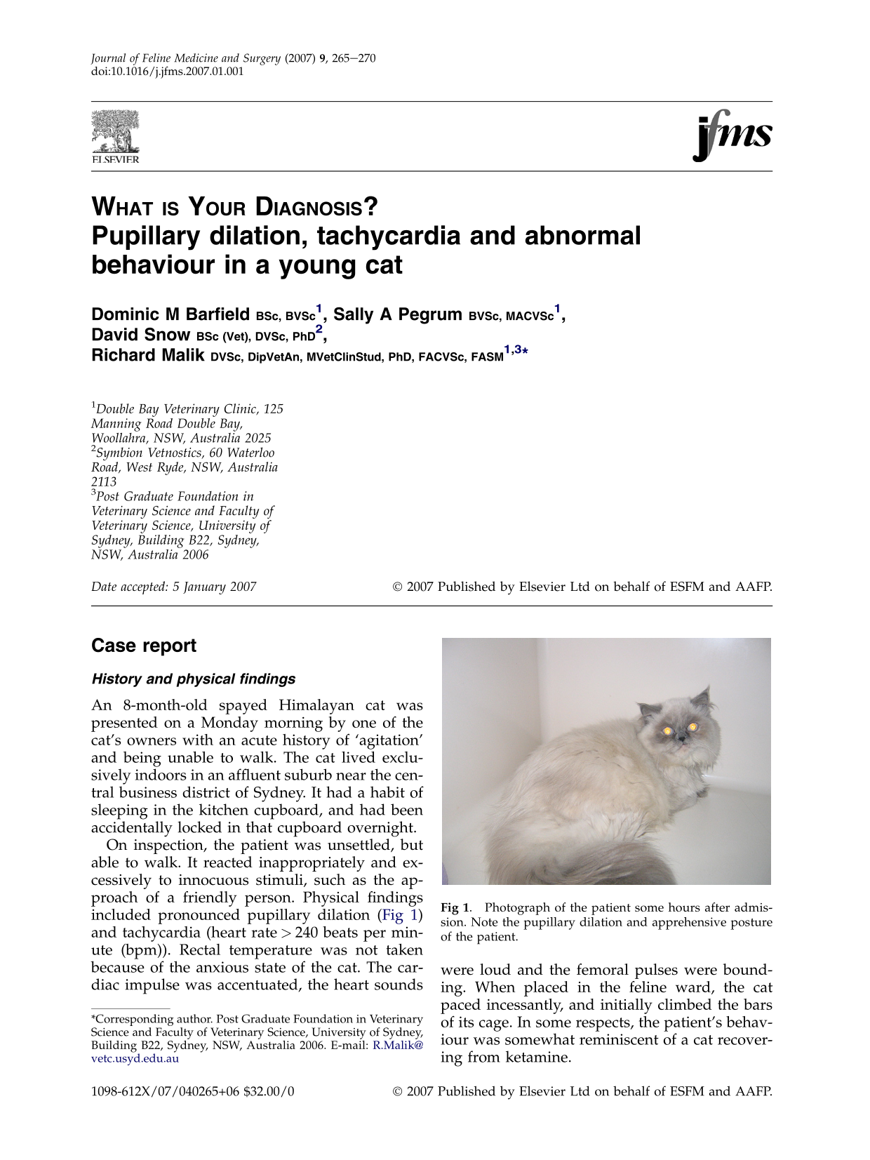
On inspection, the patient was unsettled, but able to walk. It reacted inappropriately and excessively to innocuous stimuli, such as the approach of a friendly person. Physical findings included pronounced pupillary dilation (Fig 1) and tachycardia (heart rate>240 beats per minute (bpm)). Rectal temperature was not taken because of the anxious state of the cat. The cardiac impulse was accentuated, the heart sounds were loud and the femoral pulses were bounding. When placed in the feline ward, the cat paced incessantly, and initially climbed the bars of its cage. In some respects, the patient's behaviour was somewhat reminiscent of a cat recovering from ketamine.

Photograph of the patient some hours after admission. Note the pupillary dilation and apprehensive posture of the patient.
The owner was quizzed about possible access to toxicants, including spoiled food, exotic plants, ‘party’ drugs and prescription medication; he denied these various possibilities. The cat was hospitalised in a cool quiet ward for observation while plans for investigation and treatment were formulated.
Questions
What is your assessment? How would you investigate this case further? How would you manage the patient?
Answers on page 267
Answers to What is Your Diagnosis? on page 265 Pupillary dilation, tachycardia and abnormal behaviour in a young cat
1. What is your assessment?
The physical findings suggested that, potentially, there were two distinct problems: (i) a cardiovascular problem to account for the tachycardia and increased cardiac impulse; and (ii) a neurological and/or behavioural problem that accounted for the abnormal demeanor of the cat and its pupillary dilation. The most parsimonious way to explain both problems would be to implicate the action of a toxicant or toxicants that would cause sympathetic activation and behavioural aberrations concurrently.
Possibilities considered included endogenous toxins (eg, those giving rise to uraemic or hepatic encephalopathy) and exogenous toxicants. Endogenous toxins seemed unlikely given the peracute development of signs. Furthermore, hepatic encephalopathy is usually associated with hypersialism in the cat, and usually other findings are evident such as stunted growth, copper-coloured irises, and intermittent gastrointestinal signs. Similarly, uraemic encephalopathy usually is accompanied by other signs, eg, polyuria, polydipsia, depression, poor appetite and anaemia.
The possibility of an acute intoxication therefore seemed most cogent. The list of potential exogenous toxicants included plants (eg, cannabis and the fruit of Duranta erecta (Scanlan et al 2006)), fungal toxins (eg, penetrem A (Arp and Richard 1979) on moldy food, poisonous mushrooms), naphthalene, prescription medication inadvertently given to or ingested accidentally by the cat or ‘recreational’ drugs (tetrahydrocannabinol, phencyclidine, ketamine, cocaine, amphetamines, opiates) ingested accidentally.
2. How would you investigate this case further?
Philosophically, two different conceptual approaches could have been adopted in the patient of this report: (i) a conservative one, based on observation and minimally invasive diagnostics and symptomatic care, and (ii) an aggressive approach, with heavy sedation, collection of blood and urine specimens for extensive non-discretionary testing, gastric lavage under anaesthesia, intense cardiovascular monitoring (indirect Doppler or oscillometric blood pressure, continuous electrocardiography) and so on. We elected to follow the first approach, at least in part because the case was seen in a first opinion practice.
Although haematology, serum biochemistries and urinalysis findings would have provided useful additional data to rule in or rule out certain diagnostic possibilities, it was considered that the restraint required to permit safe venepuncture was not sufficiently justified given the agitated state of the patient. Further consultation with the owners was considered the most expedient way to determine the underlying aetiology.
In a telephone conversation with the owner's wife, it became clear that the cat may have had access to cocaine, as it was available in the owner's house over the weekend. Apparently the illicit material had been available ‘on plates’ on the Saturday night, 2 days before the cat was admitted. It was not possible to determine whether the cat had licked a surface contaminated with cocaine, or if powder had somehow been deposited in its coat, and was ingested subsequently.
As the cat's signs were compatible with cocaine intoxication, it was treated on that basis (see below). It seemed likely that the patient had received only a small dose of cocaine on an mg/kg basis given the accidental nature of the intoxication.
To confirm the tentative diagnosis, polystyrene packing beads were used in place of cat litter to obtain a urine specimen for toxicological studies. Such a specimen was obtained overnight, and submitted to a veterinary laboratory (Symbion Vetnostics) for a urine ‘drug screen’ using enzyme immunoassay technology (Clonal Enzyme Doctor Immunoassay; Microgenics). The primary screen was positive for cocaine metabolites and benzodiazepines, but negative for methadone, opiates, sympathomimetics, cannabinoids and barbiturates. The presence of benzoylecgonine and ecgonine methyl ester (cocaine metabolites) and oxazepam and temazepam were confirmed subsequently by gas chromatography/mass spectrometry (using an Agilent 5973 N GCMS instrument) following hydrolysis using β-glucuronidase (at a pH of 5), buffering to pH 9 and extraction into an organic solvent.
3. How would you manage this patient?
We elected to initially try a minimalist approach, given the mild nature of the cat's symptomatology. Shortly after a tentative diagnosis of cocaine intoxication was made, the cat was given atenolol (6.25 mg orally; 1/4 of a 25 mg tablet) and placed in a cool quiet environment with a blanket over the front of its cage to reduce ambient visual and auditory stimuli. The rationale for giving atenolol was that it was a cardioselective β-adrenoreceptor antagonist, and would therefore reduce the chronotropic and inotropic effects of cocaine on the myocardium. Provided peripheral vascular resistance was unchanged, this was likely to result in reduced blood pressure.
The cat was substantially improved after several hours (with a heart rate of 160 bpm) and was unremarkable in all respects the following morning, when it was discharged. The owners were counselled about dangers of ‘party’ drugs being accidentally ingested, in an attempt to prevent further similar episodes occurring.
Had we known more about cocaine intoxication when we were managing this case, we would have probably administered acepromazine (0.05 mg/kg) as well, for its sedative and hypotensive qualities, and drawn up (but not administered) a therapeutic dose (0.3 mg/kg) of midazolam in a needle and syringe, to have at the ready in case the cat developed convulsions.
Discussion
Cocaine is a stimulant and a local anaesthetic with potent vasoconstrictor properties. The drug produces physiological and behavioural effects following oral, intranasal, intravenous or inhalation following pyrolysis (smoking). The drug has potent and complex pharmacological effects on dopaminergic, noradrenergic and serotoninergic neurons within the central nervous system (Mendelson and Mello 1994). Because cocaine inhibits reuptake of catecholamines at adrenergic nerve endings, it potentiates sympathetic nervous system activity centrally and peripherally. Cocaine also blocks voltage-gated sodium channels in excitable tissues including the myocardium (Rezvani and Hartfield 2006).
Cocaine produces a brief stimulation and enhancement of mood (in humans) and dose-related increases in heart rate, blood pressure and body temperature. High doses may induce lethal pyrexia and/or hypertension. Cocaine has a short plasma half-life (in the order of 60–90 min in man) and is metabolised primarily by liver and plasma esterases, resulting in excretion of metabolites in the urine (Mendelson and Mello 1994). It is rapidly absorbed across oral mucous membranes and from the stomach following ingestion. In humans, 20% of an oral dose is absorbed. In patients with cocaine intoxication, the rapid uptake of the drug limits the value of procedures designed to prevent absorption, such as emesis, gastric lavage, and dosing with activated charcoal. These would in any case be difficult to implement in an affected feline patient, without recourse to general anaesthesia. Furthermore, by the time clinical signs have developed, inducing emesis risks precipitation of a seizure, with the concomitant risk of aspiration (Llera and Volmer 2006).
Although the recreational use of cocaine is said to be relatively ‘safe’, there are numerous reports of human death from respiratory depression, cardiac arrhythmias and convulsions after snorting or intravenous administration. Toxicity is especially common in people who transport cocaine within their body – the so-called ‘body packer syndrome’ (Wetli and Mittlemann 1981). Symptoms include mydriasis, seizures, acute toxic psychosis, and coma in various combinations.
Treatment of cocaine overdose is a medical emergency (Shanti and Lucas 2003). Severe intoxication can result in pyrexia, hypertension, tachycardia, tonic–clonic seizures, dyspnoea and ventricular arrhythmias. The effects on the heart are especially worrisome, as increases in myocardial oxygen consumption due to increased rate and strength of cardiac contractions occur in the setting of coronary arterial vasoconstriction and increased afterload. Intravenous diazepam (0.5 mg/kg) or lorazepam (0.05 mg/kg) have been shown to be effective in controlling seizures in human patients, including children (Rezvani and Hartfield 2006). Cardiac issues have been managed successfully using intravenous propranolol (Mendelson and Mello 1994). There is experimental work suggesting that administration of butyrylcholinesterase may be helpful in reducing serum concentrations of cocaine and its active metabolites (Mattes et al 1997).
Few reports document cocaine intoxication in companion animals. Intoxication has been reported in police dogs used for drug detection (Dumonceaux and Beasley 1990), and in a dog that lived in an illicit cocaine manufacturing facility (Frazier et al 1998). It should be borne in mind that the LD50 in the dog is said to be 13 mg/kg for intravenous administration, with the oral LD50 2–4 times higher. Even if the cat is more sensitive to cocaine than the dog, as has been suggested (Coppock et al 1989), it is unlikely that a feline patient would be exposed to such doses due to accidental ingestion of recreational quantities of the drug.
In the present case, clinical signs were considered relatively mild and likely self-limiting. Accordingly, atenolol was administered orally in an attempt to reduce the rate and strength of cardiac contractions, thereby reducing myocardial strain and indirectly reducing blood pressure. It could be argued that diltiazem (1/8 of 60 mg tablet) would have been a better choice, as it produces coronary and systemic vasodilation in addition to reducing the inotropic and chronotropic state of the heart. We elected not to administer a phenothiazine sedative such as acepromazine, as we were concerned that this might precipitate convulsions by lowering the seizure threshold; however, the experimental literature does not bear out this concern (Catravas and Waters 1981), and in dogs chlorpromazine pretreatment prevented cardiovascular effects, minimised seizures and prevented hyperthermia. We also considered sedating the cat using phenobarbitone, but elected not to do so, as time (ie, sufficient for biotransformation) and the atenolol seemed to be producing a satisfactory effect. Although the cat's temperature was not measured, it was fortunate that our cat ward was air-conditioned, as a warm ambient environment would have been inadvisable given that pyrexia is commonly seen with cocaine intoxication.
We can only speculate about the presence of benzodiazepines in the urine specimen submitted for analysis. Certainly, they were not administered by the veterinary team. Perhaps the cocaine supply had been ‘cut’ with temazepam powder, which was subsequently metabolised hepatically to oxazepam and excreted in the urine. Apparently, cocaine and amphetamine users have found that benzodiazepines are effective at blunting some of the more unpleasant effects of these stimulants (Sheehan et al 1991). Incorporating a drug like temazepam in a mixture with cocaine might produce a more acceptable balance of positive and negative effects, and is compatible with the toxicology results in our patient (Sheehan et al 1991). The possibility of such drug combinations should be borne in mind when assessing the signs evident in a cat exposed to ‘party drugs’, and in determining a drug regimen for therapy. Cocaine can also be ‘cut’ with a variety of other substances, including mannitol, inositol, sucrose, and cornstarch, which may indeed render the resulting mixture more palatable for cats. Additional adulterants which may also be present include lignocaine, procaine, tetracaine, caffeine, amphetamine and quinine (Kisseberth and Trammel 1990).
The legal implications in this case were of some concern. Without doubt, exposure of the cat to cocaine was accidental, and that the patient's owners were disturbed and puzzled by the symptomatology and remorseful following the diagnosis. Apparently there is no legal obligation in Australia to report a case such as this to law enforcement authorities. Accordingly, we did not pursue the matter further. In contradistinction, reporting of an accidental death of a pet dog resulted in arrest of the owners for drug manufacturing and trafficking in a case reported from the United States (Frazier et al 1998).
Importantly, this case illustrates the value of urine drug screening in the evaluation of patients with unexplained neurological or cardiovascular signs. Although the authors have diagnosed neurological dysfunction in a dog attributable to ingestion of ‘hash butter’ on the basis of astute history taking and compatible neurological signs, laboratory confirmation of such cases is sensitive, specific and inexpensive using urine collected within 48 h of an episode. Recently, we were able to confirm a presumptive diagnosis of clonazepam intoxication in a young dog presented for ataxia and somnolence by collecting a cystocentesed urine specimen shortly after its presentation.