
Abstract

Overview
These investigators report on the continuing development of a microfluidic-based, bioseparation technology intended to enable the direct, in situ detection and measurement of biological analytes on a microchip. As part of the so-called ‘laβ-on-a-chip’ efforts currently being developed in the US and worldwide, this widely pursued emerging technology has the potential to revolutionize how, to what extent, and where analytical and clinical chemistry analyses are performed.
Integrated sample preparation is one of the remaining obstacles to the commercialization of this promising technology. 1,2 These investigators have focused their efforts on the development of a unique microfilter technology aimed at providing an ‘on-chip’ sample preparation step for the clinical analysis of complex fluids, such a blood and the like.
In preliminary studies previously reported by these investigators 1,3 the feasibility of microfilter blood separation on chip with controlled hemolysis was demonstrated. In this study, we report on the efficacy of an on-chip micro-bioseparation device design operating entirely on capillary action.
A series of microfilter device designs, conceptualized and fabricated by these investigators, were evaluated experimentally for their ability to fractionate whole blood, in situ. The results of this preliminary study indicate that it is feasible to separate complex, labile fluids, such as whole blood, on-chip. To date, we have been able to harvest nano-liter volumes of plasma from a drop of whole blood, while extending the microdevice operational times from a few seconds to several minutes using a novel microfluidic design strategy. These results will help identify key microdevice design parameters relevant to the development of on-chip bioseparation microdevice strategies.
Introduction
The miniaturization of instrumentation currently used in chemical laboratory and clinical analysis is envisioned to lead to large-scale, massively parallel analyses of a wide variety of fluids of interest to the chemical, biotechnology, defense, and health care industries. One added advantage of miniaturization, i.e., portability, would also make feasible the so-called ‘point-of-use’ applications. In health care, for example, laβ-on-a-chip, point-of-use clinical analyses would permit the direct analysis of a complex fluid, such as, blood from a finger stick, without the requirement of an additional sample preparation step. Of the conventional methods used to separate whole blood, i.e., centrifugation, sedimentation, or microporous membrane filtration technologies, the latter was deemed the most suitable technique adaptable for on-chip bioseparation purposes. More details on this rationale are reported elsewhere. 4
SPECIFIC GOALS OF MICROFILTER BLOOD SEPARATION RESEARCH
The research goals of this study were: (a) the development of microdevice design, fabrication, and test methodologies, (b) the assessment of totally passive, ‘on-chip’ microfilter blood separation, (c) the evaluation of a novel microcapillary device feature designed to extend microfilter operating time, and (d) the characterization of selected microfilter device operational variables (i.e., shear rate, blood hematocrit, etc) on plasma flux.
Methods
Planar microfilter devices were fabricated in silicon and glass wafers using the clean room facilities of the Center for Solid State Electronics Research at ASU. Devices were patterned in silicon using photolithography and SF6 plasma etching. Fluid flow channels were enclosed by anodically bonding a glass wafer to the patterned silicon substrate. On-chip microfilter devices were fabricated with cross-flow operation, weir-type filters, and an ‘expanded’ microcapillary layout located downstream of the main channel/filter assembly to extend microdevice operation time (Figure 1 and 2). A 100um wide by 10um deep filtration channel was connected in series to the expanded channel, consisting of 45um wide parallel flow channels, 10um deep, and 15 mm long. Microdevice filtration channels were fabricated at lengths of 2, 4, and 15mm to allow control of the average blood flow velocity and wall shear rate. The expanded channel width varied between 0.8, 3, or 6mm. Weir filters were fabricated as a series of 200um wide by 0.5um high rectangular “pores” placed along the filtration channel separated by predetermined ‘dead spaces’.
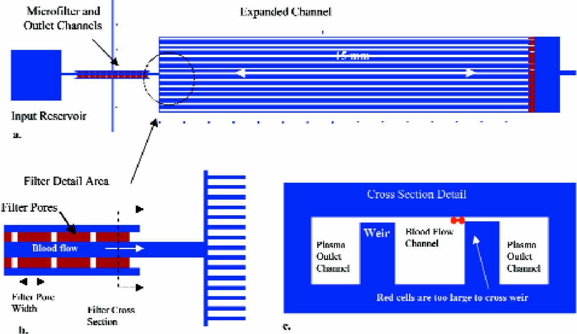
Microfilter device design and detail: a) top view of generic device design with narrow and expanded channels, b) filter detail area showing filter pores and expanded channel layout, c) weir style filter cross section.
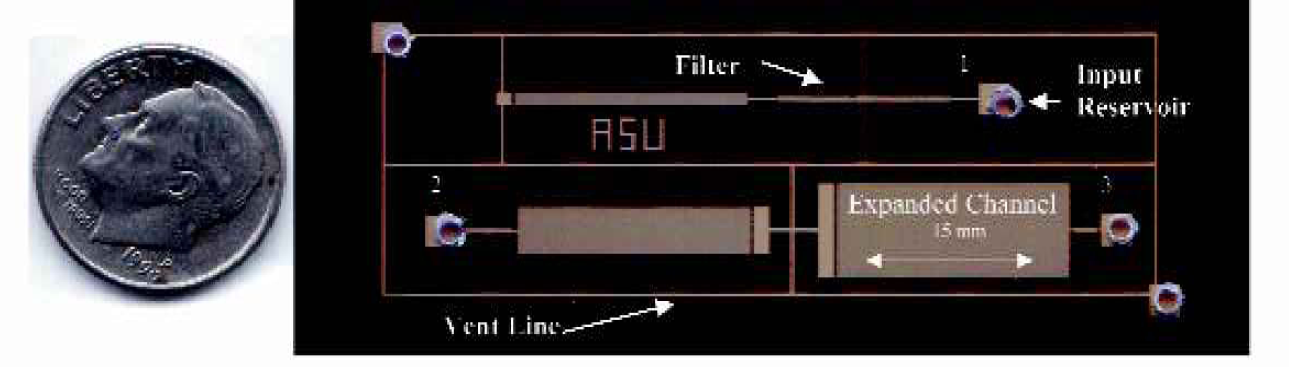
Optical micrograph of shear rate test devices integrated into a single test cell. 1) 15mm long main channel, 2) 4 mm long main channel, 3) 2mm long main channel.
On chip blood filtration tests were performed at room temperature using citrated whole bovine blood run at two levels of hematocrit, 19 and 40%. Device operation was digitally recorded using a stereoscope, CCD camera, and a digital video recorder. Digital video analysis of device operation was utilized to determine the blood flow rate in the expanded channel, device operation time, and volume of plasma extracted. Average velocity, and wall shear rate in the filtration channel were computed using flow rate data from the digital video analysis.
Results
Eighteen microfilter blood fractionation devices using the on-chip design footprint depicted in Figure 2, operated solely by passive capillary forces, provided plasma volumes between 10 and 50 nl without significant hemolysis (Figure 3). Plasma filter flux, Jf, was found to be a strong function of wall shear rate, γw in the filtration channel, but was not significantly influenced by blood hematocrit, H%, as verified by the simple regression model shown in Figure 4a.
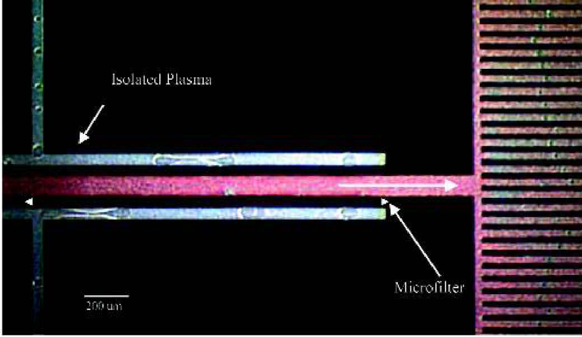
Optical micrograph of passive microfilter device showing plasma isolated from whole bovine blood.

(a) Plasma flux dependence on wall shear rate for two levels of blood hematocrit (H%). Each point represents an average of three devices tested. Shear rate was varied by altering the length of the filtration channel (L). Trend line is a power law fit to all data, R2 = 0.964. (b) Idealized depiction of blood separation concept with microporous membrane.
It was also determined that the pseudo-steady state microfiltration operation time increased ∼50 fold over single channel designs from ∼1–2 seconds to 30–100 seconds by the incorporation of the downstream expansion channel. This feature essentially ‘amplifies’ the capillary driving force, which effectively extends the operation time. From an investigational viewpoint, the capillary expander permits experimental evaluation at practical time scales and quasi steady-state conditions. From a more practical on-chip perspective, it is envisioned to provide important operational capabilities, e.g. improved fractionation capability of which there is much practical utility for laβ-on-a-chip applications.
Discussion
Previous studies of blood fractionation using large-scale microfiltration membrane technology (plasmapheresis) demonstrated that plasma filter flux is a function of transmembrane pressure, wall shear rate, and hematocrit. 5,6 In membrane plasmapheresis applications, the filtration flux models are based on a steady-state, convective-diffusive mass transfer balance between cellular (mainly red blood cells) accumulation at the filter wall due to bulk plasma flow and cellular migration from the filter due to shear enhanced “effective diffusion” of cells, Figure 4b. These earlier plasmapheresis mass transfer models demonstrated a similar power law relationship between flux and shear rate, Equation 1 in Figure 4a, as that obtained in this study using on-chip microfilter devices. However, unlike the earlier plasmapheresis models, our ‘micro-plasmapheresis’ model was found to be insensitive to hematocrit. This suggests the possibility of small length scale effects that, to date, are not fully appreciated, and warrant further investigation.
Conclusions
This study demonstrates that it is possible to perform on-chip selective bioseparation of complex fluids, such as blood. Miniaturized microfilter devices operating totally by passive capillary forces can be designed for flexible integration with numerous laβ-on-a-chip technologies currently under development. A novel microfluidic design layout has been shown to extend the microfiltration operating time of passively driven microdevice systems from a few seconds to several minutes. Although the proposed on-chip microfilter device and operational features have many similarities with the larger-scale microporous membrane systems, apparent differences in device performance exist which warrant closer inspection.
Acknowledgments
This research was funded, in part, by the ASASU Graduate Research Support Program, the NSF Research Training Grant in Optical Bimolecular Devices (#9602258), and the ASU Multidisciplinary Initiative on Engineering Novel Biomaterials, Biointerfaces, and Small-Scale Biohybrid Devices. Microdevice fabrication performed in the ASU Center for Solid State Electronics Research (CSSER), and evaluation performed in the Goldwater Materials Visualization Facility, and the ASU Bioengineering Laboratories is gratefully acknowledged.